北京大学学报(医学版) ›› 2020, Vol. 52 ›› Issue (1): 129-137. doi: 10.19723/j.issn.1671-167X.2020.01.021

• 论著 • 上一篇    下一篇

三款口内三维扫描仪获取无牙颌红膏初印模精度的对比评价

曹悦,陈俊锴,邓珂慧,王勇,孙玉春(),赵一姣()   

  1. 北京大学口腔医学院·口腔医院,口腔医学数字化研究中心,口腔修复教研室 口腔数字化医疗技术和材料国家工程实验室 卫生部口腔医学计算机应用工程技术研究中心 口腔数字医学北京市重点实验室 国家口腔疾病临床医学研究中心,北京 100081
  • 收稿日期:2019-10-08 出版日期:2020-02-18 发布日期:2020-02-20
  • 通讯作者: 孙玉春,赵一姣 E-mail:kqsyc@bjmu.edu.cn;kqcadcs@bjmu.edu.cn
  • 基金资助:
    国家重点研发计划(2018YFB1106900);首都卫生发展科研专项(首发)(2018-2-41031);北京大学口腔医学院新技术新疗法(PKUSSNCT-18G01);教育部联合基金(6141A020226XX);宁夏自治区重点研发计划重点项目课题(2018BEG02012)

Accuracy of three intraoral scans for primary impressions of edentulous jaws

Yue CAO,Jun-kai CHEN,Ke-hui DENG,Yong WANG,Yu-chun SUN(),Yi-jiao ZHAO()   

  1. Center of Digital Dentistry, Faculty of Prosthodontics, Peking University School and Hospital of Stomatology & National Engineering Laboratory for Digital and Material Technology of Stomatology & Research Center of Engineering and Technology for Digital Dentistry of Ministry of Health & Beijing Key Laboratory of Digital Stomatology & National Clinical Research Center for Oral Diseases, Beijing 100081, China
  • Received:2019-10-08 Online:2020-02-18 Published:2020-02-20
  • Contact: Yu-chun SUN,Yi-jiao ZHAO E-mail:kqsyc@bjmu.edu.cn;kqcadcs@bjmu.edu.cn
  • Supported by:
    Supported by the National Key R&D Program of China(2018YFB1106900);Capital’s Funds for Health Improvement and Research(2018-2-41031);Program for New Clinical Techniques and Therapies of Peking University School and Hospital of Stomatology(PKUSSNCT-18G01);Joint Fund of Ministry of Education of China for Equipment(6141A020226XX);Key R&D Program of Ningxia Hui Autonomous Region(2018BEG02012)

摘要:

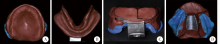
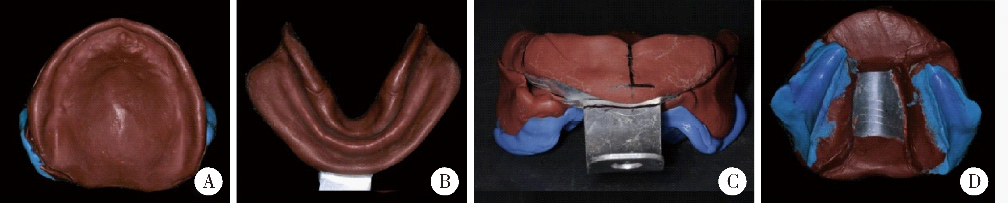
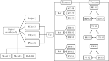
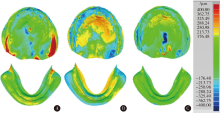
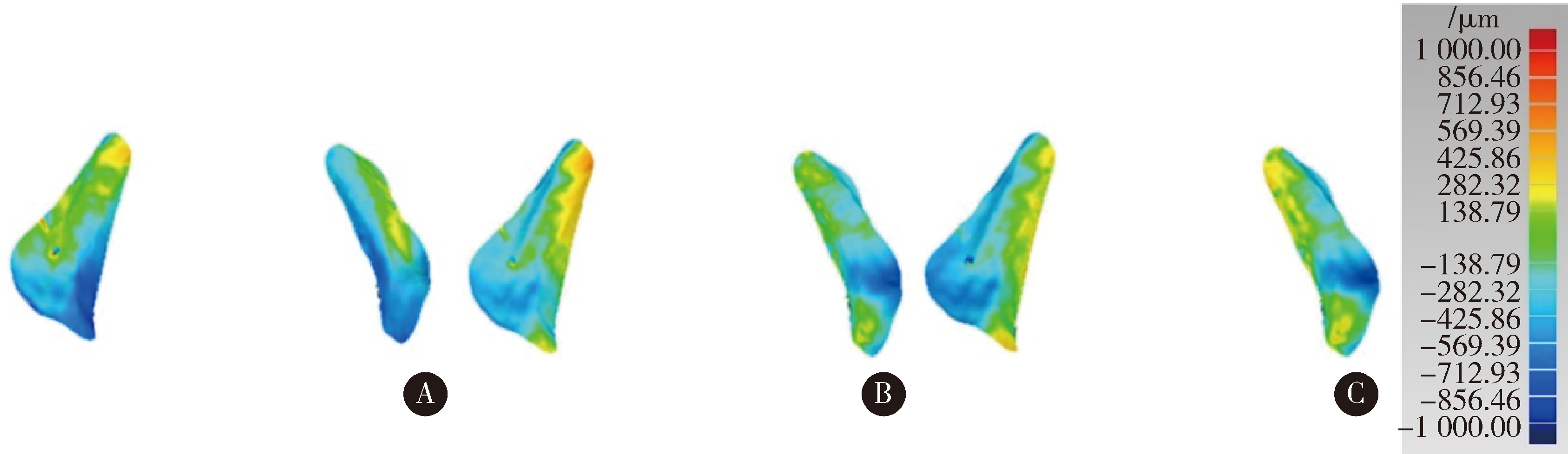
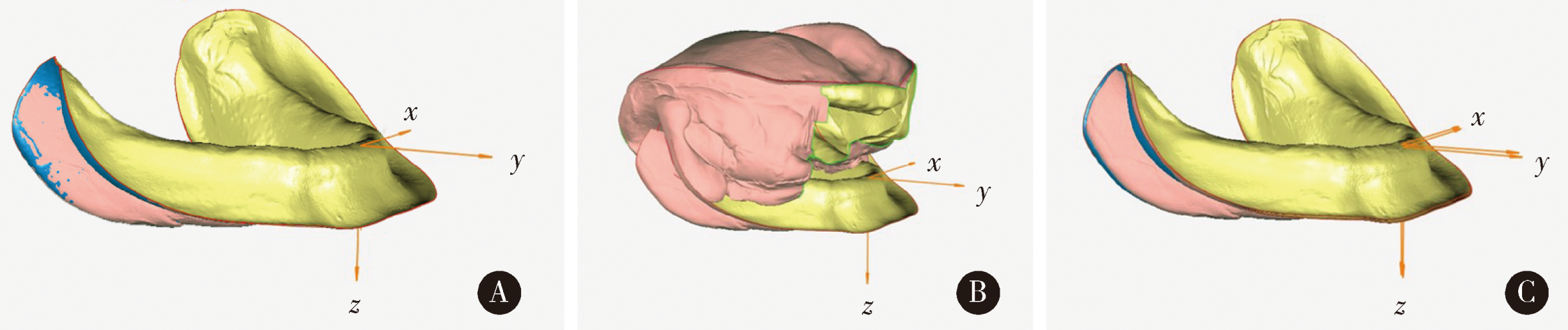
目的:分析比较了三款口内三维扫描仪扫描无牙颌红膏初印模及颌位记录的精度,为口内三维扫描仪应用于扫描无牙颌红膏初印模及颌位记录、制作临床诊断义齿提供参考。方法:共纳入无牙颌患者红膏初印模6副,每副印模包含上颌初印模、下颌初印模和上颌背面的颌位记录。使用Dentscan Y500牙颌模型三维扫描仪扫描红膏初印模获取三角网格数据(stereolithography,STL)格式数据作为参考模型。分别使用i500,Trios 3和CEREC Primescan三款口内三维扫描仪扫描红膏初印模获取STL数据,重复3次作为实验组。在Geomagic Studio 2013软件中,将口内三维扫描仪扫描获得的数据与参考模型比较评价正确度,同一扫描仪三次扫描的数据互相比较评价精密度。将口内三维扫描仪扫描的上颌数据与参考模型上颌数据配准,评价颌位记录扫描的形态误差。将口内三维扫描仪扫描的下颌与颌位记录配准,评价下颌颌位的偏差。采用SPSS 20.0统计软件,对三款口内三维扫描仪的正确度、精密度和颌位记录的形态误差分别进行独立样本t检验和Mann-Whitney U非参数检验。采用Bland-Altman图示法对三款口内三维扫描仪测量的下颌颌位的一致性进行两两评价,判断其在垂直方向、前后方向和左右方向偏移量的差异。结果: i500、Trios 3和CEREC Primescan三款扫描仪的正确度分别为:上颌(182.34±101.21) μm,(145.21±71.73) μm,(78.34±34.79) μm;下颌(106.42±21.63) μm,95.08(63.08) μm,(78.45±42.77) μm。扫描上、下颌时三款扫描仪的正确度差异无统计学意义(P>0.05)。三款扫描仪的精密度分别为:上颌147.65(156.30) μm,(147.54±83.33) μm,40.30(32.80) μm;下颌(90.96±30.77) μm,(53.73±23.56) μm,37.60(93.93) μm。扫描上颌时CEREC Primescan扫描仪精密度显著优于另两款扫描仪(P<0.05),扫描下颌时Trios 3和CEREC Primescan 扫描仪的精密度显著优于i500扫描仪(P<0.05),i500和Trios 3扫描仪扫描下颌的精密度优于上颌(P<0.05)。三款扫描仪扫描上、下颌初印模的正确度和精密度的95%置信区间的上、下限均在±300 μm范围内。扫描颌位记录的形态误差,i500扫描仪(337.68±128.54) μm,Trios 3扫描仪(342.89±195.41) μm,CEREC Primescan扫描仪(168.62±88.35) μm,i500扫描仪和Trios 3扫描仪的95%置信区间上限大于300 μm,CEREC Primescan扫描仪显著优于i500扫描仪(P<0.05)。扫描测量的下颌颌位,垂直差异:i500扫描仪(0.83±0.56) mm,Trios 3扫描仪(0.80±0.45) mm,CEREC Primescan扫描仪(0.91±0.75) mm;水平前后差异:i500扫描仪(0.79±0.58) mm,Trios 3扫描仪(0.62±0.18) mm,CEREC Primescan扫描仪(0.53±0.53) mm;水平左右差异:i500扫描仪(0.95±0.59) mm,Trios 3扫描仪(0.69±0.45) mm,CEREC Primescan扫描仪(0.60±0.22) mm。Bland-Altman图示法三款扫描仪扫描获得的下颌颌位在垂直方向、前后方向和左右方向的偏移量两两之间比较均位于95%一致性界限内。结论:三款口内三维扫描仪扫描上、下颌红膏初印模表现出较好的正确度和精密度, i500和Trios 3扫描仪记录的颌位记录的误差较大,但仅作为初始颌位记录使用,提示临床医生可以使用这三种口内三维扫描仪应用于数字化全口义齿前期数据获取来制作诊断义齿和个别托盘,减少将红膏初印模长途寄运至技工室导致的红膏碰撞破损及高温变形。

关键词: 口内扫描, 无牙颌, 数字印模, 精度

Abstract:

Objective: To provide a reference for using intraoral scanners for making clinical diagnostic dentures of edentulous jaws by comparing the accuracy of three intraoral scanners for primary impression and jaw relation record of edentulous jaws. Methods: This study contained 6 primary impressions of the edentulous patients. Each of the impressions consisted of the maxillary primary impression, the mandibular primary impression and the jaw relation record. For each of them, a dental cast scanner (Dentscan Y500) was used to obtain stereolithography(STL)data as reference scan, and then three intraoral scanners including i500, Trios 3 and CEREC Primescan were used for three times to obtain STL data as experiment groups. In Geomagic Studio 2013 software, trueness was obtained by comparing experiment groups with the reference scan, and the precision was obtained from intragroup comparisons. Registered maxillary data of the intraoral scan with reference scan, the morphological error of jaw relation record was obtained by comparing jaw relation record of the intraoral scan with the reference scan. Registered mandibular data with jaw relation record of intraoral scan and the displacement of the jaw position were evaluated. Independent samples t test and Mann-Whitney U test in the SPSS 20.0 statistical software were used to statistically analyze the trueness, precision and morphological error of jaw relation record of three intraoral scanners. The Bland-Altman diagram was used to evaluate the consistency of the jaw relationship measured by the three intraoral scanners. Results: The trueness of i500, Trios 3 and CEREC Primescan scanners was (182.34±101.21) μm, (145.21±71.73) μm, and (78.34±34.79) μm for maxilla; (106.42±21.63) μm, and 95.08 (63.08) μm, (78.45±42.77) μm for mandible. There was no significant difference in trueness of the three scanners when scanning the maxilla and mandible(P>0.05). The precision of the three scanners was 147.65 (156.30) μm, (147.54±83.33) μm, and 40.30 (32.80) μm for maxilla; (90.96±30.77) μm, (53.73±23.56) μm, and 37.60 (93.93) μm for mandible. The precision of CEREC Primescan scanner was significantly better than that of the other two scanners for maxilla(P<0.05). Trios 3 and CEREC Primescan scanners were significantly better than i500 scanner for mandible(P<0.05). The precision of the i500 and Trios 3 scanners for mandible was superior to maxilla(P<0.05). The upper limit of 95% confidence intervals of trueness and precision of three scanners for both maxilla and mandible were within ±300 μm which was clinically accepted. The morphological error of jaw relation record of the three scanners was (337.68±128.54) μm, (342.89±195.41) μm, and (168.62±88.35) μm. The 95% confidence intervals of i500 and Trios 3 scanners were over 300 μm. CEREC Primescan scanner was significantly superior to i500 scanner(P<0.05).The displacement of the jaw position of the three scanners was (0.83±0.56) mm, (0.80±0.45) mm, and (0.91±0.75) mm for vertical dimension; (0.79±0.58) mm, (0.62±0.18) mm, and (0.53±0.53) mm for anterior and posterior directions; (0.95±0.59) mm, (0.69±0.45) mm, and (0.60±0.22) mm for left and right directions. The displacement of the jaw position of the three scanners in vertical dimension, anterior and posterior directions and the left and right directions were within the 95% consistency limit. Conclusion: Three intraoral scanners showed good trueness and precision. The i500 and Trios 3 scanners had more errors in jaw relation record, but they were used as primary jaw relation record. It is suggested that three intraoral scanners can be used for obtaining digital data to make diagnostic dentures and individual trays, reducing possible deforming or crack when sending impressions from clinic to laboratory.

Key words: Intraoral scanner, Edentulous jaws, Digital impression, Accuracy

中图分类号: 

  • R783.6

图1

上、下颌红膏初印模及初始颌位记录"

表1

三款口内三维扫描仪及牙颌模型三维扫描仪的参数"

System Manufacture Software Country Acquisition technology
i500 MEDIT 2.0.3 Korea Triangulation
Trios 3 3 SHAPE 1.4.7.5 Denmark Confocal measurement
CEREC Primescan Sirona 5.0.1.16682 Germany Triangulation
Dentscan Y500 Nanjing Geosmart 3D China Blue grating

图2

使用DentscanY500模型三维扫描仪和三款口内三维扫描仪扫描上颌初印模(Model U)、背面咬合记录(Model O)和下颌初印模(Model L)的流程图,实验数据(Exp)与参考数据(Ref)进行比较检验正确度,同一扫描仪三次扫描模型(定义为S1、S2、S3)两两比较检验精密度"

图3

Model U 和 Model L 模型的3D偏差分析色阶图"

图4

Model O模型的 3D偏差分析色阶图"

图5

在Model L Ref上建立坐标系Ref"

图6

下颌颌位偏差的分析"

表2

三款扫描仪的正确度"

Items Model U Model L
MD TR PS MD TR PS
Trueness/μm,
x?±s/Median (IQR)
182.34±101.21 145.21±71.73 78.34±34.79 106.42±21.63 95.08(63.08) 78.45±42.77
95%CI (76.13,288.55) (69.94,220.49) (41.83,114.86) (83.72,129.12) (55.28,175.36) (33.56,123.34)

表3

正确度的统计学测量值"

Items Model U Model L Model U versus Model L
MD-TRa TR-PSa MD-PSa MD-TRb TR-PSb MD-PSa MDa TRb PSa
Difference value 0.733 2.055 2.380 0.320 1.363 1.430 1.793 0.801 0.005
P value 0.480 0.067 0.054 0.749 0.173 0.183 0.127 0.423 0.996

表4

三款扫描仪的精密度"

Items Model U Model L
MD TR PS MD TR PS
Precision/(μm),
x?±s/Median (IQR)
147.65(156.30) 147.54±83.33 40.30(32.80) 90.69±30.77 53.73±23.56 37.60(93.93)
95%CI (120.90,243.77) (106.11,188.98) (57.39,105.99) (75.39,105.99) (42.01,65.44) (37.28,94.57)

表5

精密度的统计学测量值"

Items Model U Model L Model U versus Model L
MD-TRb TR-PSb MD-PSb MD-TRa TR-PSb MD-PSb MDb TRa PSb
Difference value 0.601 4.176 4.651 4.046 1.155 2.247 2.816 4.597 0.174
P value 0.548 0.000* 0.000* 0.000* 0.248 0.025* 0.005* 0.000* 0.862

表6

三款扫描仪的颌位记录的形态误差"

Items MD TR PS
Morphological error of jaw relation record/μm, x?±s 337.68±128.54 342.89±195.41 168.62±88.35
95%CI (202.78,472.57) (137.82,547.96) (75.90,261.34)

表7

颌位记录形态误差的统计学测量值"

Items MD-TR TR-PS MD-PS
Difference value 0.055 1.991 2.655
P value 0.958 0.075 0.024*

表8

下颌颌位的偏差"

Items Group z y x
Displacement of jaw relationship/mm, x?±s MD 0.83±0.56 0.79±0.58 0.95±0.59
TR 0.80±0.45 0.62±0.18 0.69±0.45
PS 0.91±0.75 0.53±0.53 0.60±0.22
95%CI MD (0.24,1.41) (0.18,1.39) (0.33,1.57)
TR (0.34,1.27) (0.44,0.81) (0.22,1.16)
PS (0.38,1.69) (-0.30,1.09) (0.38,0.83)

图7

下颌颌位偏差的Bland-Altman图示"

[1] Baba NZ, Alrumaih HS, Goodacre BJ , et al. Current techniques in CAD/CAM denture fabrication[J]. Gen Dent, 2016,64(6):23.
[2] 孙玉春, 孙儒, 邓珂慧 , 等. 全口义齿数字化修复技术的研发和应用进展[J]. 中华口腔医学杂志, 2018,53(1):60-65.
[3] Deng KH, Wang Y, Zhou YS , et al. Functionally suitable digital removable complete dentures: A dental technique[J]. J Prosthet Dent, 2019, 10(2019-10-04)[2019-10-06].
[4] Patzelt SB, Vonau S, Stampf S , et al. Assessing the feasibility and accuracy of digitizing edentulous jaws[J]. J Am Dent Assoc, 2013,144(8):914-920.
[5] Osnes CA, Wu JH, Venezia P , et al. Full arch precision of six intraoral scanners in vitro[J]. J Prosthodont Res, 2019, 6 ( 2019 -06-18)[2019-08-08]. .
[6] Goodacre BJ, Goodacre CJ, Baba NZ . Using intraoral scanning to capture complete denture impressions, tooth positions, and centric relation records[J]. Int J Prosthodont, 2018,31(4):377-381.
[7] Güth JF, Runkel C, Beuer F , et al. Accuracy of five intraoral scanners compared to indirect digitalization[J]. Clin Oral Investig, 2017,21(5):1445-1455.
[8] Nedelcu RG, Persson AS . Scanning accuracy and precision in 4intraoral scanners: An in vitro comparison based on 3-dimensional analysis[J]. J Prosthet Dent, 2014,112(6):1461-1471.
[9] 徐明明, 刘峰 . CAD/CAM技术在口腔修复中的应用, 数字印模技术[J]. 中国实用口腔科杂志, 2013,6(6):321-326.
[10] Chia VA, Esguerra RJ, Teoh KH , et al. In vitro three-dimen-sional accuracy of digital implant impressions: The effect of implant angulation[J]. Int J Oral Maxillofac Implants, 2017,32(2):313-321.
[11] Lee SJ, Betensky RA, Gianneschi GE , et al. Accuracy of digital versus conventional implant impressions[J]. Clin Oral Implants Res, 2015,26(6):715-719.
[12] Gan N, Xiong YY, Jiao T . Accuracy of intraoral digital impressions for whole upper jaws, including full dentitions and palatal soft tissues[J]. PLoS One, 2016,11(7):e0158800.
[13] Kattadiyil MT, Mursic Z, Alrumaih H , et al. Intraoral scanning of hard and soft tissues for partial removable dental prosjournal fabrication[J]. J Prosthet Dent, 2014,112(3):444-448.
[14] Goodacre BJ, Goodacre CJ . Using intraoral scanning to fabricate complete dentures: First experiences[J]. Int J Prosthodont, 2018,31(2):166-170.
[15] Lo Russo L, Caradonna G, Troiano G , et al. Three-dimensional differences between intraoral scans and conventional impressions of edentulous jaws: A clinical study[J]. J Prosthet Dent, 2019, 5 (2019-05-29)[2019-09-10]. .
[16] Zimmermann M, Mehl A, Mörmann WH , et al. Intraoral scanning systems:A current overview[J]. Int J Comput Dent, 2015,18(2):101-129.
[17] Renne W, Ludlow M, Fryml J , et al. Evaluation of the accuracy of 7 digital scanners: An in vitro analysis based on 3-dimensional comparisons[J]. J Prosthet Dent, 2016,118(1):36-42.
[18] 张成藩, 欧阳官, 冯海兰 , 等. 总义齿颌位探讨[J]. 华西口腔医学杂志, 1988,6(2):88-91.
[19] 刘建彰, 徐军 . 不同垂直距离下肌力闭合道终点位与正中关系位的关系[J]. 北京大学学报 (医学版), 2010,42(1):56-59.
[20] Li WW, Xie QF, Wang Y , et al. A pilot study of digital recording of edentulous jaw relations using a handheld scanner and specially designed headgear[J]. Sci Rep, 2018,8(1):8975.
[1] 国丹妮,潘韶霞,衡墨笛,屈健,魏秀霞,周永胜. 应用于无牙颌种植修复设计的三维面部扫描配准方法的对比[J]. 北京大学学报(医学版), 2021, 53(1): 83-87.
[2] 姜楠,包旭东,岳林. 全冠预备体终止线局部扫描正确度对整体的影响[J]. 北京大学学报(医学版), 2021, 53(1): 102-108.
[3] 徐迪,魏冬豪,张亚池,邸萍,林野. 以苄索氯胺和异丙醇为主要有效成分的消毒剂对牙科印模精度的影响[J]. 北京大学学报(医学版), 2020, 52(6): 1112-1116.
[4] 罗佳,张宇,崔宏燕,祝宁,沈惠丹,邸萍,林野. 锥度固位结合数字化技术在后牙连续多牙种植即刻修复中的应用[J]. 北京大学学报(医学版), 2020, 52(5): 964-970.
[5] 孙玉春,王勇,邓珂慧,陈虎,李伟伟,赵一姣,潘韶霞,叶红强,周永胜. 功能易适数字化全口义齿的自主创新研发[J]. 北京大学学报(医学版), 2020, 52(2): 390-394.
[6] 游浪,邓珂慧,李伟伟,赵一姣,孙玉春,周永胜. 无牙颌患者鼻唇角变化侧面观的视觉敏感阈值[J]. 北京大学学报(医学版), 2020, 52(1): 107-112.
[7] 刘潇倩,陈秋雯,冯海兰,王兵,屈健,孙振,衡墨迪,潘韶霞. 无牙颌患者locator附着体种植覆盖义齿修复后口腔卫生维护的纵向研究[J]. 北京大学学报(医学版), 2019, 51(1): 136-144.
[8] 赵一姣,熊玉雪,杨慧芳,王勇. 3种不同原理颜面部扫描仪测量精度的评价[J]. 北京大学学报(医学版), 2014, 46(1): 76-80.
[9] 宋杨, 孙玉春, 赵一姣, 王勇, 吕培军. 牙颌模型三维扫描仪精度定量评价[J]. 北京大学学报(医学版), 2013, 45(1): 140-144.
Viewed
Full text


Abstract

Cited

  Shared   
  Discussed   
[1] 张三. 中文标题测试[J]. 北京大学学报(医学版), 2010, 42(1): 1 -10 .
[2] 赵磊, 王天龙 . 右心室舒张末期容量监测用于肝移植术中容量管理的临床研究[J]. 北京大学学报(医学版), 2009, 41(2): 188 -191 .
[3] 万有, , 韩济生, John E. Pintar. 孤啡肽基因敲除小鼠电针镇痛作用增强[J]. 北京大学学报(医学版), 2009, 41(3): 376 -379 .
[4] 张燕, 韩志慧, 钟延丰, 王盛兰, 李玲玲, 郑丹枫. 骨骼肌活组织检查病理诊断技术的改进及应用[J]. 北京大学学报(医学版), 2009, 41(4): 459 -462 .
[5] 赵奇, 薛世华, 刘志勇, 吴凌云. 同向施压测定自酸蚀与全酸蚀粘接系统粘接强度[J]. 北京大学学报(医学版), 2010, 42(1): 82 -84 .
[6] 林红, 王玉凤, 吴野平. 学校生活技能教育对小学三年级学生行为问题影响的对照研究[J]. 北京大学学报(医学版), 2007, 39(3): 319 -322 .
[7] 丰雷, 程嘉, 王玉凤. 注意缺陷多动障碍儿童的运动协调功能[J]. 北京大学学报(医学版), 2007, 39(3): 333 -336 .
[8] 李岳玲, 钱秋瑾, 王玉凤. 儿童注意缺陷多动障碍成人期预后及其预测因素[J]. 北京大学学报(医学版), 2007, 39(3): 337 -340 .
[9] . 书讯[J]. 北京大学学报(医学版), 2007, 39(3): 225 -328 .
[10] 牟向东, 王广发, 刁小莉, 阙呈立. 肺黏膜相关淋巴组织型边缘区B细胞淋巴瘤一例[J]. 北京大学学报(医学版), 2007, 39(4): 346 -350 .