北京大学学报(医学版) ›› 2023, Vol. 55 ›› Issue (4): 708-715. doi: 10.19723/j.issn.1671-167X.2023.04.023
股前外侧皮瓣修复上颌骨缺损术后面部软组织对称性感观分级
黄莹,吴志远,周行红,蔡志刚,张杰*( )
)
- 北京大学口腔医学院·口腔医院口腔颌面外科, 国家口腔医学中心, 国家口腔疾病临床医学研究中心, 口腔生物材料和数字诊疗装备国家工程研究中心, 口腔数字医学北京市重点实验室, 国家卫生健康委员会口腔医学计算机应用工程技术研究中心, 国家药品监督管理局口腔材料重点实验室, 北京 100081
Category of facial symmetry perception after maxillary reconstruction using anterolateral thigh flap
Ying HUANG,Zhi-yuan WU,Xing-hong ZHOU,Zhi-gang CAI,Jie ZHANG*()
- Department of Oral and Maxillofacial Surgery, Peking University School and Hospital of Stomatology & National Center for Stomatology & National Clinical Research Center for Oral Diseases & National Engineering Research Center of Oral Biomaterials and Digital Medical Devices & Beijing Key Laboratory of Digital Stomatology & NHC Research Center of Engineering and Technology for Computerized Dentistry & NMPA Key Laboratory for Dental Materials, Beijing 100081, China
摘要:
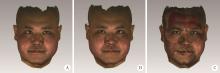
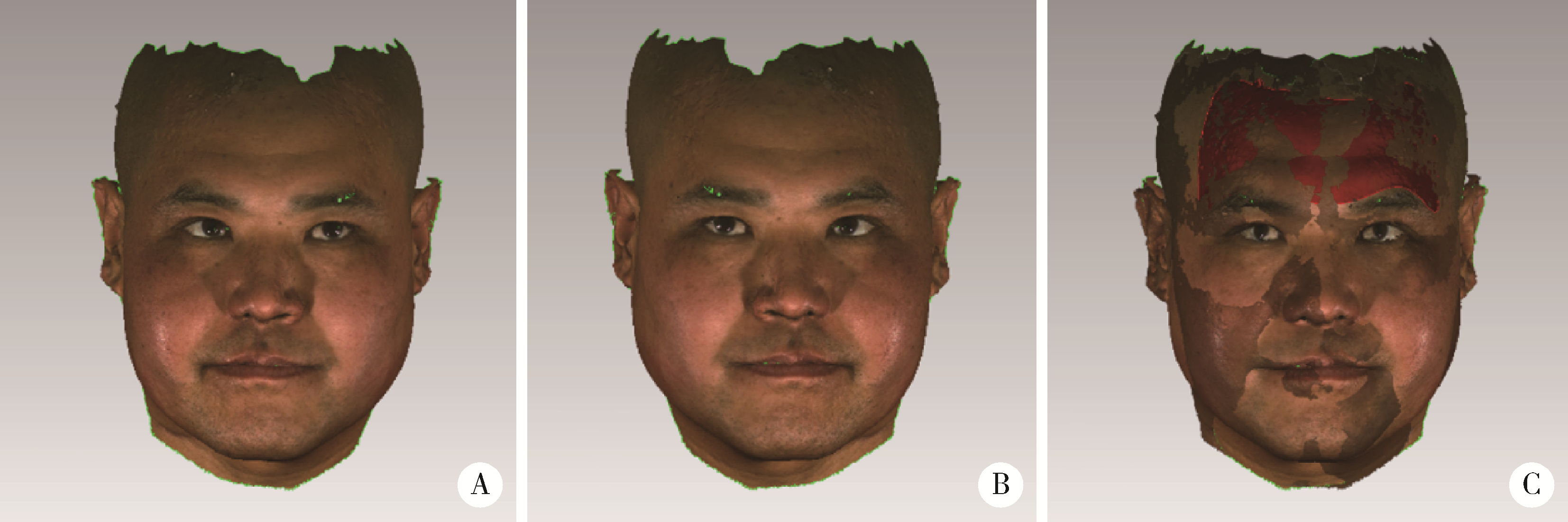
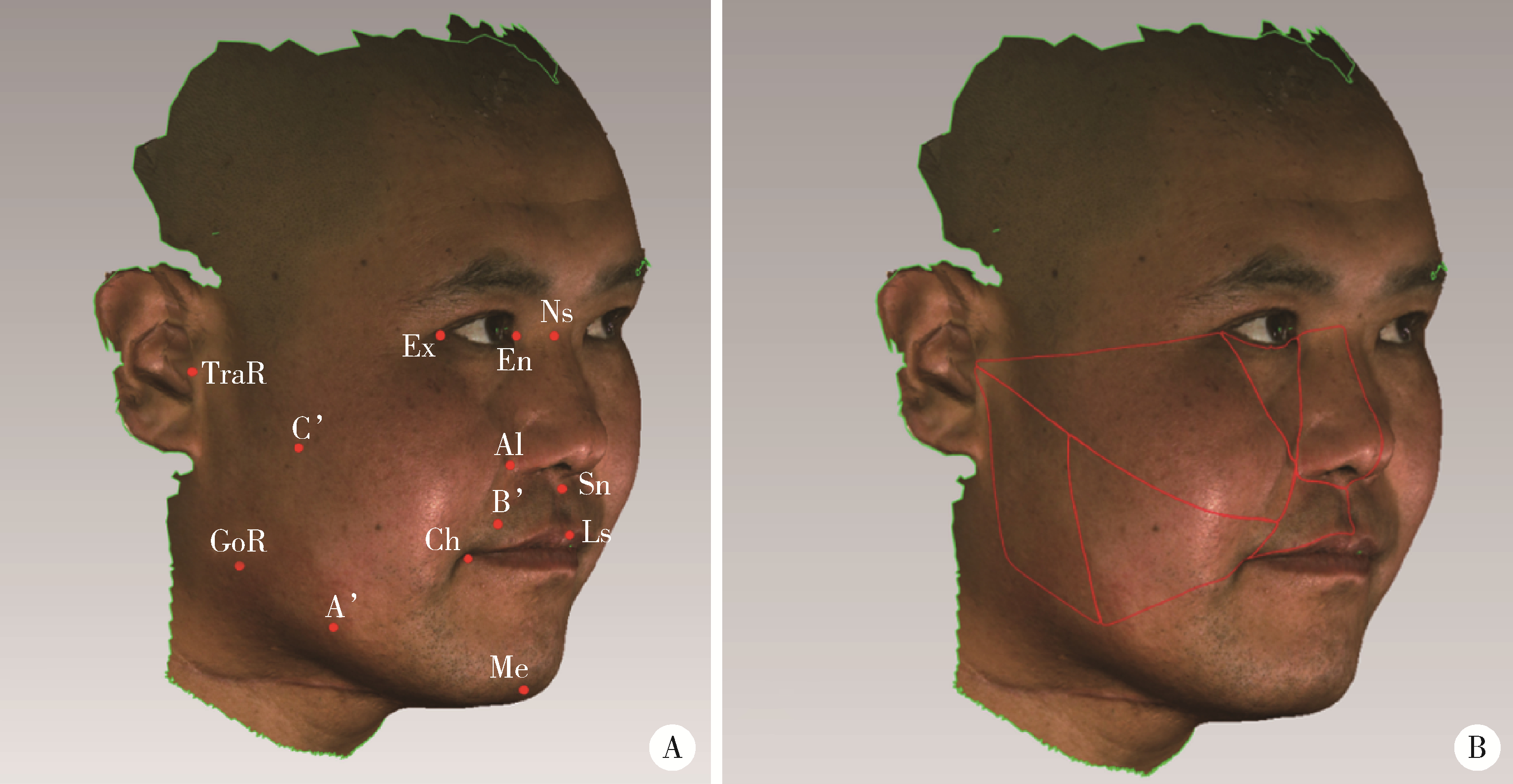
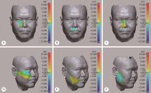
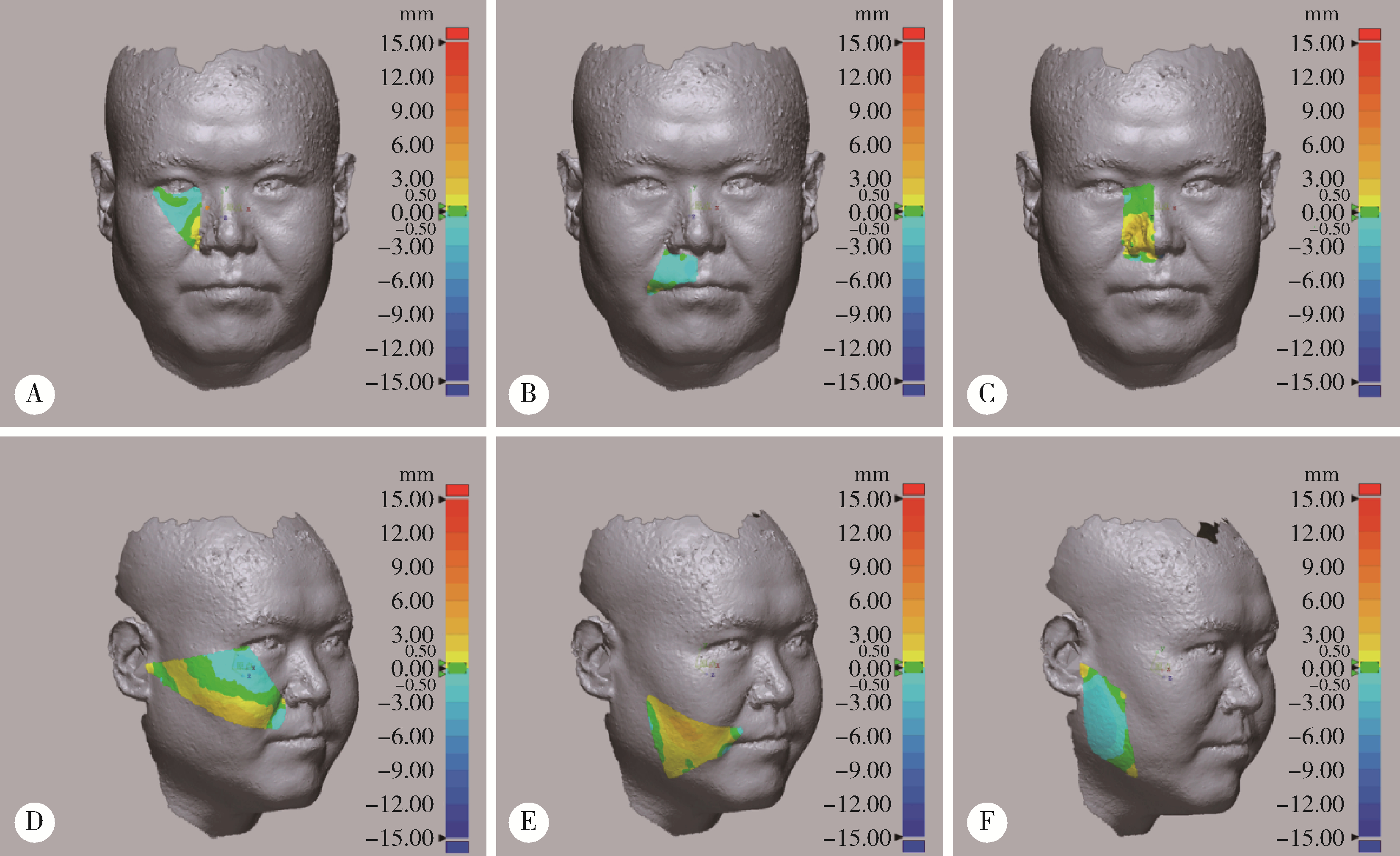
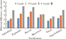
目的: 采用三维立体摄影测量与主观评价相结合的方法初步建立股前外侧皮瓣修复上颌骨缺损术后面部软组织对称性的感观分级系统。方法: 患者上颌骨因肿瘤侵犯而被行不同范围的切除, 同期行股前外侧皮瓣(anterolateral thigh flap, ALTF)修复。术后采用三维立体摄影技术获取患者三维面相, 以任意平面为对称面创建原始三维面相的镜像, 重叠配准原始面相与镜像后, 将患侧面部分为6个区域, 采用基于表面的色谱分析法测量患侧面部软组织三维变化。同时请20名非医学专业人员作为评价者对患者的三维面相进行主观评价, 并在Likert 5分量表中进行评分, 根据主观评分将面部软组织的对称性进行分级。应用SPSS 24.0统计软件对三维立体摄影测量结果与主观评价分级进行统计学分析。结果: 共有44例患者符合入组条件, 其中男性21例, 女性23例, 年龄19~79岁。按照主观评价得分将所有患者面部软组织对称性分为Ⅰ、Ⅱ、Ⅲ级。Ⅰ级患者面部基本对称, Ⅱ级患者面部略不对称, Ⅲ级患者面部明显不对称。Ⅰ、Ⅱ、Ⅲ级患者在眶下区及颧区的差异均有统计学意义(P < 0.05), Ⅰ级与Ⅲ级患者相比, 除眶下区与颧区外, 颊区与上唇区的差异也具有统计学意义(P < 0.05)。上颌骨缺损范围越大, ALTF修复后的患者面部对称性越差。结论: 不同范围上颌骨切除后经过单纯ALTF修复的患者其面部软组织依然会存在不同程度的不对称, 所引起的感观不适可分为三个等级, 等级越高表明面部软组织对称性越差; 眶下区与颧区是影响面部对称性感知的重要区域, 其次为颊区与上唇区, 临床医师在进行上颌骨缺损修复尤其是累及眶底的大范围缺损修复时, 应当重视这些区域的软组织支撑。
中图分类号:
- R782.4

| 1 |
Piazza C , Paderno A , Del Bon F , et al. Palato-maxillary reconstruction by the angular branch-based tip of scapula free flap[J]. Eur Arch Otorhinolaryngol, 2017, 274 (2): 939- 945.
doi: 10.1007/s00405-016-4266-0 |
| 2 | 于森, 王洋, 毛驰, 等. 1107例上颌骨缺损的临床分类及修复方法分析[J]. 北京大学学报(医学版), 2015, 47 (3): 509- 513. |
| 3 |
Bianchi B , Ferri A , Ferrari S , et al. The free anterolateral thigh musculocutaneous flap for head and neck reconstruction: One surgeon's experience in 92 cases[J]. Microsurgery, 2012, 32 (2): 87- 95.
doi: 10.1002/micr.20952 |
| 4 | Lavadera P , Schultz J , Sinkin J . Orbital floor and maxillary reconstruction with titanium mesh and anterolateral thigh free flap[J]. Eplasty, 2019, 19 (7): 126- 130. |
| 5 | Eitezaz FA , Rashid M , Yousaf S , et al. Can the anterolateral thigh flap replace the rectus abdominis free flap in the reconstruction of complex maxillary defects?[J]. J Ayub Med Coll Abbottabad, 2018, 30 (1): 74. |
| 6 | 刘志荣, 彭歆, 章文博. 游离腓骨瓣和股前外侧穿支皮瓣修复单侧上颌骨缺损患者的生存质量问卷调查研究[J]. 中华整形外科杂志, 2018, 34 (8): 644- 647. |
| 7 |
Springer IN , Wannicke B , Warnke PH , et al. Facial attractiveness: Visual impact of symmetry increases significantly towards the midline[J]. Ann Plast Surg, 2007, 59 (2): 156- 162.
doi: 10.1097/01.sap.0000252041.66540.ec |
| 8 | Berssenbrugge P , Lingemann-Koch M , Abeler A , et al. Mea-suring facial symmetry: A perception-based approach using 3D shape and color[J]. Biomed Tech (Berl), 2015, 60 (1): 39- 47. |
| 9 |
Philipp MM , Angelika SE , Ute B , et al. Three-dimensional perception of facial asymmetry[J]. Eur J Orthod, 2011, 33 (6): 647- 653.
doi: 10.1093/ejo/cjq146 |
| 10 | Patel A , Islam SM , Murray K , et al. Facial asymmetry assessment in adults using three-dimensional surface imaging[J]. Prog Orthod, 2015, 16 (10): 36- 44. |
| 11 |
Hallac RR , Feng J , Kane AA , et al. Dynamic facial asymmetry in patients with repaired cleft lip using 4D imaging (video stereophotogrammetry)[J]. J Craniomaxillofac Surg, 2017, 45 (1): 8- 12.
doi: 10.1016/j.jcms.2016.11.005 |
| 12 |
Cassi D , Battistoni G , Magnifico M , et al. Three-dimensional evaluation of facial asymmetry in patients with hemifacial microsomia using stereophotogrammetry[J]. J Craniomaxillofac Surg, 2019, 47 (1): 179- 184.
doi: 10.1016/j.jcms.2018.11.011 |
| 13 |
Brown J , Shaw RJ . Reconstruction of the maxilla and midface: Introducing a new classification[J]. Lancet Oncol, 2010, 11 (10): 1001- 1008.
doi: 10.1016/S1470-2045(10)70113-3 |
| 14 | Krzych J , Lach M , Joniec M , et al. The Likert scale is a powerful tool for quality of life assessment among patients after minimally invasive coronary surgery[J]. Kardiochir Torakochirurgia Pol, 2018, 15 (2): 130- 134. |
| 15 |
Naini FB , Donaldson A , Cobourne MT , et al. Assessing the influence of mandibular prominence on perceived attractiveness in the orthognathic patient, clinician, and layperson[J]. Eur J Orthod, 2012, 34 (6): 738- 746.
doi: 10.1093/ejo/cjr098 |
| 16 | 邢亚彬, 马爱霞. 欧洲五维健康量表EQ-5D-3L和EQ-5D-5L中文版比较的实证研究[J]. 上海医药, 2013, 25 (7): 31- 35. |
| 17 | 萧宁, 王勇, 赵一姣. 三维颜面部软组织正中矢状面确定方法的研究进展[J]. 中华口腔医学杂志, 2018, 53 (7): 495- 499. |
| 18 | Verhoeven TJ , Coppen C , Barkhuysen R , et al. Three dimen-sional evaluation of facial asymmetry after mandibular reconstruction: Validation of a new method using stereophotogrammetry[J]. Int J Oral Maxillofac Surg, 2013, 42 (1): 19- 25. |
| 19 | Schoot RA , Hol MLF , Merks JHM , et al. Facial asymmetry in head and neck rhabdomyosarcoma survivors[J]. Pediatr Blood Cancer, 2017, 64 (10): 26508. |
| 20 | 牛百平, 叶湘玉. 对面部畸形忍受程度的测量研究[J]. 中华口腔医学杂志, 1994, 29 (4): 213- 216. |
| 21 | Hohman MH , Kim SW , Heller ES , et al. Determining the threshold for asymmetry detection in facial expressions[J]. Laryngoscope, 2014, 124 (4): 860- 865. |
| 22 | Kim SW , Heller ES , Hohman MH , et al. Detection and percep-tual impact of side-to-side facial movement asymmetry[J]. JAMA Facial Plast Surg, 2013, 15 (6): 411- 416. |
| 23 | Alqattan M , Djordjevic J , Zhurov AI , et al. Comparison between landmark and surface-based three-dimensionalanalyses of facial asymmetry in adults[J]. Eur J Orthod, 2015, 37 (1): 1- 12. |
| 24 | Wang SJ , Zhang WB , Yao YM , et al. Factors Affecting volume change of anterolateral thigh flap in head and neck defect reconstruction[J]. J Oral Maxillofac Surg, 2020, 78 (11): 2090- 2098. |
| 25 | Brown J , Rogers SN , Mcnally DN , et al. A modified classification for the maxillectomy defect[J]. Head Neck, 2000, 22 (1): 17- 26. |
| 26 | 柯杰, 陈华, 林珠. 西安市正常牙合的青少年软组织X线头影测量研究: 计算机辅助测量[J]. 华西口腔医学杂志, 1994, 12 (3): 188- 191. |
| [1] | 康一帆,单小峰,张雷,蔡志刚. 游离腓骨瓣修复重建上颌骨术后腓骨瓣位置变化[J]. 北京大学学报(医学版), 2020, 52(5): 938-942. |
| [2] | 王顺吉,章文博,于尧,谢晓艳,杨宏宇,彭歆. 术前虚拟设计在股前外侧皮瓣修复口腔颌面部缺损中的应用[J]. 北京大学学报(医学版), 2020, 52(1): 119-123. |
| [3] | 章文博,于尧,王洋,刘筱菁,毛驰,郭传瑸,俞光岩,彭歆. 数字化外科技术在上颌骨缺损重建中的应用[J]. 北京大学学报(医学版), 2017, 49(1): 1-005. |
|
||
