北京大学学报(医学版) ›› 2025, Vol. 57 ›› Issue (5): 884-894. doi: 10.19723/j.issn.1671-167X.2025.05.012

• 论著 • 上一篇    下一篇

HDAC2介导H3K27ac修饰促进肝癌细胞的增殖和迁移

唐少海1, 杨宝明1, 李建坤1, 赵丽丽2, 王仪凡3, 王顺祥1,*()   

  1. 1. 河北医科大学第四医院肝胆外科, 石家庄 050000
    2. 河北医科大学药学院, 石家庄 050000
    3. 河北医科大学第四医院结直肠外科, 石家庄 050000
  • 收稿日期:2024-06-28 出版日期:2025-10-18 发布日期:2025-06-18
  • 通讯作者: 王顺祥
  • 基金资助:
    河北省自然科学基金(H2020206455); 河北省2023年度医学科学研究课题计划(20230875); 河北省2018年度医学科学研究重点课题计划(20180537); 以及2015年和2016年河北省政府资助临床医学优秀人才培养和基础课题研究项目计划(361006)

HDAC2-mediated H3K27 acetylation promotes the proliferation and migration of hepatocellular carcinoma cells

Shaohai TANG1, Baoming YANG1, Jiankun LI1, Lili ZHAO2, Yifan WANG3, Shunxiang WANG1,*()   

  1. 1. Department of Hepatobiliary Surgery, the Fourth Hospital of Hebei Medical University, Shijiazhuang 050000, China
    2. College of Pharmacy, Hebei Medical University, Shijiazhuang 050000, China;
    3. Department of Colorectal Surgery, the Fourth Hospital of Hebei Medical University, Shijiazhuang 050000, China
  • Received:2024-06-28 Online:2025-10-18 Published:2025-06-18
  • Contact: Shunxiang WANG
  • Supported by:
    the Natural Science Foundation Project of Hebei Province(H2020206455); the 2023 Annual Medical Science Research Project Plan of Hebei Province(20230875); the Key Medical Science Research Project Plan of Hebei Province in 2018(20180537); the 2015 and 2016 Hebei Provincial Government' s Plan for Funding the Cultivation of Outstanding Clinical Medicine Talents and Basic Research Projects(361006)

RICH HTML


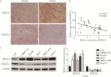
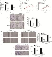
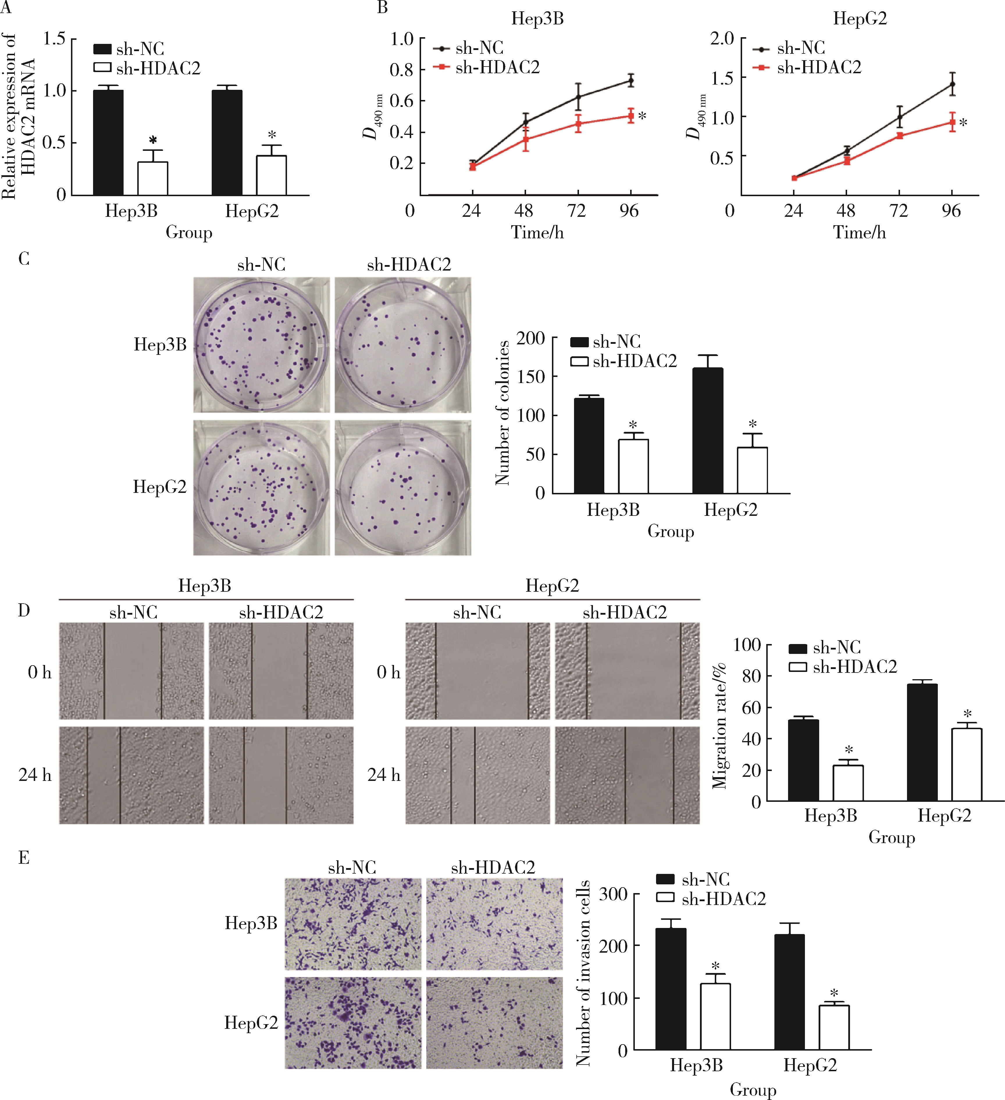
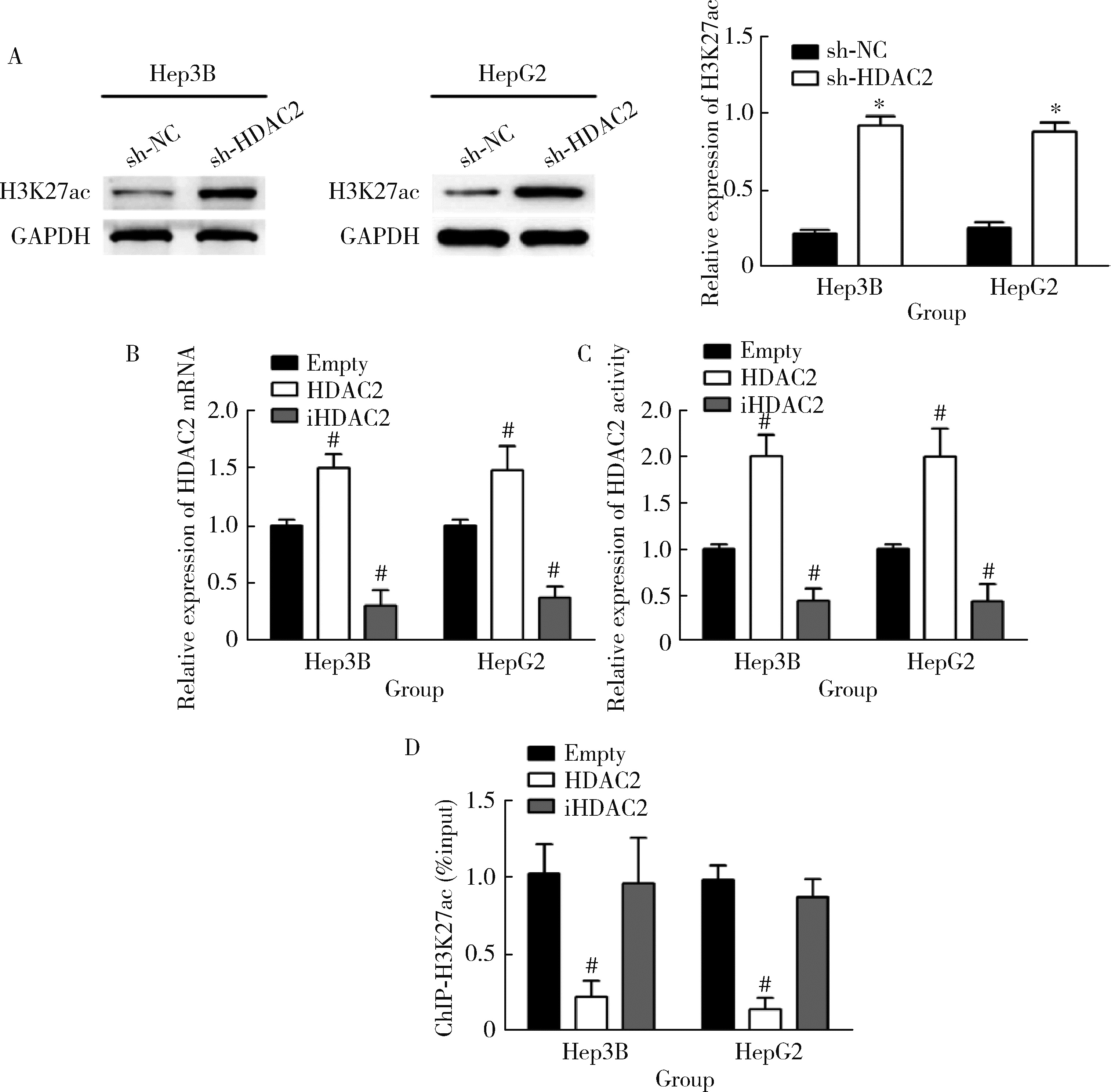
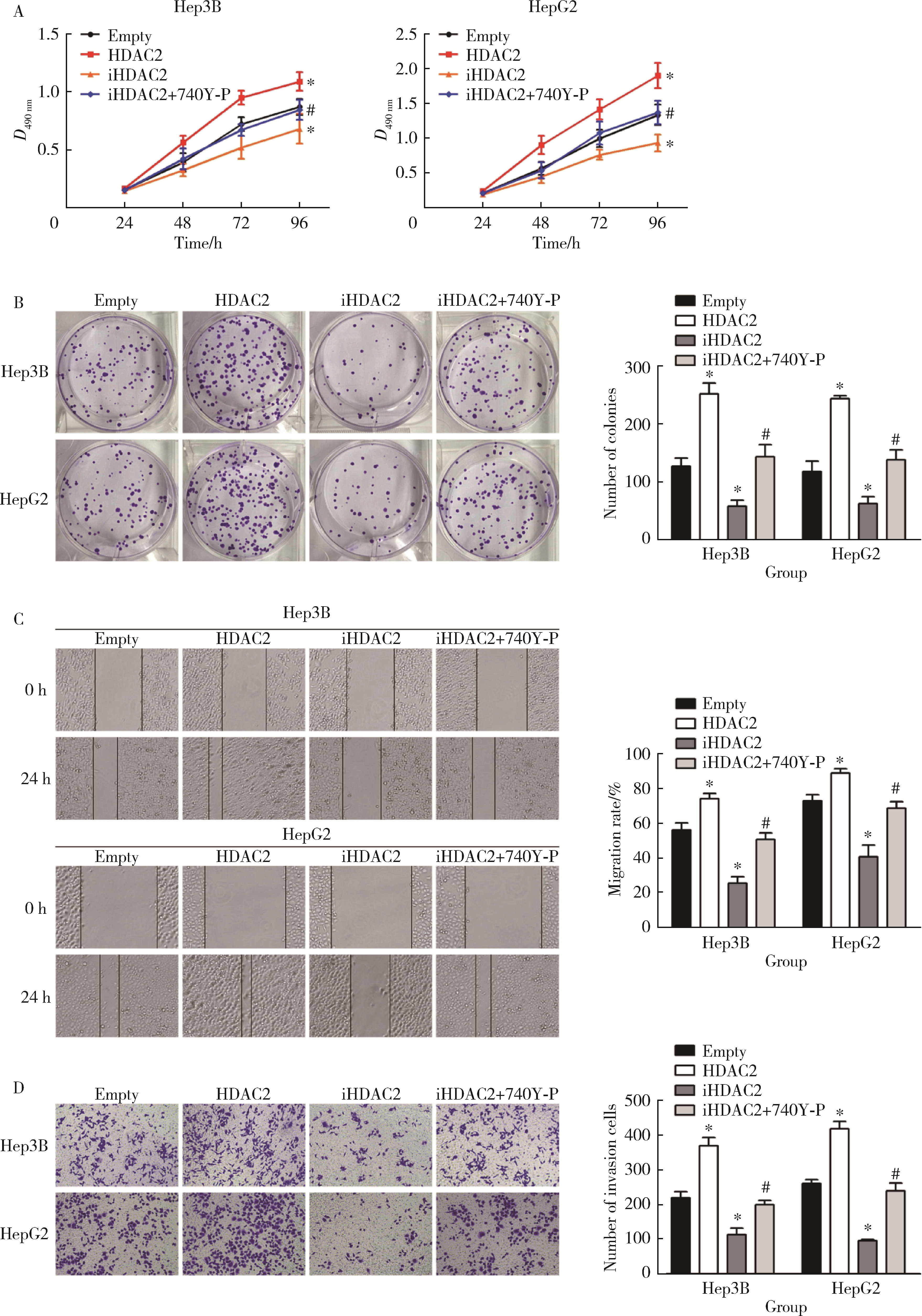
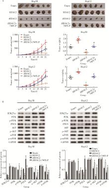
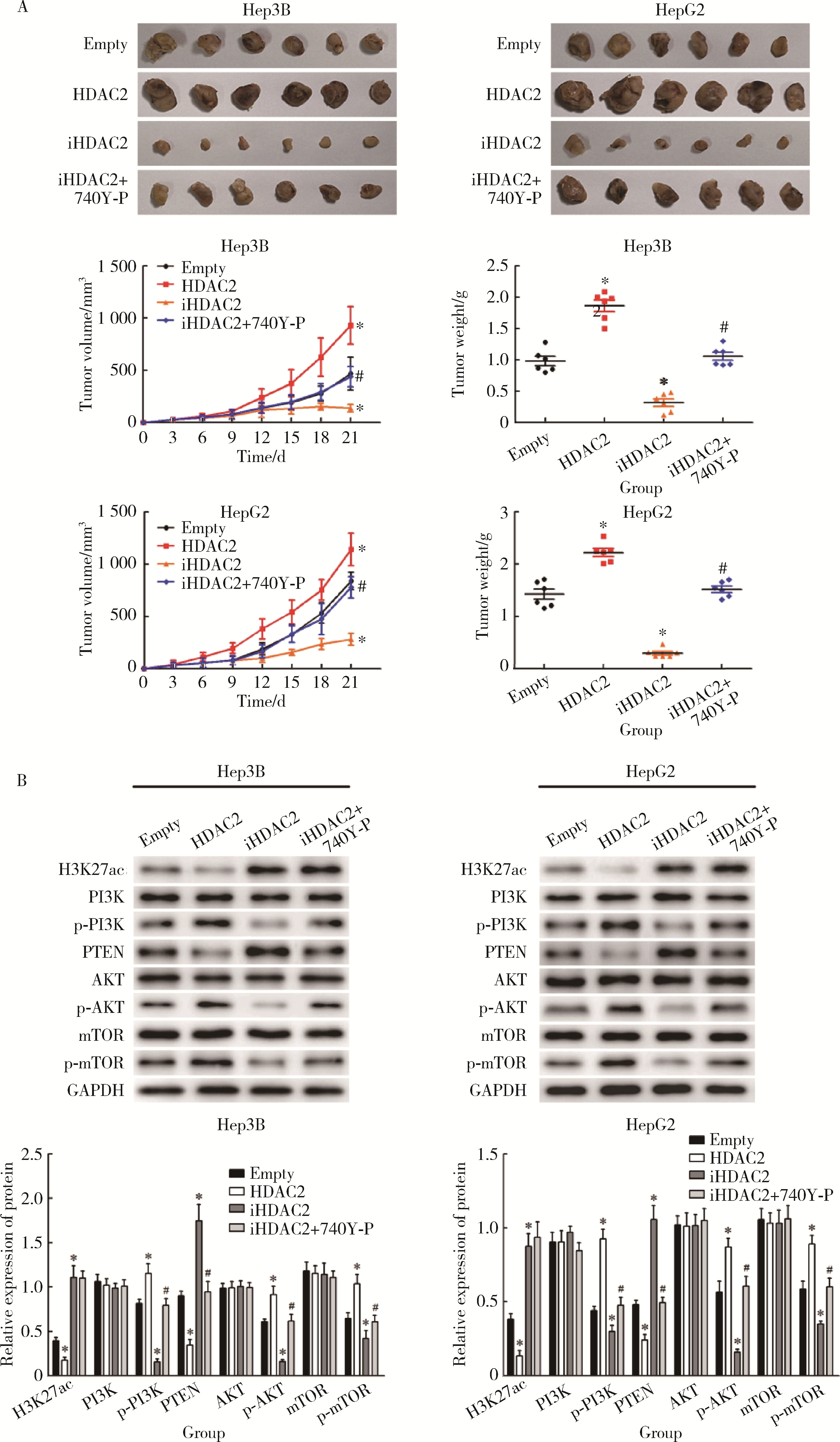
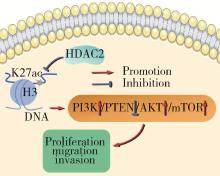
摘要: 目的: 探究组蛋白去乙酰化酶2(histone deacetylase 2, HDAC2)介导组蛋白H3第27位赖氨酸乙酰化(histone H3 lysine 27 acetylation, H3K27ac)修饰促进肝癌细胞增殖、迁移的作用机制。方法: 收集2021年1月至2023年1月经手术切除的40例肝癌、癌旁组织样本, 免疫组化和Western blotting检测肝癌、癌旁组织、细胞系HDAC2、H3K27ac表达, 分析HDAC2与H3K27ac表达水平的相关性, 以及HDAC2表达与肝癌患者临床病理特征的关系。Hep3B、HepG2细胞分为sh-NC组(转染sh-NC)、sh-HDAC2组(转染sh-HDAC2)、空质粒组(转染dCas9-空质粒)、HDAC2组(转染dCas9-HDAC2)、iHDAC2组(转染dCas9-失活HDAC2)和iHDAC2+740Y-P组(转染dCas9-失活HDAC2, 培养基加入30 μmol/mL 740Y-P)。通过MTS、克隆形成、划痕、Transwell实验测定各组Hep3B、HepG2细胞增殖、迁移、侵袭能力, 通过Western blotting、实时荧光定量PCR(real-time fluorescence quantitative PCR, qRT-PCR)、HDAC2活性检测、染色质免疫共沉淀-高通量测序(chromatin immunoprecipitation high-throughput sequencing, ChIP-seq)检测验证HDAC2介导H3K27ac修饰, 体内异种移植实验测定各组细胞成瘤能力, 检测磷脂酰肌醇-3激酶/第10号染色体磷酸酶和张力蛋白同源缺失基因/蛋白激酶B/哺乳动物雷帕霉素靶蛋白(phosphoinositide 3-kinases/phosphatase and tensin homolog deleted on chromosome ten/protein kinase B/mammalian target of rapamycin, PI3K/PTEN/AKT/mTOR)信号通路相关蛋白表达。结果: 肝癌组织、细胞系HDAC2呈高表达(P<0.05), H3K27ac呈低表达(P<0.05), 二者表达水平呈负相关(r=-0.477, P=0.002)。HDAC2表达水平与肿瘤大小、感染乙肝病毒、TNM分期、门静脉癌栓相关(P<0.05)。与Hep3B、HepG2细胞sh-NC组相比, sh-HDAC2组增殖、克隆形成、迁移、侵袭能力降低(P<0.05)。与空质粒组相比, HDAC2组HDAC2表达水平、活性、细胞增殖、克隆形成、迁移、侵袭能力, 体内成瘤体积、质量, p-PI3K、p-AKT、p-mTOR表达水平均升高(P<0.05), H3K27ac富集程度、H3K27ac、PTEN表达水平降低(P<0.05), iHDAC2组HDAC2表达水平、活性、增殖、克隆形成、迁移、侵袭能力, 体内成瘤体积、质量, p-PI3K、p-AKT、p-mTOR表达水平降低(P<0.05), H3K27ac、PTEN表达水平升高(P<0.05)。引入PI3K激活剂740Y-P验证PI3K/PTEN/AKT/mTOR信号通路参与HDAC2介导H3K27ac修饰的肝癌细胞恶性行为调控, 与iHDAC2组相比, iHDAC2+740Y-P组增殖、克隆形成、迁移、侵袭能力, 体内成瘤体积、质量, p-PI3K、p-AKT、p-mTOR表达水平升高(P<0.05), PTEN表达水平降低(P<0.05)。结论: HDAC2通过介导H3K27ac修饰启动PI3K/PTEN/AKT/mTOR信号通路, 促进肝癌发生发展。

关键词: 组蛋白去乙酰化酶2, 组蛋白H3第27位赖氨酸乙酰化, 肝癌, 增殖, 迁移

Abstract: Objective: To explore the specific mechanism of histone deacetylase 2 (HDAC2) mediated histone H3 lysine 27 acetylation (H3K27ac) modification in promoting the proliferation and migration of hepatocellular carcinoma cells. Methods: Samples of 40 cases of hepatocellular carcinoma and paracancerous tissues resected from January 2021 to January 2023 were collected. The expressions of HDAC2 and H3K27ac in hepatocellular carcinoma, paracancerous tissues and cell lines were detected by immunohistochemistry and Western blotting. The correlation between the expression levels of HDAC2 and H3K27ac and the relationship between HDAC2 expression and clinicopathological characteristics of patients with hepatocellular carcinoma were analyzed. The proliferation, migration and invasion of Hep3B and HepG2 cells were determined by MTS, clone formation, scratch and Transwell experiments. The acetylation of H3K27 mediated by HDAC2 was verified by Western blotting, real-time fluorescence quantitative PCR (qRT-PCR) and chromatin immunoprecipitation high-throughput sequencing (ChIP-seq). In vivo xenotransplantation experiment, the tumorigenicity of cells in each group was measured, and the expression of proteins related to phosphoinositide 3-kinases/phosphatase and tensin homolog deleted on chromosome ten/protein kinase B/mammalian target of rapamycin (PI3K/PTEN/AKT/mTOR) signal pathway was detected. Results: High expression of HDAC2 and low expression of H3K27ac were found in hepatocellular carcinoma tissues and cell lines (P < 0.05), and there was a negative correlation between them (r=-0.477, P=0.002). The expression of HDAC2 was related to tumor size, hepatitis B virus infection, TNM stage and portal vein tumor thrombus (P < 0.05). Compared with the sh-NC group of Hep3B and HepG2 cells, the proliferation, clone formation, migration and invasion ability of sh-HDAC2 group were decreased (P < 0.05). Compared with the Empty group, the HDAC2 group exhibited increased expression levels and activity of HDAC2, as well as enhanced cell proliferation, clone formation, migration, invasion ability, tumor volume and mass in vivo, and elevated expression levels of p-PI3K, p-AKT, and p-mTOR (P < 0.05). Conversely, the enrichment and expression levels of H3K27ac, along with the expression level of PTEN, were decreased (P < 0.05). In the iHDAC2 group, the expression levels and activity of HDAC2, as well as the proliferation, clone formation, migration, invasion ability, tumor volume and mass in vivo, and expression levels of p-PI3K, p-AKT, and p-mTOR were reduced (P < 0.05). Additionally, the expression levels of H3K27ac and PTEN were increased (P < 0.05). To validate the involvement of the PI3K/PTEN/AKT/mTOR signaling pathway in HDAC2-mediated regulation of malignant behaviors in liver cancer cells through H3K27ac, the PI3K activator 740Y-P was introduced. Compared with the iHDAC2 group, the iHDAC2+740Y-P group exhibited increased proliferation, clone formation, migration, invasion ability, tumor volume and mass in vivo, and elevated expression levels of p-PI3K, p-AKT, and p-mTOR (P < 0.05). Conversely, the expression level of PTEN was decreased (P < 0.05). Conclusion: HDAC2 initiates PI3K/PTEN/AKT/mTOR signal pathway by mediating H3K27 acetylation, which promotes the occurrence and development of hepatocellular carcinoma.

Key words: Histone deacetylase 2, Histone H3 lysine 27 acetylation, Hepatocellular carcinoma, Proliferation, Migration

中图分类号: 

  • R735.7

表1

引物序列"

Gene Primer sequences (5′-3′)
HDAC2 Forward:ATGGCGTACAGTCAAGGAGGC
Reverse:AAATCAGAACAGCTCAGCAAC
GAPDH Forward:ATGGGGAAGGTGAAGGTCGG
Reverse:TTACTCCTTGGAGGCCATGT

图1

HDAC2、H3K27ac在肝癌中的表达情况"

表2

HDAC2表达水平与肝癌患者临床病理特征间的关系"

Items Total HDAC2 expression χ2 P
High (n=20) Low (n=20)
Age/years
  ≥59 25 (62.50) 14 (35.00) 11 (27.50) 0.960 0.327
  <59 15 (37.50) 6 (15.00) 9 (22.50)
Gender
  Male 21 (52.50) 13 (32.50) 8 (20.00) 2.506 0.113
  Female 19 (47.50) 7 (17.50) 12 (30.00)
Tumor size/cm
  ≥3 28 (70.00) 17 (42.50) 11 (27.50) 4.286 0.038
  <3 12 (30.00) 3 (7.50) 9 (22.50)
HBV infection
  Positive 30 (75.00) 19 (47.50) 11 (27.50) 8.533 0.003
  Negative 10 (25.00) 1 (2.50) 9 (22.50)
Alpha-fetoprotein/(μg/L)
  ≥400 27 (67.50) 12 (30.00) 15 (37.50) 1.026 0.311
  <400 13 (32.50) 8 (20.00) 5 (12.50)
Histologic grade
  Well and moderate 25 (62.50) 10 (25.00) 15 (37.50) 2.667 0.102
  Low 15 (37.50) 10 (25.00) 5 (12.50)
TNM stage
  Ⅰ to Ⅱ 19 (47.50) 6 (15.00) 13 (32.50) 4.912 0.027
  Ⅲ to Ⅳ 21 (52.50) 14 (35.00) 7 (17.50)
Portal vein tumor thrombus
  Yes 11 (27.50) 9 (22.50) 2 (5.00) 6.144 0.013
  No 29 (72.50) 11 (27.50) 18 (45.00)
Cirrhosis
  Yes 15 (37.50) 6 (15.00) 9 (22.50) 0.960 0.327
  No 25 (62.50) 14 (35.00) 11 (27.50)

图2

抑制HDAC2对肝癌细胞增殖、迁移和侵袭的影响"

图3

HDAC2介导H3K27ac修饰"

图4

体外细胞实验验证HDAC2介导H3K27ac修饰促进肝癌细胞增殖、迁移和侵袭"

图5

体内细胞实验验证HDAC2介导H3K27ac修饰启动PI3K/PTEN/AKT/mTOR信号通路增强肝癌细胞成瘤能力"

图6

HDAC2介导H3K27ac修饰促进肝癌进展的作用机制"

1
Li X, Ramadori P, Pfister D, et al. The immunological and metabolic landscape in primary and metastatic liver cancer[J]. Nat Rev Cancer, 2021, 21(9): 541- 557.

doi: 10.1038/s41568-021-00383-9
2
Huang DQ, Singal AG, Kono Y, et al. Changing global epidemiology of liver cancer from 2010 to 2019:NASH is the fastest growing cause of liver cancer[J]. Cell Metab, 2022, 34(7): 969- 977.

doi: 10.1016/j.cmet.2022.05.003
3
Konyn P, Ahmed A, Kim D. Current epidemiology in hepatocellular carcinoma[J]. Expert Rev Gastroenterol Hepatol, 2021, 15(11): 1295- 1307.

doi: 10.1080/17474124.2021.1991792
4
Donne R, Lujambio A. The liver cancer immune microenvironment: Therapeutic implications for hepatocellular carcinoma[J]. Hepatology, 2023, 77(5): 1773- 1796.

doi: 10.1002/hep.32740
5
Chrysavgis L, Giannakodimos I, Diamantopoulou P, et al. Non-alcoholic fatty liver disease and hepatocellular carcinoma: Clinical challenges of an intriguing link[J]. World J Gastroenterol, 2022, 28(3): 310- 331.

doi: 10.3748/wjg.v28.i3.310
6
Luo XY, Wu KM, He XX. Advances in drug development for hepatocellular carcinoma: Clinical trials and potential therapeutic targets[J]. J Exp Clin Cancer Res, 2021, 40(1): 172.

doi: 10.1186/s13046-021-01968-w
7
Malik IA, Rajput M, Werner R, et al. Differential in vitro effects of targeted therapeutics in primary human liver cancer: Importance for combined liver cancer[J]. BMC Cancer, 2022, 22(1): 1193.

doi: 10.1186/s12885-022-10247-6
8
Arechederra M, Recalde M, Gárate-Rascón M, et al. Epigenetic biomarkers for the diagnosis and treatment of liver disease[J]. Cancers (Basel), 2021, 13(6): 1265.

doi: 10.3390/cancers13061265
9
Espiritu D, Gribkova AK, Gupta S, et al. Molecular mechanisms of oncogenesis through the lens of nucleosomes and histones[J]. J Phys Chem B, 2021, 125(16): 3963- 3976.

doi: 10.1021/acs.jpcb.1c00694
10
Beacon TH, Delcuve GP, López C, et al. The dynamic broad epigenetic (H3K4me3, H3K27ac) domain as a mark of essential genes[J]. Clin Epigenetics, 2021, 13(1): 138.

doi: 10.1186/s13148-021-01126-1
11
Rithanya P, Ezhilarasan D. Sodium valproate, a histone deacetylase inhibitor, provokes reactive oxygen species-mediated cyto-toxicity in human hepatocellular carcinoma cells[J]. J Gastrointest Cancer, 2021, 52(1): 138- 144.

doi: 10.1007/s12029-020-00370-7
12
Bouyahya A, El Hachlafi N, Aanniz T, et al. Natural bioactive compounds targeting histone deacetylases in human cancers: Recent updates[J]. Molecules, 2022, 27(8): 2568.

doi: 10.3390/molecules27082568
13
Uddin MH, Al-Hallak MN, Philip PA, et al. Aberrant transcription factors in the cancers of the pancreas[J]. Semin Cancer Biol, 2022, 86(Pt2): 28- 45.
14
Prvanovic'M, Nedeljkovic'M, Tanic'N, et al. Role of PTEN, PI3K, and mTOR in triple-negative breast cancer[J]. Life (Basel), 2021, 11(11): 1247.
15
Rodrigues DA, Pinheiro PSM, Fraga CAM. Multitarget inhibition of histone deacetylase (HDAC) and phosphatidylinositol-3-kinase (PI3K): Current and future prospects[J]. ChemMedChem, 2021, 16(3): 448- 457.

doi: 10.1002/cmdc.202000643
16
姜健, 王维, 崔羽楠, 等. 基于2018版肝脏影像报告及数据系统评估CT及MRI对小于等于3 cm肝细胞性肝癌的诊断价值[J]. 磁共振成像, 2021, 12(9): 25-29, 44.
17
Majchrzak-Celińska A, Warych A, Szoszkiewicz M. Novel approaches to epigenetic therapies: From drug combinations to epigenetic editing[J]. Genes (Basel), 2021, 12(2): 208.

doi: 10.3390/genes12020208
18
Kim J, Lee H, Yi SJ, et al. Gene regulation by histone-modifying enzymes under hypoxic conditions: A focus on histone methylation and acetylation[J]. Exp Mol Med, 2022, 54(7): 878- 889.

doi: 10.1038/s12276-022-00812-1
19
Chen YC, Koutelou E, Dent SYR. Now open: Evolving insights to the roles of lysine acetylation in chromatin organization and function[J]. Mol Cell, 2022, 82(4): 716- 727.

doi: 10.1016/j.molcel.2021.12.004
20
Hogg SJ, Motorna O, Cluse LA, et al. Targeting histone acetylation dynamics and oncogenic transcription by catalytic P300/CBP inhibition[J]. Mol Cell, 2021, 81(10): 2183- 2200.
21
Shvedunova M, Akhtar A. Modulation of cellular processes by histone and non-histone protein acetylation[J]. Nat Rev Mol Cell Biol, 2022, 23(5): 329- 349.

doi: 10.1038/s41580-021-00441-y
22
Du X, Wang H, Xu J, et al. Profiling and integrated analysis of transcriptional addiction gene expression and prognostic value in hepatocellular carcinoma[J]. Aging (Albany NY), 2023, 15(8): 3141- 3157.
23
Ma F, Huang J, Li W, et al. microRNA-455-3p functions as a tumor suppressor by targeting HDAC2 to regulate cell cycle in hepatocellular carcinoma[J]. Environ Toxicol, 2022, 37(7): 1675- 1685.

doi: 10.1002/tox.23516
24
Zhao LN, Yuan HF, Wang YF, et al. IFN-α inhibits HBV transcription and replication by promoting HDAC3-mediated de-2-hydroxyisobutyrylation of histone H4K8 on HBV cccDNA minichromosome in liver[J]. Acta Pharmacol Sin, 2022, 43(6): 1484- 1494.

doi: 10.1038/s41401-021-00765-7
25
Almaimani RA, Aslam A, Ahmad J, et al. In vivo and in vitro enhanced tumoricidal effects of metformin, active vitamin D3, and 5-fluorouracil triple therapy against colon cancer by modulating the PI3K/Akt/PTEN/mTOR network[J]. Cancers (Basel), 2022, 14(6): 1538.

doi: 10.3390/cancers14061538
26
Xun G, Hu W, Li B. PTEN loss promotes oncogenic function of STMN1 via PI3K/AKT pathway in lung cancer[J]. Sci Rep, 2021, 11(1): 14318.

doi: 10.1038/s41598-021-93815-3
27
Lin Z, Huang L, Li SL, et al. PTEN loss correlates with T cell exclusion across human cancers[J]. BMC Cancer, 2021, 21(1): 429.

doi: 10.1186/s12885-021-08114-x
28
Wang Z, Cui X, Hao G, et al. Aberrant expression of PI3K/AKT signaling is involved in apoptosis resistance of hepatocellular carcinoma[J]. Open Life Sci, 2021, 16(1): 1037- 1044.

doi: 10.1515/biol-2021-0101
29
Liu YR, Wang JQ, Huang ZG, et al. Histone deacetylase-2:A potential regulator and therapeutic target in liver disease (Review)[J]. Int J Mol Med, 2021, 48(1): 131.

doi: 10.3892/ijmm.2021.4964
30
Mao D, Jiang H, Zhang F, et al. HDAC2 exacerbates rheumatoid arthritis progression via the IL-17-CCL7 signaling pathway[J]. Environ Toxicol, 2023, 38(7): 1743- 1755.

doi: 10.1002/tox.23802
31
Gui L, Zhang S, Xu Y, et al. UBE2S promotes cell chemoresistance through PTEN-AKT signaling in hepatocellular carcinoma[J]. Cell Death Discov, 2021, 7(1): 357- 367.

doi: 10.1038/s41420-021-00750-3
[1] 张瑶,郭金鑫,战世佳,洪恩宇,杨慧,贾安娜,常艳,郭永丽,张璇. 富含半胱氨酸和甘氨酸蛋白2在神经母细胞瘤恶性进展中的功能和机制[J]. 北京大学学报(医学版), 2024, 56(3): 495-504.
[2] 曹钟,岑红兵,赵建红,梅俊,秦灵芝,廖伟,敖启林. 胰腺神经内分泌肿瘤和实性假乳头状肿瘤中INSM1和SOX11的表达及意义[J]. 北京大学学报(医学版), 2023, 55(4): 575-581.
[3] 王磊,韩天栋,江卫星,李钧,张道新,田野. 主动迁移技术与原位碎石技术在输尿管软镜治疗1~2 cm输尿管上段结石中的安全性和有效性比较[J]. 北京大学学报(医学版), 2023, 55(3): 553-557.
[4] 刘媛,原婉琼,李婷,王平章,吕平,吴利新,阮国瑞,韩文玲,莫晓宁. 敲减CMTM3增加急性B淋巴细胞白血病细胞对伊马替尼敏感性[J]. 北京大学学报(医学版), 2022, 54(6): 1238-1243.
[5] 杨朵,周心娜,王硕,王小利,袁艳华,杨化兵,耿会珍,彭兵,李子博,李彬,任军. 树突状细胞疫苗特异肿瘤多肽联合树突状细胞体外刺激淋巴细胞功能评估[J]. 北京大学学报(医学版), 2021, 53(6): 1094-1098.
[6] 王子乔,刘燕鹰,张霞,刘田,任立敏,沈丹华,王屹,栗占国. 17例误诊为IgG4相关疾病患者的临床特点及误诊原因分析[J]. 北京大学学报(医学版), 2019, 51(6): 1025-1031.
[7] 姚心韵,高晓敏,邹晓英,岳林. 胞内转运对根尖牙乳头干细胞表面CXC趋化因子受体4表达的影响[J]. 北京大学学报(医学版), 2019, 51(5): 893-899.
[8] 谢静,赵玉鸣,饶南荃,汪晓彤,方滕姣子,李晓霞,翟越,李静芝,葛立宏,王媛媛. 3种口腔颌面部来源的间充质干细胞成血管内皮分化潜能的比较研究[J]. 北京大学学报(医学版), 2019, 51(5): 900-906.
[9] 张帆,燕太强,郭卫. Rasfonin抑制骨肉瘤细胞143B的增殖和迁移[J]. 北京大学学报(医学版), 2019, 51(2): 234-238.
[10] 季兰岚,郝燕捷,张卓莉. 原发性骨髓纤维化引起的继发性痛风1例[J]. 北京大学学报(医学版), 2018, 50(6): 1117-1119.
[11] 王子成,程立,吕同德,苏黎,林健,周利群. 炎症因子预处理的脂肪干细胞可明显抑制外周血单个核细胞增殖[J]. 北京大学学报(医学版), 2018, 50(4): 590-594.
[12] 唐旭,赵卫红,宋琴琴,殷华奇,杜依青,盛正祚,王强,张晓威,李清,刘士军,徐涛. SOX10对前列腺癌细胞增殖及侵袭的影响[J]. 北京大学学报(医学版), 2018, 50(4): 602-606.
[13] 汪晓彤,饶南荃,方腾姣子,赵玉鸣,葛立宏. 乳牙牙髓干细胞CD146阳性/阴性细胞亚群生物学特性的比较[J]. 北京大学学报(医学版), 2018, 50(2): 284-292.
[14] 陈玮, 胡凡磊, 刘洪江, 徐丽玲, 李英妮, 栗占国. 类风湿关节炎患者髓系来源的抑制细胞促进自身B细胞增殖[J]. 北京大学学报(医学版), 2017, 49(5): 819-823.
[15] 蔡燚,郭浩,李汉忠,王文达,张玉石. 结节性硬化症细胞株TSC2-/- MEFs和正常细胞株TSC2+/+ MEFs微小RNA表达谱的差异分析[J]. 北京大学学报(医学版), 2017, 49(4): 580-584.
Viewed
Full text


Abstract

Cited

  Shared   
  Discussed   
No Suggested Reading articles found!