论著

原发灶局限于胆囊壁内胆囊癌大体分型及其与预后和癌前病变的相关性

  • 张铃福 1, * ,
  • 陈明 2, * ,
  • 赵小宇 1 ,
  • 王港 1 ,
  • 崔龙 1 ,
  • 凌晓锋 1 ,
  • 王立新 1 ,
  • 徐智 1 ,
  • 郭丽梅 , 3, * ,
  • 侯纯升 , 1, *
展开
  • 1. 北京大学第三医院普通外科, 北京 100191
  • 2. 北京大学第三医院放射科, 北京 100191
  • 3. 北京大学第三医院病理科, 北京 100191

* These authors contributed equally to this work

收稿日期: 2025-06-10

  网络出版日期: 2025-10-10

版权

版权所有,未经授权,不得转载。

Gross classification of gallbladder cancer with primary lesion limited to the gallbladder wall and its correlation with prognosis and precancerous lesions

  • Lingfu ZHANG 1 ,
  • Ming CHEN 2 ,
  • Xiaoyu ZHAO 1 ,
  • Gang WANG 1 ,
  • Long CUI 1 ,
  • Xiaofeng LING 1 ,
  • Lixin WANG 1 ,
  • Zhi XU 1 ,
  • Limei GUO , 3, * ,
  • Chunsheng HOU , 1, *
Expand
  • 1. Department of General Surgery, Peking University Third Hospital, Beijing 100191, China
  • 2. Department of Radiology, Peking University Third Hospital, Beijing 100191, China
  • 3. Department of Pathology, Peking University Third Hospital, Beijing 100191, China
GUO Limei, e-mail,
HOU Chunsheng, e-mail,

Received date: 2025-06-10

  Online published: 2025-10-10

Copyright

All rights reserved. Unauthorized reproduction is prohibited.

摘要

目的: 探讨原发灶局限于胆囊壁内胆囊癌的大体分型及其与预后和癌前病变相关性。方法: 回顾性纳入2006年1月至2020年12月北京大学第三医院收治的123例术前影像提示原发灶局限于胆囊壁内、且术后病理为腺癌患者。依据CT、MRI或大体标本分为如下4型:1型,局限性凸向腔内肿物,不伴肿物附着处胆囊壁增厚;2型,局限性凸向腔内肿物,伴肿物附着处胆囊壁增厚和/或增厚处浆膜皱褶;3型,局限于两个连续部分的胆囊壁环形增厚;4型,延续至两个以上连续部分的胆囊壁环形增厚。比较各分型之间的临床病理特征、癌前病变类型及生存情况。结果: 术前CT/MRI及术中大体标本均可以作为大体分型的依据,大体标本准确率最高。123例患者中13例无法分型,剩余110例完成大体分型。胆囊癌大体分型与T分期(P<0.001,rs=0.682)呈高强度相关,与淋巴结转移(P<0.001,rs=0.478)、组织分化程度(P<0.001,rs=0.484)、神经浸润(P<0.001,rs=0.490)以及脉管瘤栓(P<0.001,rs=0.334)等组织病理学参数呈中等强度相关,分型越高,不良组织病理学参数越多。另外,胆囊癌大体分型与患者手术治疗后残余病灶(P<0.001,rs=0.328)和术后复发(P<0.001,rs=0.619)呈中等强度和高等强度相关性。生存分析显示,分型越高,患者中位生存时间越短,1型96个月,2型73个月,3型30个月,4型14个月,P<0.001。多因素Cox回归表明胆囊癌大体分型是患者预后的独立影响因素(HR=3.609,95%CI:2.177~5.983,P<0.001)。在肿瘤生物学行为异质性最强的T2期患者中,胆囊癌大体分型同样与预后密切相关,中位生存时间分别为72个月、70个月、29个月和16个月, P<0.001。多因素Cox回归亦表明胆囊癌大体分型是患者预后的独立影响因素(HR=2.723,95%CI:1.566~4.736,P<0.001)。在肿瘤起源方面,胆囊癌大体分型与癌前病变类型显著相关:1型主要源自胆囊内乳头状肿瘤,3型与4型则多为高级别胆管上皮内瘤变或无癌前病变。肿瘤自然病程分析提示:1型进展缓慢,4型进展迅速,2型表现出较大异质性。结论: 原发灶局限于胆囊壁内胆囊癌大体分型与预后及癌前病变密切相关,可作为手术决策与分层管理的重要参考依据。

本文引用格式

张铃福 , 陈明 , 赵小宇 , 王港 , 崔龙 , 凌晓锋 , 王立新 , 徐智 , 郭丽梅 , 侯纯升 . 原发灶局限于胆囊壁内胆囊癌大体分型及其与预后和癌前病变的相关性[J]. 北京大学学报(医学版), 2026 , 58(1) : 184 -189 . DOI: 10.19723/j.issn.1671-167X.2026.01.024

Abstract

Objective: To explore the gross classification of gallbladder cancer with primary lesion confined within the gallbladder wall, and its correlation with prognosis and precancerous lesions. Methods: A retrospective study was conducted on 123 patients who were admitted to Peking University Third Hospital from January 2006 to December 2020. These patients had preoperative imaging findings suggesting that the primary lesion was confined within the gallbladder wall and had postoperative pathology of adenocarcinoma. Based on CT, MRI, or gross specimens, they were divided into the following four types: Type 1, simple intraluminal lesion: Intraluminal lesions without focal thickening of the gallbladder wall; Type 2, complex intraluminal lesion: Intraluminal lesions associated with focal thickening of the gallbladder wall and/or outer surface dimpling at the tumor base; Type 3, focal wall thickening: Circumferential focal wall thickening with heterogeneous enhancement within 2 continuous parts of the gallbladder; Type 4, diffuse wall thickening: Circumferential diffuse wall thickening extending more than 2 continuous parts of the gallbladder with heterogeneous enhancement. The clinical pathological characteristics, types of precancerous lesions, and survival status were compared among the different types. Results: Both preoperative CT/MRI and intraoperative gross specimens could serve as the basis for gross classification, with gross specimens demonstrating the highest accuracy rate. Among the 123 patients, 13 could not be classified, while the remaining 110 underwent gross classification. The gross classification of gallbladder cancer was strongly or moderately correlated with histopathological parameters such as T-stage (P < 0.001, rs=0.682), lymph node metastasis (P < 0.001, rs=0.478), tissue differentiation degree (P < 0.001, rs=0.484), nerve infiltration (P < 0.001, rs=0.490), and vascular invasion (P < 0.001, rs=0.334). The higher the classification, the more adverse histopathological parameters were observed. Additionally, the gross classification of gallbladder cancer was moderately strongly and highly strongly correlated with residual lesions after surgical treatment (P < 0.001, rs=0.328) and postoperative recurrence (P < 0.001, rs=0.619) in the patients. Survival analysis revealed that the higher the classification, the shorter the median survival time of the patients (Type 1: 96 months, Type 2: 73 months, Type 3: 30 months, Type 4: 14 months, P < 0.001). Multivariate Cox regression indicated that the gross classification of gallbladder cancer was an independent prognostic factor (HR=3.609, 95%CI: 2.177-5.983, P < 0.001). In the patients with the most heterogeneous biological behavior in stage T2, the gross classification of gallbladder cancer was also closely associated with prognosis (median survival times were 72, 70, 29, and 16 months, respectively, P < 0.001). Multivariate Cox regression further demonstrated that the gross classification of gallbladder cancer was an independent prognostic factor (HR=2.723, 95%CI: 1.566-4.736, P < 0.001). In terms of tumor origin, the gross classification of gallbladder cancer was significantly correlated with the type of precancerous lesions: Type 1 mainly originated from intracholecystic papillary neoplasm of the gallbladder, while Types 3 and 4 were mostly high-grade biliary intraepithelial neoplasm or without precancerous lesions. Analysis of the natural history of the tumor suggested that Type 1 progressed slowly, Type 4 progressed rapidly, and Type 2 exhibited greater heterogeneity. Conclusion: The gallbladder cancer with primary lesion confined within the gallbladder wall is closely related to prognosis and precancerous lesions, and can serve as an important reference for surgical decision-making and stratified management.

胆囊癌是最常见的胆道系统恶性肿瘤,随着体检的普及,越来越多的胆囊癌发现时是局限于胆囊壁内,根治手术是此类胆囊癌最主要的治疗方法[1]。虽然大多数胆囊癌术后效果欠佳,然而仍有很多患者术后长期存活,表明不同的胆囊癌生物学行为存在异质性。既往研究表明,基于术前增强CT的胆囊癌大体分型与可切除性胆囊癌进展和预后关系密切[2-3],提示其可能反映胆囊癌的异质性,尤其是反映肿瘤起源的差异,既往研究提示不同癌前病变起源的胆囊癌预后差异明显[4-5]。本研究收集术前影像考虑原发灶局限于胆囊壁内、且术后病理为胆囊腺癌的患者资料,以术前CT/MRI及术中大体标本进行分型,进一步验证胆囊癌大体分型与原发灶局限于胆囊壁内胆囊腺癌的预后关系,并分析其与癌前病变的相关性。

1 资料与方法

1.1 一般资料

选取2006年1月至2020年12月就诊于北京大学第三医院的130例术前影像原发灶局限于胆囊壁内、且术后病理为胆囊腺癌的患者,其临床资料均录入普通外科胆囊癌数据库,排除7例失访患者,其余123例患者纳入本研究。123例患者中男性39例,女性84例,年龄23~86岁(中位年龄62岁)。 本研究经北京大学第三医院医学科学研究伦理委员会批准(批准号:M2019060),所有患者均在术前签署手术知情同意书。基于回顾性研究的特点,所有患者豁免本研究的知情同意。

1.2 胆囊癌大体分型分类

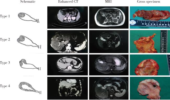
参考既往标准[3],依据增强CT、MRI或术中大体标本将胆囊癌分为如下4型:1型,单纯腔内肿物型:局限性凸向腔内肿物,不伴肿物附着处胆囊壁增厚;2型,复杂腔内肿物型:局限性凸向腔内肿物,伴肿物附着处胆囊壁增厚和/或增厚处浆膜皱褶;3型,区域厚壁型:局限于两个连续部分的胆囊壁环形增厚;4型,弥漫厚壁型:延续至两个以上连续部分的胆囊壁环形增厚(图 1)。
图1 胆囊癌大体分型

Figure 1 Each type of gallbladder cancer gross appearance

Type 1, simple intraluminal lesion: Intraluminal lesions without focal thickening of the gallbladder wall; Type 2, complex intraluminal lesion: Intraluminal lesions associated with focal thickening of the gallbladder wall and/or outer surface dimpling at the tumor base; Type 3, focal wall thickening: Circumferential focal wall thickening with heterogeneous enhancement within 2 continuous parts of the gallbladder; Type 4, diffuse wall thickening: Circumferential diffuse wall thickening extending more than 2 continuous parts of the gallbladder with heterogeneous enhancement. The gallbladder is composed of 4 parts: fundus, body, neck and cystic duct.

本组123例患者中,13例无法分型,剩余110例可分型。110例可分型患者中17例无增强CT资料,93例有增强CT资料的患者中,10例存在相邻分型纠结;14例无MRI资料,96例有MRI资料的患者中,13例存在相邻分型纠结;47例无大体标本资料,63例有大体标本资料的患者中,3例存在相邻分型纠结。上述结果表明基于大体标本的胆囊癌大体分型准确率最高。110例患者中,92例有两种以上分型依据,其中4例存在分型不一致。

1.3 术前影像原发灶局限于胆囊壁内胆囊癌的治疗情况

110例患者中,98例行根治手术(考虑既往研究证实T1b期胆囊癌单纯胆囊切除术基本可达根治效果[6-7],将本组3例行单纯胆囊切除术的T1b期胆囊癌亦纳入根治手术组),8例因远处转移无法根治行姑息切除术,4例术后石蜡病理切缘阳性,未进一步追加手术。

1.4 术前影像原发灶局限于胆囊壁内胆囊癌的临床病理学特点

110例患者中,T分期为原位癌(Tis)14例、T1a期5例、T1b期10例、T2期75例、T3期6例(显微镜下突破浆膜层或浸润胆囊床肝组织);31例有淋巴结转移(其中5例经淋巴结活检证实)。术后病理学高分化腺癌40例,中分化腺癌45例,低分化腺癌25例。110例患者中脉管内癌栓26例,神经浸润23例。

1.5 癌前病变定义

根据术后标本的病理切片,分析癌灶周边的癌前病变类型与大体分型的相关性。参考Nakanuma等[5]的癌前病变定义:胆囊内乳头状肿瘤(intracholecystic papillary neoplasm of the gallbladder, ICPN)指胆囊腔内最大病变直径超过1 cm, 并和周围黏膜相比高度超过5 mm的息肉样肿瘤;高级别胆管上皮内瘤变(biliary intraepithelial neoplasm, BilIN)指上皮内肿瘤出现高度异型性改变,但和周围黏膜相比高度<5 mm;无癌前病变指浸润性癌组织周围未见上述癌前病变组织。

1.6 统计学分析

采用SPSS 21.0软件进行统计分析。分类变量采用Pearson卡方检验或Fisher确切检验,等级变量采用Spearman相关性分析,连续变量采用单因素方差分析。使用Kaplan-Meier法行组间生存时间的分析,使用Log-rank法进行生存率检验。多因素生存分析采用Cox模型(Enter法)。以双侧P<0.05为差异有统计学意义。

2 结果

2.1 最终大体分型分类情况及其与常规临床病理学参数相关性

最终分型综合CT、MRI及大体标本决定,鉴于参考大体标本对胆囊癌进行大体分型过程中歧义少,有大体标本的以大体标本为准。本组2例依据增强CT分型为2型,但依据大体标本提示为1型,进一步证明大体标本检查的必要性。最终分型1型19例,2型56例,3型26例,4型9例。
胆囊癌大体分型与T分期(P<0.001,rs=0.682)呈高强度相关,与淋巴结转移(P<0.001,rs=0.478)、组织分化程度(P<0.001,rs=0.484)、神经瘤栓(P<0.001,rs=0.490)以及脉管瘤栓(P<0.001,rs=0.334)等组织病理学参数呈中等强度相关,分型越高,不良组织病理学参数越多。另外,胆囊癌大体分型与患者手术治疗后残余病灶(P<0.001,rs=0.328)和术后复发(P<0.001,rs=0.619)呈中等强度和高强度相关性(表 1)。
表1 胆囊癌不同大体分型患者的临床病理特征比较

Table 1 Comparison of clinical pathological characteristics among patients with different gross tumor types

Parameters Type 1 (n=19) Type 2 (n=56) Type 3 (n=26) Type 4 (n=9) rs value P value
Baseline demographics
  Age/years, $\bar x \pm s$ 59.8±12.9 65.9±10.7 63.6±10.6 62.9±12.8 0.221
  Male/female, n 2/17 19/37 9/17 4/5 0.157
Clinicopathological characteristics
  Pancreaticobiliary maljunction, n 3 13 3 2 0.630
  Gallstone, n 6 19 9 5 0.628
  CA199 elevation*, n 4 10 5 4 0.089 0.371
  CEA elevation*, n 0 6 3 2 0.167 0.090
  T stage, n 0.682 <0.001
    Tis 12 2 0 0
    T1 5 10 0 0
    T2 2 44 23 6
    T3 0 0 3 3
  Lymph node metastasis, n (%) 0 (0) 12 (21) 15 (58) 5 (56) 0.478 <0.001
  Histology grade, n 0.484 <0.001
    Well 16 20 3 1
    Moderate 2 26 14 3
    Poor 1 10 9 5
  Neural invasion, n (%) 0 (0) 5 (9) 13 (50) 5 (56) 0.490 <0.001
  Lymphovascular invasion, n (%) 1 (5) 10 (18) 10 (19) 5 (56) 0.334 <0.001
  Residual disease, n (%) 0 (0) 3 (5) 6 (23) 3 (33) 0.328 <0.001
  Recurrence, n (%) 0 (0) 12 (21) 18 (69) 9 (100) 0.619 <0.001

* indicates 6 cases did not undergo relevant examinations. rs indicates rank-size related correlation coefficient. CA199, carbohydrate antigen 199; CEA, carcinoembryonic antigen.

2.2 大体分型与预后

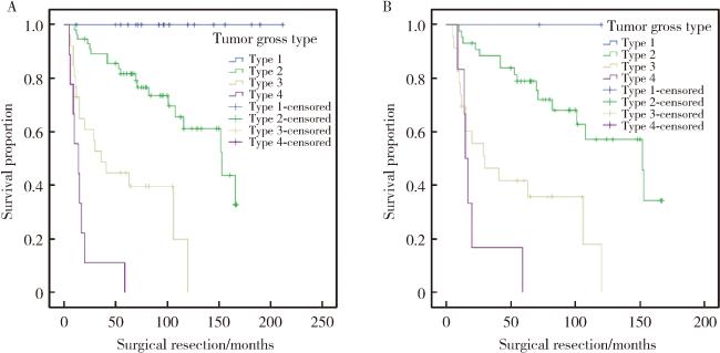
110例患者随访时间5~212个月(中位随访时间67个月)。65例患者实际生存时间长于5年。除外8例姑息手术的患者,31例患者术后出现复发转移。分型低组的生存时间优于分型高组,中位生存时间分别为96、73、30和14个月(χ2=76.9,P<0.001,图 2A)。多因素Cox回归分析结果显示胆囊癌大体分型是患者预后的独立影响因素(HR=3.609, 95%CI 2.177~5.983, P<0.001)。在生物学行为异质性明显的T2期胆囊癌中,该分型也与预后明显相关,中位生存时间分别为72、70、29和16个月(χ2=30.9,P<0.001),多因素Cox回归分析结果也显示胆囊癌大体分型是T2期患者预后的独立影响因素(HR=2.723, 95%CI 1.566~4.736, P<0.001,图 2B)。
图2 基于不同胆囊癌肿瘤大体分型的累积生存率比较

Figure 2 Comparing cumulative survival based on different tumour gross type

A, Kaplan-Meier survival curves comparing cumulative survival of cases among all 110 gallbladder cancers based on tumour gross type (Log-rank test, P < 0.001); B, Kaplan-Meier survival curves comparing cumulative survival of cases among 75 T2 gallbladder cancers based on tumour gross type (Log-rank test, P < 0.001).

表 2所示,1型癌前病变基本为ICPN,未见无癌前病变情况,3型和4型癌前病变无ICPN,2型癌前病变为ICPN 23例,高级别BilIN 26例,无癌前病变6例,上述各型与癌前病变分类相关性差异有统计学意义(P<0.001)。
表2 胆囊癌大体分型与癌前病变之间的相关性

Table 2 Correlation between gross classification of gallbladder cancer and precancerous lesions

Gross classification and precancerous lesions Type 1 (n=19) Type 2 (n=56) Type 3 (n=26)* Type 4 (n=9)
ICPN 18 23 0 0
High-grade BilIN 1 26 12 2
No precancerous lesion 0 6 12 7

* indicates 2 cases of Type 3 patients cannot be assessed for precancerous lesion type. ICPN, intracholecystic papillary neoplasm of the gallbladder.

2.3 大体分型与胆囊癌自然病程相关性

本组5例患者有胆囊癌自然病程CT资料,包括1例1型(癌前病变为ICPN),3例2型(癌前病变为2例ICPN和1例高级别BilIN)及1例4型(无癌前病变)患者。从该5例患者来看,1型进展慢,4型进展迅速,2型异质性较大(表 3)。
表3 基于胆囊癌大体分型的肿瘤自然病程

Table 3 Natural course of the tumor based on gallbladder cancer gross classification

Cases Tumor gross type Precancerous lesions Fist CT scan charateristics Interval of CTscan/months Second CT scan charateristics Operation date T stage
Case 1 Type 2 High-grade BilIN No polyps or localized thickening of the gallbladder wall 24 Intraluminal single polyp without focal thickening of the gallbladder wall 2010-11-5 Tis
Case 2 Type 3 ICPN Intraluminal single polyp without focal thickening of the gallbladder wall 12 Lesion diameter no change 2019-3-19 Tis
Case 3 Type 2 ICPN No polyps or localized thickening of the gallbladder wall 48 Intraluminal single polyp with focal thickening of the gallbladder wall 2013-7-18 T1b
Case 4 Type 2 ICPN No polyps or localized thickening of the gallbladder wall 12 Intraluminal single polyp with focal thickening of the gallbladder wall 2013-1-15 T2
Case 5 Type 4 No precancerous lesion No polyps or localized thickening of the gallbladder wall 9 Diffuse thickening of the gallbladder wall 2015-11-24 T3

ICPN, intracholecystic papillary-tubular neoplasm.

3 讨论

胆囊癌是最常见的胆道系统恶性肿瘤,异质性明显[8-10]。本研究从大体分型角度探讨其异质性,一方面进一步证实大体分型在胆囊癌预后中的作用,另一方面初步表明大体分型与癌前病变及胆囊癌自然病程相关性明显,为进一步深入研究胆囊癌生物学行为提供了分层依据。
原发灶局限于胆囊壁内胆囊癌,其基于术前影像及大体标本的分型依据可重复性及兼容性良好,对手术决策制定有一定指导意义。本中心在既往研究中提出并证实,基于术前增强CT的可切除胆囊癌可分为4型,该分型与胆囊癌进展和预后相关[3]。本研究进一步证实该分型方法的可重复性,并将其拓展到依据术前MRI和术中大体标本。本组123例患者中,110例可分型,接近90%,表明该分型可应用性良好。110例患者中,92例有两种以上分型依据,仅4例存在不一致,说明该分型方法兼容性良好。鉴于参考大体标本对胆囊癌进行大体分型过程中歧义少,而且本组2例增强CT为2型,大体标本修正为1型,进一步证明大体标本检查对胆囊癌大体分型的必要性。进一步深入分析发现,1型19例中17例为原位癌和T1期,无淋巴结转移患者,3型和4型35例患者中均为T2期及以上,淋巴结转移率高达50%以上,且有接近30%患者无法行根治手术。2型56例中T分期异质性明显,20%左右患者存在淋巴结转移,5%患者存在切缘阳性和无法行根治手术。这提示1型基本通过单纯胆囊切除即可根治,3/4型淋巴结转移率高,要重视淋巴结清扫的彻底性,且存在探查无法根治的风险。另外,生存分析表明,分型低组的生存时间优于分型高组,在异质性明显的T2期胆囊癌中该分型也与预后明显相关。上述数据表明,该分类对手术决策制定有一定指导意义。
胆囊癌大体分型除与手术决策相关的T分期和淋巴结转移显著相关外,从组织病理学角度分析发现,该分型越高,不良病理学参数的发生率也越高。本组数据表明,组织分化、脉管瘤栓、神经浸润等提示生物学行为不良的组织病理学指标与分型升高明显相关,进一步表明该分型与肿瘤生物学行为有相关性。
从肿瘤起源角度看,该分型与癌前病变相关性明显,且与肿瘤自然病程有一定相关性。既往研究表明,不同癌前病变与胆囊癌预后相关[4-5],本研究通过大体分型与癌前病变建立相关性,进一步证实了癌前病变与胆囊癌预后的相关性。这也在一定程度上表明,胆囊癌生物学行为在肿瘤起源阶段即相对确定,也就是说胆囊癌异质性很大程度上与肿瘤起源相关。从有限的5例有自然病程的患者资料来看,1型进展缓慢,4型进展迅速,2型异质性明显。我们提供的大体分型与癌前病变相关性的线索可以为进一步深入研究胆囊癌生物学行为提供简洁的分层依据。
本研究存在以下不足:(1)为回顾性研究,在标本收集及病理切片制备过程中可能存在信息不全面,尤其是对于癌前病变的评估,存在癌周组织取材不足的隐患,未来将进一步通过建立数据收集标准和前瞻性研究完善上述不足;(2)大体分型及组织病理学层面的癌前病变仅为形态学信息,缺乏分子生物学证据,为精确揭示各型胆囊癌背后的起源细胞克隆动力学和癌的演进,未来我们拟运用空间转录组学和拟时序分析,进行正常胆囊黏膜上皮、癌前病变到癌细胞的谱系追踪分析,探索胆囊癌的起源细胞分类,包括干细胞样和不同分化层次的高分辨率单细胞的图谱,进而构建出癌组织大体形成的动态模型,分析出每种类型的特征性组织和分子水平病理学标志物,以利于临床上不同类型胆囊癌患者的个体化治疗决策和预后预测。
本研究进一步证实了胆囊癌大体分型的可行性及其与预后的相关性,探索了该分型与癌前病变的相关性,为深入研究胆囊癌异质性提供了简单、可行的分层依据。

利益冲突  所有作者均声明不存在利益冲突。

作者贡献声明  张铃福:提出研究思路,影像阅片,病理阅片,撰写论文;陈明:参与研究,影像阅片,修改论文;赵小宇、王港、崔龙、凌晓锋、王立新、徐智:参与研究,审定论文;郭丽梅、侯纯升:病理阅片,提出研究思路,总体把关和审定论文。所有作者均参与论文修改,并对最终文稿进行审读和确认。

1
Roa JC , García P , Kapoor VK , et al. Gallbladder cancer[J]. Nat Rev Dis Primers, 2022, 8, 69.

DOI

2
张铃福, 侯纯升, 徐智, 等. 腹腔镜胆囊切除术中或术后意外胆囊癌腹腔镜手术治疗: 单中心10年回顾性分析[J]. 中华外科杂志, 2019, 57 (4): 277- 281.

3
Zhang L , Hou C , Chen M , et al. Tumour radiological appearance evaluated by enhanced CT correlates with tumour progression and survival in curable gallbladder cancer[J]. Eur J Surg Oncol, 2020, 46 (11): 2099- 2105.

DOI

4
Mochidome N , Koga Y , Ohishi Y , et al. Prognostic implications of the coexisting precursor lesion types in invasive gallbladder cancer[J]. Hum Pathol, 2021, 114, 44- 53.

DOI

5
Nakanuma Y , Sugino T , Nomura Y , et al. Association of precursors with invasive adenocarcinoma of the gallbladder: A clinicopathological study[J]. Ann Diagn Pathol, 2022, 58, 151911.

DOI

6
Wakai T , Shirai Y , Yokoyama N , et al. Early gallbladder carcinoma does not warrant radical resection[J]. Br J Surg, 2001, 88 (5): 675- 678.

DOI

7
张铃福, 侯纯升, 郭丽梅, 等. 术中冰冻或术后石蜡病理报告T1b期胆囊癌的外科治疗策略[J]. 北京大学学报(医学版), 2017, 49 (6): 1034- 1037.

DOI

8
Kohya N , Kitahara K , Miyazaki K . Rational therapeutic strategy for T2 gallbladder carcinoma based on tumor spread[J]. World J Gastroenterol, 2010, 16 (28): 3567- 3572.

DOI

9
Okada KI , Kijima H , Imaizumi T , et al. Clinical significance of wall invasion pattern of subserosa-invasive gallbladder carcinoma[J]. Oncol Rep, 2012, 28 (5): 1531- 1536.

DOI

10
Sung MK , Lee W , Lee JH , et al. Comparing survival rate and appropriate surgery methods according to tumor location in T2 gallbladder cancer[J]. Surg Oncol, 2022, 40, 101693.

DOI

文章导航

/