1 病例资料
1.1 主诉与检查
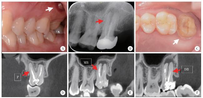
图1 左侧上颌第二磨牙术前口内照及影像学检查Figure 1 Intraoral examination, preoperative radiograph and cone-beam CT images of left maxillary second molar A, preoperative intraoral view of apical swelling (white arrow); B, preoperative periapical radiograph shows a metallic high-density fractured instrument (red arrow) within the mesiobuccal canal; C, the clinical view after crown removal reveals a resin filling and a palatal fiber post (white arrow); D, the palatal post extends to the mid-root level with an apical radiolucency; E, the image confirms the separated instrument in the apical third of the mesiobuccal root, associated with an apical radiolucency communicating with the distobuccal apical lesion; F, underfilled obturation is observed in the distobuccal root canal. P, palatal; MB, mesiobuccal; DB, distobuccal. |
1.2 治疗计划的制定
1.3 治疗过程
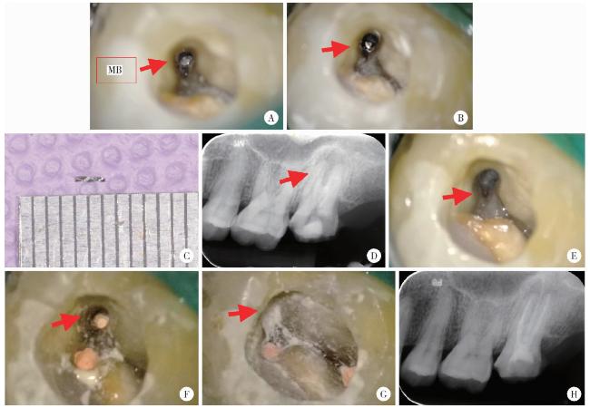
图2 左侧上颌第二磨牙器械取出、侧穿修补、根管充填图像及根尖片Figure 2 Clinical images and postoperative periapical radiograph illustrating the microscopic removal of the fractured instrument, perforation repair and root canal filling for left maxillary second molar A, fractured instrument tip observed in the mesiobuccal canal (red arrow) under dental microscope; B, instrument becomes loose (red arrow) after ultrasonic vibration; C, the retrieved 2.5 mm instrument segment; D, post-retrieval PA shows 2 mm residual in the apical region (red arrow); E, a lateral perforation is visible in the mesiobuccal canal near the furcation area after instrument retrieval (red arrow); F, the apical portion of the mesio-buccal canal is filled with gutta-percha (red arrow); G, the perforation is repaired with iRoot BP bioceramic material (red arrow); H, postoperative periapical radiograph confirms flush filling and a well-sealed perforation repair. MB, mesiobuccal. |