论著

大气压放电冷等离子体处理对人牙龈成纤维细胞生物学行为的影响

  • 郑苗 1 ,
  • 马欣蓉 1 ,
  • 陈昊 2 ,
  • 赵恒欣 3 ,
  • 张宇 4 ,
  • 谭建国 5 ,
  • 李和平 , 3, * ,
  • 王霄 , 1, *
展开
  • 1. 北京大学第三医院口腔科, 北京 100191
  • 2. 清华大学临床医学院(北京清华长庚医院), 北京 100084
  • 3. 清华大学工程物理系, 北京, 100084
  • 4. 清华大学基础医学院, 北京 100084
  • 5. 北京大学口腔医学院·口腔医院修复科, 国家口腔医学中心, 国家口腔疾病临床医学研究中心, 口腔生物材料和数字诊疗装备国家工程研究中心, 北京 100081

收稿日期: 2025-09-24

  网络出版日期: 2026-01-05

基金资助

北京市自然科学基金(L232144)

版权

版权所有,未经授权,不得转载。

Effects of cold atmosphere plasma treatment on the biological behavior of human gingival fibroblasts

  • Miao ZHENG 1 ,
  • Xinrong MA 1 ,
  • Hao CHEN 2 ,
  • Hengxin ZHAO 3 ,
  • Yu ZHANG 4 ,
  • Jianguo TAN 5 ,
  • Heping LI , 3, * ,
  • Xiao WANG , 1, *
Expand
  • 1. Department of Stomatology, Peking University Third Hospital, Beijing 100191, China
  • 2. School of Clinical Medicine, Tsinghua University (Beijing Tsinghua Changgung Hospital), Beijing 100084, China
  • 3. Department of Engineering Physics, Tsinghua University, Beijing 100084, China
  • 4. School of Basic Medical Sciences, Tsinghua University, Beijing 100084, China
  • 5. Department of Prosthodontics, Peking University School and Hospital of Stomatology & National Center for Stomatology & National Clinical Research Center for Oral Diseases & National Engineering Research Center of Oral Biomaterials and Digital Medical Devices, Beijing 100081, China
LI Heping, e-mail,
WANG Xiao, e-mail,

Received date: 2025-09-24

  Online published: 2026-01-05

Supported by

Beijing Natural Science Foundation(L232144)

Copyright

All rights reserved. Unauthorized reproduction is prohibited.

摘要

目的: 探究大气压放电冷等离子体(cold atmosphere plasma, CAP)直接作用于人牙龈成纤维细胞(human gingival fibroblasts, HGFs)对其迁移及增殖能力的影响, 以及作用效果的剂量相关性。方法: 采用常压冷等离子体生物医学实验台作为CAP的发生装置, 通过固定放电电压、频率和气流量保证CAP源特性恒定, 通过调整放电时间产生不同剂量CAP并处理HGFs。实验分为未经CAP处理组及CAP处理20 s、60 s、120 s、180 s组, 检测不同剂量CAP处理后HGFs培养液的温度、酸碱度、活性氧(reactive oxygen species, ROS)含量。通过免疫荧光染色观察不同剂量CAP处理后HGFs的形貌, 并测算细胞周长及面积; 通过划痕实验检测不同剂量CAP处理后HGFs的迁移能力; 通过细胞计数试剂盒检测不同剂量CAP处理后HGFs的增殖能力。结果: 随着处理时间的延长, 常压冷等离子体生物医学实验台产生CAP的剂量为0~210.6 J。不同剂量CAP不改变HGFs培养液温度。随着CAP剂量的不断增加, HGFs培养液pH由初始的8.18±0.06先降低至8.13±0.20, 再逐渐升高至8.63±0.15(P<0.05)。细胞培养液中H2O2浓度在CAP处理60 s组达到峰值, 为(55.96±1.51) μmol/L, 随着处理时间的进一步延长逐渐降低至(22.92±0.57) μmol/L(P<0.05)。CAP处理20 s组HGFs细胞表面积更大, 伸出伪足更多; 而CAP处理180 s组的部分HGFs呈现窄长梭形, 表面积较CAP处理20 s组减小。相较于未处理组, CAP处理20 s可显著提升HGFs的迁移及增殖能力(P<0.05), 而CAP处理180 s会抑制HGFs的迁移及增殖能力(P<0.05)。结论: 不同剂量CAP处理会改变HGFs培养液的酸碱度及ROS含量; CAP处理对HGFs生物学行为的影响存在剂量相关性, 低剂量CAP处理可增强HGFs的迁移能力及增殖能力, 而过高剂量CAP处理则会抑制HGFs的迁移及增殖能力。

本文引用格式

郑苗 , 马欣蓉 , 陈昊 , 赵恒欣 , 张宇 , 谭建国 , 李和平 , 王霄 . 大气压放电冷等离子体处理对人牙龈成纤维细胞生物学行为的影响[J]. 北京大学学报(医学版), 2026 , 58(1) : 60 -67 . DOI: 10.19723/j.issn.1671-167X.2026.01.008

Abstract

Objective: To preliminarily investigate the effects of direct treatment with cold atmosphere plasma (CAP) on the migration and proliferation capabilities of human gingival fibroblasts (HGFs), as well as the correlation between the doses and the effects. Methods: The CAP Bio-Med Platform was used to generate the CAP in this study. The characteristics of the CAP source were kept constant by fixing the discharge voltage, frequency, and gas flow rate. Different CAP doses were generated by adjusting the discharge time (20 s, 60 s, 120 s, 180 s) and used to treat HGFs. The temperature, pH, and reactive oxygen species (ROS) levels in the HGFs culture medium were measured following treatment with different CAP doses. The morphology of the HGFs after treatment was observed via immunofluorescence staining, and the cell perimeter and area were calculated. The migration ability of the HGFs after treatment was assessed using a scratch assay, and their proliferation ability was evaluated using a cell counting kit. Results: As the treatment duration increased, the CAP dose generated by the platform ranged from 0 J to 210.6 J. Different CAP doses did not affect the temperature of the HGFs culture medium. As the CAP dose increased, the pH of the HGFs culture medium first decreased from an initial 8.18±0.06 to 8.13±0.20, then gradually increased to 8.63±0.15 (P < 0.05). The concentration of H2O2 in the culture medium peaked at (55.96±1.51) μmol/L in the 60 s CAP treatment group. With an extension in treatment time, the concentration decreased gradually to (22.92±0.57) μmol/L (P < 0.05). Following low-dose CAP treatment (20 s), HGFs exhibited a larger surface area and more pseudopodia extensions. In contrast, following excessively high-dose CAP treatment(180 s), some HGFs displayed a narrow, elongated spindle shape with a smaller surface area than the low-dose group. Compared with the untreated group, low-dose CAP treatment significantly enhanced the migration and proliferation abilities of HGFs (P < 0.05), whereas excessively high-dose CAP treatment inhibited HGFs migration and proliferation (P < 0.05). Conclusion: Treatment with different doses of CAP alters the pH and ROS levels of the HGFs culture medium. CAP treatment has a dose-dependent effect on the biological behavior of HGFs: Low-dose treatment enhances migration and proliferation, while excessively high-dose treatment inhibits these abilities.

种植体周病是一种炎症性疾病,包含种植体周黏膜炎及种植体周炎,是口腔种植修复常见的并发症之一[1]。种植体周病发病率高、治疗周期长,已成为影响种植体存活率的重要因素之一[2]。非手术治疗是治疗种植体周病的必要环节,机械清创是种植体周病非手术治疗方法中的核心和基础。然而机械清创的治疗效果有限,难以彻底清除种植体表面螺纹凹陷处的感染物质,并可能破坏基台及种植体表面微观结构,导致菌斑更易堆积[3]。药物辅助治疗及激光治疗是非手术治疗阶段常见的辅助机械清创治疗的方法,但目前尚未见充足的临床证据显示以上治疗方法的效果明显优于单纯机械清创治疗[4]。因此,探究安全、高效、便捷的预防及治疗种植体周病的非手术治疗新方法具有重要临床意义。
大气压放电冷等离子体(cold atmosphere plasma,CAP)是一种在开放环境中产生的由电子、离子、原子和中性粒子组成的部分电离气体,其气体温度通常接近室温。CAP中丰富的化学活性粒子具有对生物材料的改性作用并可影响组织细胞的生物学行为。基于上述特点,等离子体医学作为一门将等离子体物理学、生命科学及临床医学有机结合的新兴的交叉学科应运而生,并在口腔医学领域蓬勃发展[5]。本课题组既往研究证实,使用CAP处理氧化锆基台可通过在氧化锆表面接枝含氧官能团促进人牙龈成纤维细胞(human gingival fibroblasts,HGFs)黏附并抑制种植体周病致病菌增殖,具有预防种植体周病的作用[6-8]。此外,CAP直接处理可灭活种植体周病致病菌,可尝试用于治疗种植体周病[9]。然而,当CAP直接应用于基台-软组织界面时,CAP对牙周组织及HGFs生物学行为的影响少有报道。因此,本研究通过探究不同剂量CAP直接作用于HGFs对其生物学行为的影响,从剂量依赖性角度系统评估CAP对HGFs迁移与增殖的影响,探讨CAP应用于基台-软组织界面预防及治疗种植体周病的剂量相关性及生物安全性。

1 材料与方法

1.1 等离子体射流设备
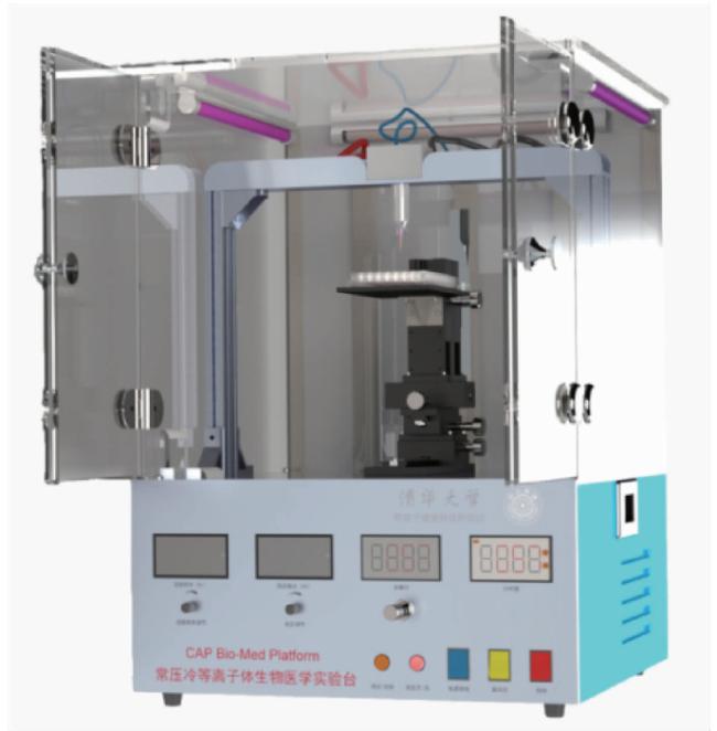
本研究使用清华大学工程物理系等离子体健康科技研究组研发的常压冷等离子体生物医学实验台(图 1),在超净台内对培养在96孔细胞培养板中的HGFs进行处理[10]。该实验台放电单元采用同轴型介质阻挡放电结构,高压电极为直径1 mm的钨针,外面套有内径1 mm、外径2 mm的玻璃管,地电极则使用铜箔胶带缠绕在外径为8 mm、内径为6 mm的玻璃管上。工作时两绝缘玻璃管间的空间中充满高纯氦气(99.999%),在外加高压交流电场的作用下电离形成等离子体。本实验中采用的高压交流电源驱动频率为23 kHz,放电电压幅值为3.5 kV,氦气流量为8 L/min,放电功率为1.17 W。
图1 常压冷等离子体生物医学实验台

Figure 1 Cold atmosphere plasma Bio-Med Platform

使用电压/电荷李萨如图形(Lissajous figure)计算CAP放电功率[11]。该方法是通过在放电回路中串联一个已知容值(Cm)的测试电容,以反映放电过程中的电荷量。利用数字示波器和两个电压探头,同步测量放电电压(U)和电容两端电压(Uc),并在U-Uc坐标系上描绘出闭合的李萨如图形。放电功率(Pin)根据该图形所包围的面积(A)计算得出,公式为Pin=f·Cm·A,其中f是交流电源驱动频率。

1.2 HGFs的体外培养及CAP处理

本研究所使用的HGFs购自中国湖北省武汉普诺赛生命科技有限公司,使用前通过波形蛋白免疫荧光进行鉴定。本实验选用第4~7代HGFs,并将细胞培养在含5%(体积分数)CO2的37 ℃恒温培养箱内。实验用培养基来自美国Gibco公司,由Dulbecco改良Eagle培养基(Dulbecco ’ s modified Eagle ’ s medium, DMEM)、10%(体积分数)胎牛血清及1%(体积分数)青霉素-链霉素组成。HGFs每2~3天更换1次培养液。
将实验台放置于超净台内对HGFs进行处理,射流装置头末端距孔板上缘1 mm。由于实验采用开放环境放电,为了避免空气对高浓度工作气体造成混杂,在处理前使用高纯氦气吹扫1 min, 以尽可能排空处理仓内电极间的空气。依据CAP处理时长进行实验分组,分为未经CAP处理组(对照组)及CAP处理20 s、60 s、120 s、180 s组。将CAP处理后的孔板置于37 ℃培养箱内孵育3 h后更换培养液。

1.3 HGFs培养液温度、酸碱度及活性氧含量检测

将100 μL HGFs培养液置于96孔细胞培养板内,将实验台放置于超净台内对HGFs培养液进行处理。使用台式酸度计(PHS-3E,雷磁,中国)在处理后即刻对培养液温度及酸碱度进行检测。向处理后的培养液中加入Amplex® Red试剂,该试剂与过氧化氢(hydrogen peroxide, H2O2)按1 ∶ 1的化学计量比反应,生成红色荧光氧化产物;该产物在波长λ=550 nm处激发,在λ=595 nm处发射荧光。采用多功能酶标仪(Infinite M1000 PRO,Tecan,瑞士)测定处理后培养液中长效活性氧(reactive oxygen species,ROS)H2O2浓度。实验重复3次, 每次每组4个平行样。

1.4 HGFs黏附形态检测

将生长状态良好的HGFs以5 000个/孔的密度接种于96孔细胞培养板内,每孔加入100 μL DMEM培养基,将培养孔板置于培养箱中培养24 h。使用实验台按实验分组对HGFs进行处理,处理后继续培养24 h,使用磷酸盐缓冲液(phosphate buffer saline,PBS)冲洗3遍(每次浸泡5 min)。使用异硫氰酸酯(fluorescein isothiocyanate,FITC)标记的鬼笔环肽(phalloidin,Sigma公司,美国)进行胞浆染色,使用4 ’, 6-二脒基-2-苯基吲哚(4 ’, 6-diamidino- 2-phenylindole,DAPI,Roche Basler公司,瑞士)进行细胞核染色。使用高内涵细胞成像系统(high content analysis system,Operetta CLS,PerkinElmer,美国)对细胞形态进行观察。使用ImageJ软件(美国国立卫生研究院)随机选取视野内10个细胞测量面积并进行组间比较。

1.5 HGFs迁移能力检测

将生长状态良好的培养至4~7代的HGFs以20 000个/孔的密度接种于96孔细胞培养板内,每孔加入100 μL DMEM培养基,将培养孔板置于培养箱中培养24 h。使用实时活细胞分析系统(IncuCyte S3 system,Sartorius公司,德国)制备划痕。使用实验台按实验分组对HGFs进行处理,更换无血清培养基后继续使用实时活细胞分析系统在处理第0 h、12 h和24 h拍摄照片,分别计算12 h及24 h的相对划痕密度(%),即测试时刻划痕区域细胞密度与初始时刻划痕区域外细胞密度的比值。实验重复3次, 每次每组5个平行样。

1.6 HGFs增殖能力检测

将生长状态良好的培养至4~7代的HGFs以5 000个/孔的密度进行接种,每孔加入100 μL DMEM培养基,将培养孔板置于培养箱中培养24 h。使用实验台按实验分组对HGFs进行处理,处理后继续培养24 h、48 h及72 h,并测定细胞增殖能力。检测时吸去原培养基及未贴壁的细胞,PBS冲洗3遍,每孔加入90 μL DMEM培养基及10 μL细胞计数试剂(cell counting kit 8,CCK-8)。在培养箱中孵育2 h,吹打混匀后将培养基移至96孔板中,使用酶标仪(ELX808,BioTek公司,美国)在波长450 nm下测定光密度值。实验重复3次, 每次每组5个平行样。

1.7 统计学分析方法

所有结果均由软件SPSS 27.0进行统计分析,数据用均值±标准差表示。本研究组间样本量相同,为了降低假阳性率,在单因素方差分析中选择Tukey方法进行比较,显著性水平设置为P=0.05。

2 结果

2.1 CAP处理剂量

随着CAP处理时间的延长,其剂量逐渐提高。CAP处理20 s、60 s、120 s和180 s的剂量依次为23.4 J、70.2 J、140.4 J和210.6 J。

2.2 CAP处理后HGFs培养液温度、酸碱度及H2O2浓度变化

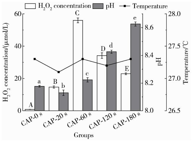
CAP处理前后,细胞培养液温度无明显变化。未经CAP处理组pH为8.18±0.06,CAP处理20 s组pH为8.13±0.20,随着处理时间从60 s延长至180 s,细胞培养液碱性显著增加,pH依次为8.22±0.15、8.43±0.01、8.63±0.15(P<0.01,图 2)。
图2 对照组及CAP处理组细胞培养液中温度,pH及H2O2浓度

Figure 2 Temperature, pH, and concentration of H2O2 in the cell culture medium of the control group and the CAP-treated groups

CAP-0 s, CAP-20 s, CAP-60 s, CAP-120 s, and CAP-180 s represent no CAP treatment, CAP treatment for 20 s, 60 s, 120 s, and 180 s respectively. Differences in H2O2 concentration among groups are indicated by uppercase letters. Differences in pH among groups are indicated by lowercase letters. Different letters indicate that the difference between groups is statistically significant (P < 0.05). CAP, cold atmosphere plasma.

使用Amplex® Red试剂检测不同剂量CAP处理后细胞培养液中H2O2浓度,结果显示,在CAP处理0~60 s,细胞培养液中H2O2浓度随着处理时间的增加而升高,其中CAP处理20 s组H2O2浓度为(14.70±0.67)μmol/L,CAP处理60 s组H2O2浓度为(55.96±1.51) μmol/L;当处理时间延长至120 s及180 s后,细胞培养液中H2O2浓度下降,分别为(34.16±1.74) μmol/L和(22.92±0.57) μmol/L (图 2)。

2.3 CAP处理改变HGFs形态

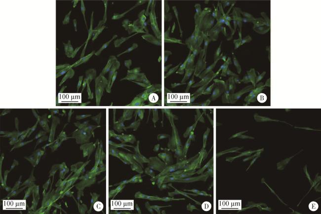
CAP处理后培养HGFs 24 h并对细胞核及细胞骨架进行染色,观察细胞形态。结果显示,CAP处理20 s、60 s及120 s组HGFs细胞形态伸展并伸出伪足,细胞间连接更多;而CAP处理180 s组细胞多呈窄长梭形,细胞间连接较少(图 3)。
图3 高内涵细胞成像系统观察对照组及CAP处理组HGFs形态(免疫荧光染色×20)

Figure 3 Observation of HGFs' morphology in control and CAP-treated groups by high content analysis system (immunofluorescence staining ×20)

A, no CAP treatment group; B, CAP treatment for 20 s group; C, CAP treatment for 60 s group; D, CAP treatment for 120 s group; E, CAP treatment for 180 s group. CAP, cold atmosphere plasma; HGFs, human gingival fibroblasts.

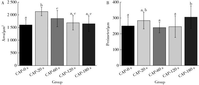
在20倍镜视野下随机选取10个细胞测量面积及周长并比较平均值,结果见 图 4。CAP处理20 s组,细胞表面积显著大于未经CAP处理组及其他各处理组(P < 0.05),CAP处理60 s组细胞表面积也较未处理组有明显增加(P < 0.05)。CAP处理180 s组细胞周长大于未经CAP处理组及CAP处理60 s和120 s组(P < 0.05)。
图4 对照组及CAP处理组HGFs的表面积(A)及周长(B)

Figure 4 Spreading area (A) and perimeter (B) of HGFs in the control and CAP-treated groups

CAP-0 s, CAP-20 s, CAP-60 s, CAP-120 s and CAP-180 s represent no CAP treatment, CAP treatment for 20 s, 60 s, 120 s and 180 s respectively. Different lowercase letters indicate that the difference between groups is statistically significant (P < 0.05). CAP, cold atmosphere plasma; HGFs, human gingival fibroblasts.

2.4 CAP处理改变HGFs迁移能力

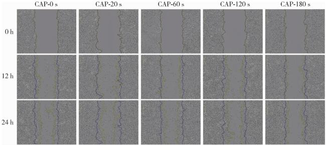
使用实时活细胞分析系统制备划痕,并在不同时长处理后0 h、12 h及24 h拍摄实时影像获得HGFs在不同时刻的迁移图像(图 5)。
图5 划痕制备后对照组及CAP处理组HGFs迁移图像

Figure 5 Representative images of HGFs migration in the control and CAP-treated groups after scratch wounding

CAP-0 s, CAP-20 s, CAP-60 s, CAP-120 s and CAP-180 s represent no CAP treatment, CAP treatment for 20 s, 60 s, 120 s and 180 s respectively. The initial wound boundaries (0 h) are outlined in blue. The migrated edges after 12 h and 24 h are outlined in yellow. CAP, cold atmosphere plasma; HGFs, human gingival fibroblasts.

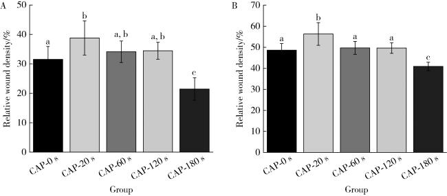
在12 h及24 h测定经不同剂量CAP处理后HGFs的相对划痕密度,比较不同剂量CAP处理对HGFs迁移能力的影响,结果见图 6。HGFs经CAP处理180 s后在12 h及24 h的相对划痕密度均显著低于未经CAP处理组及CAP处理20 s、60 s、120 s组(P<0.05),CAP处理20 s组HGFs在12 h及24 h时的相对划痕密度显著优于未经CAP处理组(P<0.05)。
图6 不同剂量CAP处理后12 h(A)和24 h(B)HGFs的迁移能力

Figure 6 Migratory ability of HGFs at 12 h (A) and 24 h (B) after treatment in control and CAP-treated groups

CAP-0 s, CAP-20 s, CAP-60 s, CAP-120 s and CAP-180 s represent no CAP treatment, CAP treatment for 20 s, 60 s, 120 s and 180 s respectively. Different lowercase letters indicate that the difference between groups is statistically significant (P < 0.05). CAP, cold atmosphere plasma; HGFs, human gingival fibroblasts.

2.5 CAP处理改变HGFs增殖能力

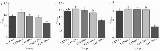
通过在不同培养时刻测定经不同剂量CAP处理后的HGFs数量,比较不同剂量CAP对HGFs黏附及增殖能力的影响,结果如图 7所示。HGFs经CAP处理180 s后培养24 h、48 h及72 h,细胞数量均显著低于未经CAP处理组及CAP处理20 s、60 s、120 s组(P<0.05)。CAP处理20 s可在培养24 h及48 h时显著提高HGFs的数量(P<0.05),且效果显著优于CAP处理60 s和120 s组(P<0.05)。随着HGFs培养时间延长至72 h,CAP处理对HGFs数量的促进作用逐渐减小,但在CAP处理20 s、60 s、120 s组间差异无统计学意义(P>0.05)。
图7 对照组及CAP处理组HGFs培养24 h(A)、48 h(B)、72 h(C)细胞增殖能力(CCK-8)

Figure 7 The proliferation ability of HGFs in the control and CAP-treated groups measured by CCK-8 assay after 24 h (A), 48 h (B) and 72 h (C) of culture

CAP-0 s, CAP-20 s, CAP-60 s, CAP-120 s and CAP-180 s represent no CAP treatment, CAP treatment for 20 s, 60 s, 120 s and 180 s respectively. Different lowercase letters indicate that the difference between groups is statistically significant (P < 0.05). CAP, cold atmosphere plasma; HGFs, human gingival fibroblasts; CCK-8, cell counting kit 8.

3 讨论

大气压放电冷等离子体在口腔医学领域中有着广泛的应用。CAP处理陶瓷、金属、树脂等生物材料后,可通过在材料表面接枝活性基团增加口腔生物材料的亲水性,提高口腔生物材料的生物相容性及粘接强度[12-13]。CAP对口腔颌面部感染性疾病(龋病、牙髓及根尖周病、牙周病、种植体周病等)的多种致病菌均具有良好的杀灭作用,具备去除龋坏牙体组织、根管消毒、消毒义齿等功能[14]。此外,CAP还可用于牙齿漂白。然而,CAP直接应用于口腔内环境时,其对牙龈、口腔黏膜等组织细胞生物学行为的影响鲜有文献报道,亟待论证。CAP直接作用于细胞可通过修饰代谢酶、调控细胞代谢通路、影响线粒体功能等方式影响细胞代谢,其作用效果与CAP剂量相关[15]。生理水平的CAP具有促进成纤维细胞迁移、增殖、再血管化等功能,而病理水平的CAP可损伤细胞DNA,引起细胞衰老、凋亡、坏死及其他免疫反应[16-17]。因此,本研究使用不同剂量CAP直接作用于HGFs,从剂量依赖性角度系统评估CAP对HGFs迁移与增殖的影响。
CAP与细胞培养体系间存在复杂的相互作用,是涉及多重物理、化学和生物过程的非线性、多变量耦合。鉴于此,本研究参照本课题组提出的剂量学评估方法[10],通过固定放电电压、频率和气流量等物理参数,确保放电功率(Pin)一致,保证CAP源特性恒定。在此恒定工况下,仅调节处理时间(t),可有效地控制等离子体在培养基中的累积效应和持续作用时间。在这种特定条件下,作用剂量可通过输入功率乘以时间进行量化(即放电能量Weff=Pin×t),从而实现了在恒定CAP源特性下的精准剂量调节。
本研究结果显示,CAP对HGFs生物学行为的影响同样存在剂量相关性,低剂量CAP(20 s)处理HGFs后,细胞表现出更大的表面积(P<0.05),迁移能力及增殖能力也较未经CAP处理及中等剂量CAP(60 s、120 s)处理组有显著提升(P<0.05)。当CAP剂量过高时(180 s),HGFs在24 h、48 h和72 h的增殖能力均显著低于未经CAP处理组,相对细胞密度亦显著低于未经CAP处理组,表现为迁移及增殖能力均被抑制。
有关等离子体活化培养基的研究显示,等离子体内各类带电粒子、光子以及ROS、活性氮等化学活性基团溶解于培养液,可改变培养液温度、酸碱度及ROS含量,进而影响细胞生物学行为[18-19]。本实验过程严格控制等离子体射流温度,结果显示,经不同剂量CAP处理后的培养液温度均接近室温且差异无统计学意义。对培养液pH测定的结果显示,含10%胎牛血清及1%青霉素-链霉素的DMEM培养基pH为8.18,当其置于5%CO2恒温培养箱中平衡后,pH稳定至中性,有利于细胞生长。ROS是等离子体作用于水/培养基等溶液后产生的一类化学性质非常活泼、含氧的化学反应分子和自由基的总称[20],常见液相ROS主要包括·O、·OH、1O2、H2O2、O3、O2-等,其中,H2O2是最常见的长效ROS[21]。因此,本研究通过测定H2O2浓度来表征不同剂量CAP处理培养液后培养液中ROS含量。
本实验中,低剂量CAP(CAP-20 s)处理可为培养液注入能量并发生系列反应,引起培养液中H2O2浓度的升高(P<0.05);培养液pH变化幅度较小,推测是培养液中缓冲物质尚充足。此时,低剂量CAP产生的适宜剂量的ROS可诱导细胞产生轻度氧化应激,激活细胞的抗氧化防御系统,进而促进细胞增殖、迁移和组织再生[22]。培养液H2O2浓度在CAP处理60 s组达到峰值,既往研究认为当ROS超过某一阈值后,过量的ROS会导致细胞氧化还原稳态失衡,引发不可逆的氧化损伤[23],从而触发细胞凋亡、坏死、焦亡乃至铁死亡等多种细胞死亡模式[24],这可能是CAP处理60 s组HGFs增殖及迁移能力低于CAP处理20 s组的原因。等离子体活化水中活性成分的相关研究显示,随着CAP剂量的进一步增加,大量H2O2在其他离子、紫外线辐射等的作用下发生分解,并与溶液中其他有机物反应,消耗速率大于生成速率,浓度逐渐降低[25],这与本研究中CAP处理120 s及180 s组的结果一致。此时,培养液中的缓冲物质逐渐消耗,碱性物质累积,导致培养液pH显著增大(P<0.01)。推测过高剂量(180 s)CAP处理后H2O2浓度出现回落,但HGFs迁移及增殖能力仍被抑制的原因可与培养液过高的pH有关[26]
本研究仍存在一定的局限性。第一,CAP在与培养基作用时会产生大量的含有氧元素及氮元素的活性物质,上述物质对细胞的生物学行为均可能产生影响,本研究仅检测了一种长效ROS物质H2O2,但由于不同氧化物的时效性及生物学作用不同,仅测定H2O2浓度难以反映ROS的生物学效应,故应在后续实验中进一步对ROS的其他代表性指标进行检测;同时在分子生物学层面应完善有关ROS作为信号分子是否能激活细胞内的增殖和迁移信号通路,加速HGFs的迁移等相关研究内容。第二,本研究初步从剂量依赖性角度探讨了CAP直接作用对HGFs生物学行为的影响,虽在一定程度上为CAP预防及治疗种植体周病提供了参考价值,但在临床转化前仍须进行动物实验、临床实验等对相关结论进行进一步验证。第三,有关过高的pH是高剂量CAP抑制HGFs增殖迁移能力这一推断缺乏相关的实验验证,应进一步完善有关培养液酸碱度对HGFs生物学行为影响的检测以进行验证。
综上所述,本研究发现CAP直接处理可改变HGFs培养液酸碱度及ROS含量。CAP直接处理对HGFs的增殖、迁移能力的影响具有剂量相关性,即低剂量CAP直接处理对HGFs的增殖及迁移能力具有促进作用,而过高剂量CAP直接处理则会抑制HGFs的增殖及迁移能力。

利益冲突  所有作者均声明不存在利益冲突。

作者贡献声明  郑苗:提出研究思路,撰写论文;郑苗、马欣蓉:设计研究方案;郑苗、马欣蓉、陈昊、赵恒欣:收集、分析、整理数据;王霄、李和平、谭建国、张宇:总体把关和审定论文。所有作者均参与论文修改,并对最终文稿进行审读和确认。

1
Berglundh T , Armitage G , Araujo MG , et al. Peri-implant diseases and conditions: Consensus report of workgroup 4 of the 2017 World Workshop on the Classification of Periodontal and Peri-Implant Diseases and Conditions[J]. J Clin Periodontol, 2018, 45 (Suppl 20): S286- S291.

2
Galarraga-Vinueza ME , Pagni S , Finkelman M , et al. Prevalence, incidence, systemic, behavioral, and patient-related risk factors and indicators for peri-implant diseases: An AO/AAP systematic review and meta-analysis[J]. J Periodontol, 2025, 96 (6): 587- 633.

DOI

3
Barootchi S , Wang HL . Peri-implant diseases: Current understanding and management[J]. Int J Oral Implantol (Berl), 2021, 14 (3): 263- 282.

4
Hakkers J , Vangsted TE , van Winkelhoff AJ , et al. Do systemic amoxicillin and metronidazole during the non-surgical peri-implantitis treatment phase prevent the need for future surgical treatment? A retrospective long-term cohort study[J]. J Clin Periodontol, 2024, 51 (8): 997- 1004.

DOI

5
Laroussi M , Bekeschus S , Keidar M , et al. Low-temperature plasma for biology, hygiene, and medicine: Perspective and roadmap[J]. IEEE Trans Radiat Plasma Med Sci, 2022, 6 (2): 127- 157.

DOI

6
Yang Y , Zheng M , Jia YN , et al. Time-dependent reactive oxygen species inhibit Streptococcus mutans growth on zirconia after a helium cold atmospheric plasma treatment[J]. Mater Sci Eng C Mater Biol Appl, 2021, 120, 111633.

DOI

7
Yang Y , Zheng M , Yang Y , et al. Inhibition of bacterial growth on zirconia abutment with a helium cold atmospheric plasma jet treatment[J]. Clin Oral Investig, 2020, 24 (4): 1465- 1477.

DOI

8
Zheng M , Ma XR , Tan JG , et al. Enhancement of biocompatibility of high-transparency zirconia abutments with human gingival fibroblasts via cold atmospheric plasma treatment: An in vitro study[J]. J Funct Biomater, 2024, 15 (7): 200.

DOI

9
Alqutaibi AY , Aljohani A , Alduri A , et al. The effectiveness of cold atmospheric plasma (CAP) on bacterial reduction in dental implants: A systematic review[J]. Biomolecules, 2023, 13 (10): 1528.

DOI

10
Li J , Zhao LX , He T , et al. A novel method for estimating the dosage of cold atmospheric plasmas in plasma medical applications[J]. Appl Sci, 2021, 11 (23): 11135.

DOI

11
Kogelschatz U . Dielectric-barrier discharges: Their history, discharge physics, and industrial applications[J]. Plasma Chem Plasma Process, 2003, 23 (1): 1- 46.

DOI

12
Matthes R , Jablonowski L , Miebach L , et al. In-vitro biofilm removal efficacy using water jet in combination with cold plasma technology on dental titanium implants[J]. Int J Mol Sci, 2023, 24 (2): 1606.

DOI

13
Matthes R , Jablonowski L , Pitchika V , et al. Training in the use of the water jet and cold atmospheric plasma jet for the decontamination of dental implants[J]. Clin Oral Investig, 2024, 28 (6): 355.

DOI

14
El Shishiny SA , Morad YO , Hindi RI , et al. Efficacy of non-thermal pressure plasma versus other modalities for disinfection of primary root canals[J]. BMC Oral Health, 2025, 25 (1): 54.

DOI

15
Hahn O , Waheed TO , Sridharan K , et al. Cold atmospheric pressure plasma-activated medium modulates cellular functions of human mesenchymal stem/stromal cells in vitro[J]. Int J Mol Sci, 2024, 25 (9): 4944.

DOI

16
Wu Y , Yu S , Zhang X , et al. The regulatory mechanism of cold plasma in relation to cell activity and its application in biomedical and animal husbandry practices[J]. Int J Mol Sci, 2023, 24 (8): 7160.

DOI

17
Scharf C , Eymann C , Emicke P , et al. Improved wound healing of airway epithelial cells is mediated by cold atmospheric plasma: A time course-related proteome analysis[J]. Oxid Med Cell Longev, 2019, 2019, 7071536.

18
Konchekov EM , Glinushkin AP , Kalinitchenko VP , et al. Properties and use of water activated by plasma of piezoelectric direct discharge[J]. Front Phys, 2021, 8, 616385.

DOI

19
Adachi T , Tanaka H , Nonomura S , et al. Plasma-activated medium induces A549 cell injury via a spiral apoptotic cascade involving the mitochondrial-nuclear network[J]. Free Radic Biol Med, 2015, 79, 28- 44.

DOI

20
Lee JH , Jaiswal MS , Jang YS , et al. No-ozone cold plasma induces apoptosis in human neuroblastoma cell line via increased intracellular reactive oxygen species (ROS)[J]. BMC Complement Med Ther, 2024, 24 (1): 46.

DOI

21
Kaushik N , Mitra S , Baek EJ , et al. The inactivation and destruction of viruses by reactive oxygen species generated through physical and cold atmospheric plasma techniques: Current status and perspectives[J]. J Adv Res, 2023, 43, 59- 71.

DOI

22
Boeckmann L , Schäfer M , Bernhardt T , et al. Cold atmospheric pressure plasma in wound healing and cancer treatment[J]. Appl Sci, 2020, 10 (19): 6898.

DOI

23
Dai X , Bazaka K , Thompson EW , et al. Cold atmospheric plasma: A promising controller of cancer cell states[J]. Cancers (Basel), 2020, 12 (11): 3360.

DOI

24
Dai X , Wu J , Lu L , et al. Current status and future trends of cold atmospheric plasma as an oncotherapy[J]. Biomol Ther (Seoul), 2023, 31 (5): 496- 514.

DOI

25
Milhan NVM , Chiappim W , Sampaio ADG , et al. Applications of plasma-activated water in dentistry: A review[J]. Int J Mol Sci, 2022, 23 (8): 4131.

DOI

26
Sampaio ADG , Chiappim W , Milhan NVM , et al. Effect of the pH on the antibacterial potential and cytotoxicity of different plasma-activated liquids[J]. Int J Mol Sci, 2022, 23 (22): 13893.

DOI

文章导航

/