论著

异种骨与人工合成骨在磨牙拔牙同期微翻瓣牙槽嵴保存术中的临床效果比较

  • 张斯巧 1, * ,
  • 刘建 1, * ,
  • 徐涛 , 2, * ,
  • 胡文杰 , 1, * ,
  • 张浩筠 1 ,
  • 危伊萍 1
展开
  • 1. 北京大学口腔医学院·口腔医院牙周科, 国家口腔医学中心, 国家口腔疾病临床医学研究中心, 口腔生物材料和数字诊疗装备国家工程研究中心, 口腔数字医学北京市重点实验室, 北京 100081
  • 2. 北京大学口腔医学院·口腔医院急诊科, 国家口腔医学中心, 国家口腔疾病临床医学研究中心, 口腔生物材料和数字诊疗装备国家工程研究中心, 口腔数字医学北京市重点实验室, 北京 100081

* These authors contributed equally to this work

收稿日期: 2025-10-10

  网络出版日期: 2025-12-09

基金资助

北京大学口腔医(学)院临床研究基金(PKUSS-2023CRF505)

版权

版权所有,未经授权,不得转载。

Clinical comparison of xenograft versus synthetic bone graft materials in micro crestal flap-alveolar ridge preservation following extraction of molars

  • Siqiao ZHANG 1 ,
  • Jian LIU 1 ,
  • Tao XU , 2, * ,
  • Wenjie HU , 1, * ,
  • Haoyun ZHANG 1 ,
  • Yiping WEI 1
Expand
  • 1. Department of Periodontology, Peking University School and Hospital of Stomatology & National Center for Stomatology & National Clinical Research Center for Oral Diseases & National Engineering Research Center of Oral Biomaterials and Digital Medical Devices & Beijing Key Laboratory of Digital Stomatology, Beijing 100081, China
  • 2. Department of Oral Emergency, Peking University School and Hospital of Stomatology & National Center for Stomatology & National Clinical Research Center for Oral Diseases & National Engineering Research Center of Oral Biomaterials and Digital Medical Devices & Beijing Key Laboratory of Digital Stomatology, Beijing 100081, China
XU Tao, e-mail,
HU Wenjie, e-mail,

Received date: 2025-10-10

  Online published: 2025-12-09

Supported by

the Clinical Research Foundation of Peking University School and Hospital of Stomatology(PKUSS-2023CRF505)

Copyright

All rights reserved. Unauthorized reproduction is prohibited.

摘要

目的: 重度牙周病变的患者在磨牙拔牙同期实施微翻瓣牙槽嵴保存术(micro crestal flap-alveolar ridge preservation, MCF-ARP)时, 对采用以脱蛋白牛骨矿物基质(deproteinized bovine bone mineral, DBBM)为主要成分的异种骨移植材料和采用以羟基磷灰石(hydroxyapatite, HA)为主要成分的人工合成骨移植材料的临床效果进行比较, 为骨移植材料的临床应用推广提供参考。方法: 本研究为回顾性病例系列研究, 纳入2024年10月至2025年4月期间就诊的患者, 所有患者均在拔牙同期接受MCF-ARP手术, 分别植入DBBM或HA。使用锥形束计算机断层扫描(cone beam computed tomography, CBCT)结合口内扫描模型, 对比评估两组术前及术后6个月的硬组织变化及牙槽嵴轮廓塌陷量, 并分析术后软组织愈合过程。结果: 共纳入14例患者的14颗重度牙周病变磨牙, 术后6个月时, 两组患者在硬组织指标上的差异无统计学意义(P>0.05);术后2周和1个月时, HA组的拔牙窝中央轮廓垂直向塌陷量显著高于DBBM组[术后2周: (2.73±1.89) mm vs. (0.00±0.79) mm, P < 0.05;术后1个月: (2.74±1.13) mm vs. (0.35±2.34) mm, P < 0.05];后续愈合时间点的牙槽嵴轮廓塌陷量两组间比较差异无统计学意义(P>0.05)。创面愈合方面, HA组在术后2周和1个月时的创面未覆盖角化黏膜的面积比例显著高于DBBM组(术后2周: 47.88%±6.56% vs. 29.43%±14.25%, P < 0.05;术后1个月: 25.68%±13.06% vs. 7.19%±7.18%, P < 0.01)。结论: 本研究有限的样本分析显示, 重度牙周病变的患者在磨牙拔牙同期行MCF-ARP时, 使用DBBM和HA在术后6个月时的硬组织指标与牙槽嵴轮廓指标差异无统计学意义, 使用DBBM时早期软组织愈合更快, 未来需开展随机对照试验并结合组织学评估以进一步验证。

本文引用格式

张斯巧 , 刘建 , 徐涛 , 胡文杰 , 张浩筠 , 危伊萍 . 异种骨与人工合成骨在磨牙拔牙同期微翻瓣牙槽嵴保存术中的临床效果比较[J]. 北京大学学报(医学版), 2026 , 58(1) : 74 -83 . DOI: 10.19723/j.issn.1671-167X.2026.01.010

Abstract

Objective: To compare the clinical outcomes between xenogeneic bone graft materials primarily composed of deproteinized bovine bone mineral (DBBM) and synthetic bone graft materials primarily composed of hydroxyapatite (HA) used in micro crestal flap-alveolar ridge preservation (MCF-ARP) of periodontally compromised molars, so as to provide a reference for their application and promotion. Methods: In this retrospective case series study, patients who received treatment between October 2024 and April 2025 were enrolled. All the patients underwent MCF-ARP, using either DBBM or HA. Hard tissue changes and alveolar ridge contour collapse were evaluated and compared between the two groups preoperatively and 6 months postoperatively using cone beam computed tomography (CBCT) and intraoral scanning. Soft tissue healing process after the surgery was also assessed. Results: A total of 14 molars from 14 patients were included. No significant differences were found in hard tissue changes between the two groups 6 months after the surgery (P>0.05). Two weeks and 1 month postoperatively, the vertical collapse of the contour at the ridge center was significantly greater in the HA group compared with the DBBM group [2 weeks: (2.73±1.89) mm vs. (0.00±0.79) mm, P < 0.05; 1 month: (2.74±1.13) mm vs. (0.35±2.34) mm, P < 0.05]. No significant differences were found in other sites at any other follow-up points (P>0.05). In terms of wound healing, the HA group showed a significantly higher percentage of wound area lacking keratinized tissue coverage compared with the DBBM group both 2 weeks and 1 month after the surgery (2 weeks: 47.88%±6.56% vs. 29.43%±14.25%, P < 0.05; 1 month: 25.68%±13.06% vs. 7.19%±7.18%, P < 0.01). Conclusion: Within the limitations of this study, the analysis suggests that there is no statistically significant difference in hard tissue and alveolar ridge contour parameters 6 months after MCF-ARP following extraction of periodontally compromised molars. However, early soft tissue healing is faster when using DBBM. Future randomized controlled trials with histological analysis are warranted for further validation.

重度牙周病变的患牙在拔牙后的自然愈合过程中常伴随牙槽骨吸收,导致缺牙区严重的软硬组织缺损[1-2]。在拔牙同期行牙槽嵴保存术可减少拔牙后牙槽嵴的水平和垂直骨吸收[3-6],实现软硬组织的保存或增量,从而减少种植治疗中额外软硬组织增量手术的需求,简化治疗流程[7-11]
目前,临床用于牙槽嵴保存术的骨移植材料种类众多,其中应用最为广泛的是异种骨移植材料。脱蛋白牛骨矿物基质(deproteinized bovine bone mineral, DBBM)在临床中使用较多,其特点是骨引导作用较强,体积稳定性良好[12]。近年来,人工合成骨移植材料也逐渐应用于口腔临床实践,此种材料来源较为丰富,安全性好,具有良好的临床推广潜能。人工合成骨移植材料的主要成分一般为羟基磷灰石(hydroxyapatite,HA),该材料可制备为孔径均匀的三维立体结构,具备一定的骨引导作用。
有文献报道,在前牙及前磨牙行牙槽嵴保存术时,异种骨移植材料维持牙槽嵴宽度及高度的效果优于人工合成骨移植材料[13],但目前仍缺乏在重度牙周病变磨牙位点中使用效果的相关研究证据。本课题组既往开展了一系列聚焦于重度牙周病变磨牙位点的临床研究,并开创了微翻瓣牙槽嵴保存术式(micro crestal flap-alveolar ridge preservation, MCF-ARP)[14]。既往研究证实,使用DBBM对重度牙周病变的拔牙位点进行牙槽嵴保存术可以有效减少拔牙后的牙槽嵴宽度和高度丧失,降低种植治疗的复杂性[2, 9, 15],同时可保留更多的角化组织[16-17]。本研究拟通过锥形束计算机断层扫描(cone beam computed tomography, CBCT)和口内扫描模型数据分析,比较在重度牙周病变磨牙拔牙同期使用DBBM或HA实施MCF-ARP的临床效果。

1 资料与方法

1.1 患者一般资料

本研究为回顾性病例系列研究,纳入2024年10月至2025年4月就诊于北京大学口腔医院牙周科、因重度牙周病变而拔除磨牙并接受牙槽嵴保存术的患者,对患者的临床资料进行回顾性分析。本研究共纳入20例患者的20颗磨牙进行评估,其中6颗不符合纳入标准而排除,最终共纳入14例患者的14颗磨牙。本研究已获得北京大学口腔医院生物医学伦理委员会批准(2025-01-107-08a)。
纳入标准:(1)患者全身健康,或合并高血压/糖尿病但控制良好,其中,高血压患者要求收缩压 < 160 mmHg、舒张压 < 100 mmHg,糖尿病患者要求空腹血糖 < 7.0 mmol/L;(2)拔牙前已进行完善的牙周基础治疗,待拔磨牙已进行龈上和龈下超声清创,去除菌斑、牙结石,控制局部炎症;(3)患牙为因重度牙周病变需要拔除的磨牙,且基线特征符合:至少一个位点探诊深度(probing depth, PD)>6 mm,附着丧失(attachment loss, AL)≥5 mm,影像学检查示牙槽骨吸收超过根长的1/2及以上,多根牙有根分叉病变,牙齿松动>Ⅱ°,患牙牙槽窝存在骨缺损,至少有2个剩余骨壁高度≥3 mm(颊、舌、近远中骨壁),存在至少1颗邻牙;(4)具有拔牙前和术后6个月(±1个月)两次CBCT检查结果;(5)具有术前、术后即刻、术后2周(±5 d)、术后1个月(±10 d)、术后3个月(±10 d)、术后6个月(±1个月)完善的口内扫描数据;(6)使用DBBM(Bio-Oss®骨粉)或HA(瑞邦®骨泰松质骨粒Ⅰ型)进行MCF-ARP,根据使用的骨移植材料,分别划分进入DBBM组和HA组。
排除标准:(1)患者 < 18岁;(2)吸烟≥10支/d;(3)怀孕期或哺乳期妇女;(4)头颈部放射治疗史;(5)长期服用非甾体类抗炎药、皮质类固醇、四环素、双膦酸盐或者其他影响骨再生的药物;(6)牙列明显拥挤、异位、扭转;(7)患牙存在急性感染;(8)牙龈增生。

1.2 干预措施

1.2.1 术前准备

纳入的患者术前需已进行完善的全口牙周基础治疗,待拔磨牙已进行龈上和龈下超声清创,去除牙结石和菌斑,控制局部炎症。拔牙前需已进行口内扫描并拍摄CBCT影像。

1.2.2 拔牙同期行MCF-ARP

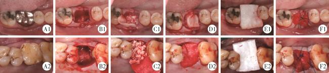
手术过程如图 1所示:常规消毒,局部麻醉下,围绕患牙距龈缘0.5~1.0 mm行内斜切口,微创拔牙,充分清创,使用P24G骨膜分离器于拔牙窝颊侧与舌/腭侧牙槽嵴顶处根向微翻瓣,暴露2 mm骨嵴,拔牙窝内植入骨移植材料,DBBM组使用Bio-Oss®骨粉(0.5 g,大颗粒,Geistlich公司,瑞士),HA组使用瑞邦®骨泰松质骨粒Ⅰ型(0.5 cm3,大颗粒,上海瑞邦生物材料有限公司,上海),骨粉稍过量,超过拔牙窝最高骨嵴顶2~3 mm,将Bio-Gide®可吸收膜(13 mm×25 mm,Geistlich公司,瑞士)修剪为合适大小后插入骨嵴顶与龈瓣间,完全覆盖骨移植材料,可吸收膜表面覆盖倍菱®可吸收胶原蛋白海绵(4.5 cm×1.5 cm×0.2 cm,益而康生物工程有限公司,北京)封闭创面,颊舌/腭侧交叉缝合固定。
图1 微创拔牙同期分别应用DBBM或HA行MCF-ARP的手术过程

Figure 1 The surgical procedure of minimally invasive tooth extraction combined with MCF-ARP using DBBM or HA

A1-F1, DBBM group; A2-F2, HA group; A1 and A2, preoperative occlusal view; B1 and B2, minimally invasive tooth extraction, thorough debridement of the socket, and minimal flap reflection to expose the bone surface; C1 and C2, bone graft material placed in the socket, absorbable membrane placed between the gingival flap and the crestal bone; D1 and D2, absorbable membrane covering the bone graft material extending to the apical aspect of the contralateral crestal bone; E1 and E2, absorbable collagen sponge covering the membrane; F1 and F2, coronally repositioned flap, fixed with 4-0 non-absorbable interrupted sutures. MCF-ARP, micro crestal flap-alveolar ridge preservation; DBBM, deproteinized bovine bone mineral; HA, hydroxyapatite.

1.2.3 随访资料的收集

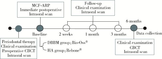
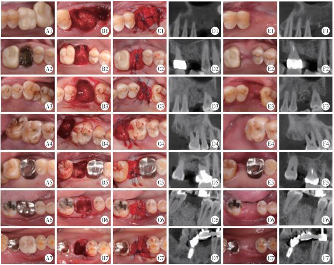
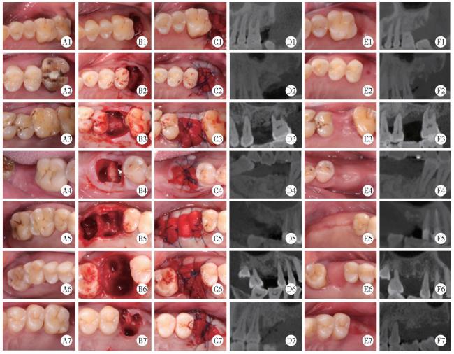
术后即刻与术后2周、1个月、3个月、6个月的口内扫描数据均被纳入分析以评估牙槽嵴轮廓,同时收集术后6个月种植前再次拍摄的CBCT影像,分析硬组织变化,整体随访及数据收集流程如图 2所示。两组患者随访过程的临床照片与CBCT影像对比如图 3图 4所示。
图2 数据收集流程

Figure 2 Data collection

MCF-ARP, micro crestal flap-alveolar ridge preservation; DBBM, deproteinized bovine bone mineral; HA, hydroxyapatite; CBCT, cone beam computed tomography.

图3 DBBM组患者不同时间点的临床照片与CBCT图像

Figure 3 Clinical photographs and CBCT cross-sectional images of DBBM group at different time points

A1-A7, preoperative occlusal view; B1-B7, tooth socket; C1-C7, immediate postoperative occlusal view; D1-D7, immediate postoperative CBCT; E1-E7, 6 months follow-up; F1-F7; CBCT taken 6 months postoperative. Abbreviations as in Figure 2.

图4 HA组患者不同时间点的临床照片与CBCT图像

Figure 4 Clinical photographs and CBCT cross-sectional images of HA group at different time points

A1-A7, preoperative occlusal view; B1-B7, tooth socket; C1-C7, immediate postoperative occlusal view; D1-D7, immediate postoperative CBCT; E1-E7, 6 months follow-up; F1-F7; CBCT taken 6 months postoperative. Abbreviations as in Figure 2.

1.3 测量指标

1.3.1 硬组织变化测量参数

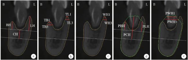
参考本课题组既往发表的影像学指标测量方法[2],使用Mimics Research 21.0软件(Materialise,Leuven,Belgium)对两组患者的术区基线参数和术后6个月的CBCT进行配准。在配准后的CBCT中,选取穿过拔牙窝中央的冠状面作为参考平面,以牙长轴作为垂直参考线,以上颌窦底或下颌管上缘为基准作水平参考线,在此平面内,测量以下硬组织参数:拔牙窝中央骨高度(height at the center of the socket,CH),颊侧骨壁高度(buccal bone height,BH),舌/腭侧骨壁高度(lingual/palatal bone height,LH),颊、舌/腭侧骨嵴顶根方1 mm、3 mm处骨壁厚度(若骨壁缺失则记为0),高骨壁嵴顶根方1 mm、3 mm处骨嵴宽度(若高骨壁与低骨壁高度差大于3 mm,则此处骨嵴宽度等于骨壁厚度)。愈合后阶段按照相同方法测量(图 5)。
图5 CBCT影像学指标的测量

Figure 5 Measurement of CBCT radiographic indicators

a, height at the center of the socket (CH), buccal bone height (BH) and lingual/palatal bone height (LH); b, bone wall thicknesses at 1 mm and 3 mm apically below the buccal and lingual/palatal bone crests (TB1, TB3, TL1, TL3); c, ridge widths at 1 mm and 3 mm apically below the higher bone crests (WH1, WH3); d, postoperative height at the center of the socket (PCH), postoperative buccal bone height (PBH) and postoperative lingual/palatal bone height (PLH); e, postoperative ridge widths at 1 mm and 3 mm apically below the higher bone crests (PWH1, PWH3). Yellow curve, baseline alveolar bone contour; Green curve, 6-month postoperative alveolar bone contour. B, buccal; L, lingual; CBCT, cone beam computed tomography.

1.3.2 牙槽嵴轮廓塌陷量的测量

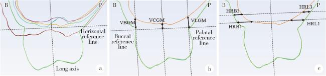
将各随访时间点的口内扫描模型导入Geomagic Wrap 2021软件(3D Systems Inc., Rock Hill, SC, USA)进行配准,将配准后的文件导入3-matic Research 13.0软件(Materialise, Leuven, Belgium)。建立穿过颊舌面中点的冠状参考平面,过颊侧、舌/腭侧游离龈缘且平行于牙长轴作垂直参考线,以术后6个月牙槽嵴轮廓最冠方点确定水平参考线。测量各随访时间点颊侧龈缘垂直向变化,舌/腭侧龈缘垂直向变化,拔牙窝中央轮廓垂直向变化,愈合后嵴顶根方1 mm、3 mm处颊侧、舌/腭侧水平向变化,如图 6所示。
图6 口内扫描牙槽嵴轮廓指标的测量

Figure 6 Measurement of alveolar ridge contour parameter by intraoral scanning

a, changes in soft tissue contour at follow-up time points; b, vertical reduction of the contour on the buccal free gingival margins (VBGM), vertical reduction of the contour on the lingual/palatal free gingival margins (VLGM) and vertical reduction of the contour on the center of gingival margins (VCGM); c, horizontal reduction of the buccal or lingual/palatal contour at 1 mm, 3 mm apically (HRB1, HRB3, HRL1, HRL3). Green curve, preoperative; Red curve, immediately postoperative; Blue curve, 2 weeks postoperative; Purple curve, 1 month postoperative; Orange curve, 3 months postoperative; Yellow curve, 6 months postoperative. B, buccal; P, palatal.

1.3.3 软组织愈合过程分析

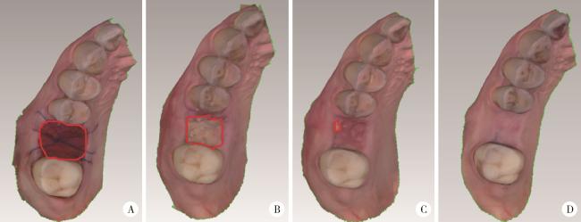
将各随访时间点的口内扫描模型以PLY格式导入Geomagic Wrap 2021软件,将模型调整至咬合面并保存该截面图像,于Image J(National Institutes of Health,Bethesda, MD)软件中选取术区未被角化黏膜完全覆盖的部分(以下简称“创面”),测量其面积,计算随访2周、1个月、3个月时创面面积与术后即刻创面面积的比值,如图 7所示。
图7 不同时间节点创面面积的测量

Figure 7 Measurement of wound area at different time points

A, immediately postoperative; B, 2 weeks postoperative; C, 1 month postoperative; D, 3 months postoperative. Red curve, wound area.

1.4 统计学分析

本研究所有数据采用SPSS 27.0(IBM Corporation, Armonk, NY, 美国)统计学软件进行分析。由一位有经验的研究者对所有患牙进行CBCT测量与牙槽嵴轮廓测量,间隔大于7天后再次测量,使用组内相关系数(intra-class correlation coefficient,ICC)评价研究者自身一致性,ICC>0.80认为信度良好,结果显示CBCT测量的ICC值为0.918~0.974,牙槽嵴轮廓测量的ICC值为0.923~0.982。
使用Shapior-Wilk进行正态性检验,分类变量使用卡方检验,当期望值< 5时进行Fisher精确检验。
对于硬组织变化及牙槽嵴轮廓塌陷量的比较,在符合正态分布时采用独立样本t检验,数据非正态分布时使用Mann-Whitney U检验。对符合正态分布的数据以均数±标准差(${\bar x}$±s)表示,非正态分布的数据以中位数(四分位数间距)[M (P25P75)]表示。使用重复测量方差分析对不同随访时间点的牙槽嵴轮廓塌陷量参数进行组内和组间比较。P < 0.05为差异有统计学意义。

2 结果

2.1 纳入患者的基线情况

本研究纳入了14例患者的14颗磨牙,DBBM组和HA组患者手术时的年龄、性别、牙位分布、基线骨壁厚度及骨壁高度差异均无统计学意义(均P>0.05,表 1)。
表1 患者基线资料的比较

Table 1 Comparison of patient baseline characteristics

Items DBBM (n=7) HA (n=7) t/Z P
Age/years 52.29±9.90 53.86±11.17 -0.28a 0.785
Gender 0.266
  Female 4 1
  Male 3 6
Tooth position >0.999
  Maxillary 5 6
  Mandibular 2 1
BH/mm 5.32±3.69 4.28±4.00 0.51a 0.618
LH/mm 5.85±3.97 4.67±3.74 0.57a 0.577
CH/mm 5.08±3.53 3.65±2.04 0.93a 0.374
TB1/mm 1.40±1.02 1.09±0.89 0.61a 0.555
TB3/mm 1.64±1.39 1.65±1.60 -0.02a 0.987
TL1/mm 1.46±0.40 1.58±1.07 -0.29a 0.780
TL3/mm 2.07±0.79 2.18±1.99 -0.13a 0.897
WH1/mm 1.83 (1.52, 2.19) 1.70 (1.31, 8.28) -0.06b 0.949
WH3/mm 6.83 (2.57, 13.26) 5.67 (1.60, 12.77) -0.06b 0.949

Data are presented as n, ${\bar x}$±s or M (P25, P75). a, represents t value; b, represents Z value. DBBM, deproteinized bovine bone mineral; HA, hydroxyapatite; BH, buccal bone height; LH, lingual/palatal bone height; CH, height at the center of the socket; TB1, TB3, TL1, TL3, bone wall thicknesses at 1 mm and 3 mm apically below the buccal and lingual/palatal bone crests; WH1, WH3, ridge widths at 1 mm and 3 mm apically below the higher bone crests.

2.2 硬组织指标的变化

两组患者基线参数和愈合6个月后的硬组织变化见表 2。愈合6个月后,两组患者的上述各项指标差异均无统计学意义(P>0.05)。愈合前后只有BH的变化量差异有统计学意义(P=0.004),其他指标的差异均无统计学意义(P>0.05)。
表2 硬组织指标变化的比较

Table 2 Comparison of changes in hard tissue parameter

Items DBBM (n=7) HA (n=7) t/Z P
PBH/mm 3.45±4.01 6.11±3.38 -1.34a 0.204
PLH/mm 5.85±3.64 5.46±2.32 0.24a 0.813
PCH/mm 13.61±4.07 10.43±5.03 1.30a 0.217
DBH/mm -1.88±1.48 1.83±2.36 -3.53a 0.004*
DLH/mm 0.00±3.07 0.89±3.34 -0.52a 0.615
DCH/mm 8.53±2.56 6.77±3.45 1.09a 0.298
PWH1/mm 8.84±2.66 8.66±3.14 0.12a 0.908
PWH3/mm 10.55±2.65 11.37±2.46 -0.60a 0.558
DWH1/mm 7.10 (4.73, 8.94) 7.22 (-0.54, 9.58) -0.19b 0.848
DWH3/mm 3.13±6.10 4.59±5.17 -0.48a 0.637

Data are presented as ${\bar x}$±s or M (P25, P75). a, represents t value; b, represents Z value. * P < 0.05. DBBM, deproteinized bovine bone mineral; HA, hydroxyapatite; PBH, postoperative buccal bone height; PLH, postoperative lingual/palatal bone height; PCH, postoperative height at the center of the socket; DBH, DLH, DCH, difference between PBH and BH, PLH and LH, PCH and CH; PWH1, PWH3, postoperative ridge widths at 1 mm and 3 mm apically below the higher bone crests; DWH1, DWH3, difference between PWH1 and WH1, PWH3 and WH3.

2.3 牙槽嵴轮廓塌陷量

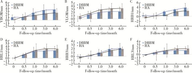
DBBM组与HA组在各随访节点的牙槽嵴轮廓塌陷量重复测量方差分析结果显示,组别主效应对拔牙窝中央轮廓塌陷量效应显著(P=0.027),对其他牙槽嵴轮廓塌陷指标效应不显著(P>0.05),随访时间点主效应对各测量指标效应均显著(P < 0.05),组别和随访时间点交互效应均不显著(P>0.05)。两组患者各随访节点牙槽嵴轮廓塌陷量见图 8。在随访2周和1个月时,HA组的拔牙窝中央轮廓塌陷量明显大于DBBM组[2周: (2.73±1.89) mm vs. (0.00±0.79) mm;1个月: (2.74±1.13) mm vs. (0.35±2.34) mm],差异有统计学意义(P < 0.05)。其余的水平及垂直向轮廓塌陷量,两组相比差异均无统计学意义(P>0.05),见图 8。牙槽嵴轮廓水平及垂直向的塌陷主要发生在术后1个月内,并在术后3~6个月中基本保持稳定。
图8 两组患者各随访节点的牙槽嵴轮廓塌陷量

Figure 8 Alveolar ridge contour reduction at follow-up time points of the two groups

A, vertical reduction of the contour on the buccal free gingival margins (VBGM); B, vertical reduction of the contour on the lingual/palatal free gingival margins (VLGM); C, horizontal reduction of the buccal contour at 1 mm apically (HRB1); D, horizontal reduction of the lingual/palatal contour at 1 mm apically (HRL1); E, horizontal reduction of the buccal contour at 3 mm apically (HRB3); F, horizontal reduction of the lingual/palatal contour at 3 mm apically (HRL3). DBBM, deproteinized bovine bone mineral; HA, hydroxyapatite.

2.4 软组织愈合过程的分析

比较不同随访时间节点软组织创面面积与基线创面面积的比值,术后2周和1个月时,HA组创面面积比例显著高于DBBM组[2周:47.88%±6.56% vs. 29.43%±14.25%,P < 0.05;1个月:25.68%±13.06% vs. 7.19%±7.18%,P < 0.01];术后3个月,两组患者的创面均完全愈合。

3 讨论

既往研究表明,针对重度牙周病变的磨牙行拔牙同期牙槽嵴保存术可以显著改善缺牙区软硬组织条件,降低种植修复的难度[2, 14, 18-21]。目前,牙槽嵴保存术中采用哪种骨移植材料更优尚无定论,对人工合成骨移植材料的研究相对较少,其临床效果有待进一步探讨。本回顾性病例系列研究旨在对比DBBM与HA在重度牙周病变磨牙拔牙同期行MCF-ARP的临床效果,以便为材料选择提供参考。

3.1 DBBM与HA在牙槽嵴保存术中的应用

目前临床可用的骨移植材料种类包括自体骨移植材料、同种异体骨移植材料、异种骨移植材料和人工合成骨移植材料。异种骨移植材料中的DBBM因其优异的成骨效果和空间维持能力成为临床应用最广泛的材料。既往研究证实,牙槽嵴保存术中应用DBBM可有效保存牙槽嵴的高度与宽度[12, 22-24]。本课题组以往多项针对牙槽嵴保存术在重度牙周病变磨牙中应用的研究结果进一步证实,应用DBBM可有效保持颊舌侧骨壁高度与骨嵴宽度[2, 24],并简化种植治疗的程序[9]。同时,本课题组开创的MCF-ARP术式与传统的纵切口术式相比,患者术后6个月时硬组织指标的变化相似,且能保存更多的角化组织[17]
人工合成骨移植材料凭借其来源稳定和成本可控等优势日益受到重视。HA为人工合成骨移植材料中的常见成分,其在体内可逐步降解为钙离子与磷酸根离子,通过提高局部离子浓度促进成骨细胞分化。有研究表明,人工合成HA在各类骨缺损修复中均能获得良好的成骨效果[25-27],当HA应用于牙槽嵴保存术时,可有效维持牙槽嵴的高度与宽度[28-29]。Kijartorn等[30]的一项随机对照临床试验中,使用HA作为牙槽嵴保存术的骨替代材料,发现在术后4个月时,牙槽骨宽度基本维持稳定,平均水平骨吸收量仅为0.59 mm。

3.2 牙槽嵴保存术后硬组织保存与增量效果

充足的牙槽骨高度与宽度是种植治疗的基础。本研究结果显示,患者术后6个月时,DBBM组与HA组在硬组织指标上无明显差异,这与部分既往研究结论一致[31-32]。Gao等[32]的一项多中心随机对照临床试验显示,在牙槽嵴保存术后的6个月,HA与DBBM在牙槽骨宽度和高度的维持上差异无统计学意义,但也有研究认为DBBM在骨量维持方面优于HA[33]。Jung等[34]在一项纳入前牙与前磨牙的随机对照临床试验中发现,人工合成骨移植材料β-磷酸三钙与DBBM相比,在拔牙后6个月时的骨吸收更严重,推测这可能是由于HA的吸收率更高[27-28],且伤口封闭与愈合过程中骨移植材料流失更多。本组针对磨牙的研究也同样发现HA组患者的软组织封闭更慢,与Jung等[34]的观察结果一致。本研究与部分既往研究得出的结论不一致的可能原因是,本研究中应用的MCF-ARP技术是在手术过程中植入骨粉稍过量于拔牙窝最高骨嵴顶2~3 mm,既考虑了牙槽嵴保存术时的增量,又用以补偿术后软组织封闭过程中可能发生的骨粉流失。同时,与既往研究纳入的研究对象多为前牙及前磨牙[33-34]有所不同,本研究纳入的患牙为重度牙周病变的磨牙,虽创口较大但效果仍能保持良好。

3.3 牙槽嵴轮廓塌陷量的评估

良好的牙槽嵴轮廓有利于以修复为导向的种植体植入,提高修复体美观程度,有利于种植修复体的口腔卫生维护与长期健康。Vignoletti等[35]的研究认为,牙槽嵴保存术的目的在于保持现有软硬组织轮廓,为实现良好的功能和美学效果提供稳定且具有一定体积的牙槽骨,以便简化未来种植修复的治疗程序。既往其他研究和本课题组以往的多项研究均证实,牙槽嵴保存术中应用DBBM与自然愈合相比,可更好地维持牙槽嵴轮廓[14, 36],然而,目前尚缺乏在牙槽嵴保存术中DBBM与HA对牙槽嵴轮廓影响的对比研究。
既往关于两种材料在牙槽嵴保存术中的对比研究多限于硬组织指标的比较,缺少对牙槽嵴整体轮廓变化的评价。本研究使用口内扫描技术,通过测量术后6个月内的多个不同随访节点的水平及垂直方向的牙槽嵴轮廓塌陷量,初步探索和评价了微创拔牙同期使用DBBM和HA行MCF-ARP的牙槽嵴整体轮廓塌陷量,结果发现,两组患者在术后6个月时的牙槽嵴轮廓差异无统计学意义,仅在术后愈合过程中,即2周和1个月时,DBBM组在拔牙窝中央的轮廓塌陷量小于HA组,提示牙槽嵴保存术中应用DBBM和HA在早期的愈合过程中存在一定差异。

3.4 软组织愈合过程的分析

既往的研究中,针对牙槽嵴保存术后软组织愈合过程的分析报道较少。牙槽嵴保存术后骨移植材料的稳定性是保证牙槽嵴保存术最终临床效果的重要影响因素,特别是在术后早期,软组织快速愈合可避免骨移植材料暴露,降低术后感染风险,减少牙槽嵴吸收。本研究使用口内扫描技术,对软组织愈合过程中不同时间节点的创面面积进行了量化分析,实现了对牙槽嵴保存术后软组织愈合过程的客观评估,结果显示,两组患者在术后3个月时均可达到软组织完全封闭,但术后早期的愈合过程有所差异。术后1个月内,DBBM组的角化黏膜覆盖速度快于HA组,创面小于HA组,这提示临床医生在使用HA进行牙槽嵴保存术后,针对患者的术后护理有更高的要求,可适当增加随访频率,及时去除溢出的骨粉颗粒,以促进软组织的愈合。有研究发现,富血小板纤维蛋白(platelet-rich fibrin,PRF)等血小板浓缩物在促进高质量骨形成和软组织愈合方面有优势,在牙槽嵴保存术中与骨移植材料联用时可显著提高新生骨密度和质量,并减少骨吸收[29]。添加了重组人骨形态发生蛋白-2(recombinant human bone morphogenetic protein-2,rhBMP-2)的人工合成骨移植材料也能显著减少牙槽骨高度和宽度的吸收[37],这为今后人工合成骨移植材料的改良提供了方向。
综上所述,本研究利用CBCT影像学与口内扫描技术,从硬组织保存、牙槽嵴轮廓塌陷及软组织愈合过程三个方面比较了DBBM与HA在重度牙周病变的磨牙拔牙同期行MCF-ARP的临床应用效果,结果表明,在术后6个月时,两种骨移植材料的硬组织指标差异无统计学意义,但DBBM组患者的软组织早期愈合更快。本研究的局限性在于未进行组织学分析,样本量有限,随访时间较短,仅为探索性分析DBBM与HA两种材料在重度牙周病变的磨牙拔牙同期行MCF-ARP的临床应用效果,未来需设计更为严谨的大样本随机对照试验,结合组织学评价,并对种植修复后的长期效果进行随访,从而更全面地评估人工合成骨移植材料在复杂临床情境下的应用特点和价值,为临床提供更为可靠的循证依据。

利益冲突  所有作者均声明不存在利益冲突。

作者贡献声明  张斯巧:研究设计,患者随访复查,分析数据,撰写文章;刘建:患者随访复查,患者管理,研究统筹;胡文杰:研究设计和统筹,患者手术操作及随访复查,论文修改;徐涛:患者管理,手术操作,论文修改;张浩筠:研究设计,分析数据,患者随访复查,论文修改;危伊萍:研究设计,患者随访复查,论文修改。所有作者均参与论文修改,并对最终文稿进行审读和确认。

1
Atieh MA , Alnaqbi M , Abdunabi F , et al. Alveolar ridge preservation in extraction sockets of periodontally compromised teeth: A systematic review and meta-analysis[J]. Clin Oral Implants Res, 2022, 33 (9): 869- 885.

DOI

2
Zhao L , Xu T , Hu W , et al. Preservation and augmentation of molar extraction sites affected by severe bone defect due to advanced periodontitis: A prospective clinical trial[J]. Clin Implant Dent Relat Res, 2018, 20 (3): 333- 344.

DOI

3
Hämmerle CHF , Araújo MG , Simion M . Evidence-based knowledge on the biology and treatment of extraction sockets[J]. Clin Oral Implants Res, 2012, 23 (Suppl 5): 80- 82.

4
Bassir SH , Alhareky M , Wangsrimongkol B , et al. Systematic review and meta-analysis of hard tissue outcomes of alveolar ridge preservation[J]. Int J Oral Maxillofac Implants, 2018, 33 (5): 979- 994.

DOI

5
Pelegrine AA , da Costa CES , Correa MEP , et al. Clinical and histomorphometric evaluation of extraction sockets treated with an autologous bone marrow graft[J]. Clin Oral Implants Res, 2010, 21 (5): 535- 542.

DOI

6
Atieh MA , Alsabeeha NH , Payne AG , et al. Interventions for replacing missing teeth: Alveolar ridge preservation techniques for dental implant site development[J]. Cochrane Database Syst Rev, 2021, 4 (4): CD010176.

7
Park SH , Song YW , Sanz-Martín I , et al. Clinical benefits of ridge preservation for implant placement compared to natural healing in maxillary teeth: A retrospective study[J]. J Clin Periodontol, 2020, 47 (3): 382- 391.

DOI

8
Cha JK , Song YW , Park SH , et al. Alveolar ridge preservation in the posterior maxilla reduces vertical dimensional change: A randomized controlled clinical trial[J]. Clin Oral Implants Res, 2019, 30 (6): 515- 523.

DOI

9
Wei Y , Xu T , Zhao L , et al. Ridge preservation in maxillary molar extraction sites with severe periodontitis: A prospective observational clinical trial[J]. Clin Oral Investig, 2022, 26 (3): 2391- 2399.

DOI

10
García-González S , Galve-Huertas A , Aboul-Hosn Centenero S , et al. Volumetric changes in alveolar ridge preservation with a compromised buccal wall: A systematic review and meta-analysis[J]. Med Oral Patol Oral Cir Bucal, 2020, 25 (5): e565- e575.

11
MacBeth N , Trullenque-Eriksson A , Donos N , et al. Hard and soft tissue changes following alveolar ridge preservation: A systematic review[J]. Clin Oral Implants Res, 2017, 28 (8): 982- 1004.

DOI

12
Lai VJ , Michalek JE , Liu Q , et al. Ridge preservation following tooth extraction using bovine xenograft compared with porcine xenograft: A randomized controlled clinical trial[J]. J Periodontol, 2020, 91 (3): 361- 368.

DOI

13
Barootchi S , Tavelli L , Majzoub J , et al. Alveolar ridge preservation: Complications and cost-effectiveness[J]. Periodontol 2000, 2023, 92 (1): 235- 262.

DOI

14
Zhang H , Xu T , Wei Y , et al. Assessment of soft and hard tissue changes following micro crestal flap-Alveolar ridge preservation and augmentation at molar extraction sites in patients with stage Ⅲ/Ⅳ periodontitis: A randomized controlled trial[J]. J Clin Periodontol, 2024, 51 (10): 1311- 1322.

DOI

15
赵丽萍, 詹雅琳, 胡文杰, 等. 不同测量方法评价磨牙拔牙位点保存术后牙槽骨的变化[J]. 北京大学学报(医学版), 2016, 48 (1): 126- 132.

DOI

16
赵丽萍, 胡文杰, 徐涛, 等. 罹患重度牙周病变磨牙拔牙后两种牙槽嵴保存方法的比较[J]. 北京大学学报(医学版), 2019, 51 (3): 579- 585.

DOI

17
Wei Y , Zhao L , Zhang H , et al. Ridge preservation in periodontally compromised molar sockets with and without primary wound closure: A comparative controlled clinical trial[J]. Clin Oral Implants Res, 2024, 35 (1): 131- 139.

DOI

18
Araújo MG , Silva CO , Misawa M , et al. Alveolar socket healing: What can we learn?[J]. Periodontol 2000, 2015, 68 (1): 122- 134.

19
Araújo M , Linder E , Lindhe J . Effect of a xenograft on early bone formation in extraction sockets: An experimental study in dog[J]. Clin Oral Implants Res, 2009, 20 (1): 1- 6.

20
Lindhe J , Cecchinato D , Donati M , et al. Ridge preservation with the use of deproteinized bovine bone mineral[J]. Clin Oral Implants Res, 2014, 25 (7): 786- 790.

DOI

21
Sbordone C , Toti P , Martuscelli R , et al. Retrospective volume analysis of bone remodeling after tooth extraction with and without deproteinized bovine bone mineral insertion[J]. Clin Oral Implants Res, 2016, 27 (9): 1152- 1159.

DOI

22
Vaia E , Nicolò M , Vaia E , et al. Alveolar ridge preservation with deproteinized bovine bone mineral and xenogeneic collagen matrix: A 12-month clinical and histomorphometric case series[J]. Int J Periodontics Restorative Dent, 2021, 41 (3): 423- 430.

23
Lee J , Yun J , Kim JJ , et al. Retrospective study of alveolar ridge preservation compared with no alveolar ridge preservation in periodontally compromised extraction sockets[J]. Int J Implant Dent, 2021, 7 (1): 23.

DOI

24
Wei Y , Xu T , Hu W , et al. Socket preservation following extraction of molars with severe periodontitis[J]. Int J Periodontics Restorative Dent, 2021, 41 (2): 269- 275.

DOI

25
Mangano C , Luongo G , Luongo F , et al. Custom-made computer-aided-design/computer-assisted-manufacturing (CAD/CAM) synthetic bone grafts for alveolar ridge augmentation: A retrospective clinical study with 3 years of follow-up[J]. J Dent, 2022, 127, 104323.

DOI

26
Luo G , Huang Y , Gu F . The osteogenesis effect of rhBMP2-loaded calcium phosphate cements in repairing dental extraction sockets[J]. Am J Transl Res, 2022, 14 (10): 7172- 7177.

27
朱磊, 刘庆成, 于洪波. 牙槽骨增量骨皮质切开术后2种植骨材料成骨的影像学分析[J]. 中国口腔颌面外科杂志, 2021, 19 (6): 525- 530.

28
Ozzo S , Kheirallah M . The efficiency of two different synthetic bone graft materials on alveolar ridge preservation after tooth extraction: A split-mouth study[J]. BMC Oral Health, 2024, 24 (1): 1040.

DOI

29
Stumbras A , Kuliesius P , Januzis G , et al. Alveolar ridge preservation after tooth extraction using different bone graft materials and autologous platelet concentrates: A systematic review[J]. J Oral Maxillofac Res, 2019, 10 (1): e2.

30
Kijartorn P , Wongpairojpanich J , Thammarakcharoen F , et al. Clinical evaluation of 3D printed nano-porous hydroxyapatite bone graft for alveolar ridge preservation: A randomized controlled trial[J]. J Dent Sci, 2022, 17 (1): 194- 203.

DOI

31
Mardas N , Chadha V , Donos N . Alveolar ridge preservation with guided bone regeneration and a synthetic bone substitute or a bovine-derived xenograft: A randomized, controlled clinical trial[J]. Clin Oral Implants Res, 2010, 21 (7): 688- 698.

DOI

32
Gao L , Zhu H , Kou Y , et al. The effects of β-TCP and/or xenogeneic bone substitute on alveolar ridge preservation: A randomized clinical trial[J]. Clin Oral Investig, 2025, 29 (4): 200.

DOI

33
Shakibaie-M B . Comparison of the effectiveness of two different bone substitute materials for socket preservation after tooth extraction: A controlled clinical study[J]. Int J Periodontics Restorative Dent, 2013, 33 (2): 223- 228.

DOI

34
Jung RE , Philipp A , Annen BM , et al. Radiographic evaluation of different techniques for ridge preservation after tooth extraction: A randomized controlled clinical trial[J]. J Clin Periodontol, 2013, 40 (1): 90- 98.

DOI

35
Vignoletti F , Matesanz P , Rodrigo D , et al. Surgical protocols for ridge preservation after tooth extraction. A systematic review[J]. Clin Oral Implants Res, 2012, 23 (Suppl 5): 22- 38.

36
Thoma DS , Bienz SP , Lim HC , et al. Explorative randomized controlled study comparing soft tissue thickness, contour changes, and soft tissue handling of two ridge preservation techniques and spontaneous healing two months after tooth extraction[J]. Clin Oral Implants Res, 2020, 31 (6): 565- 574.

DOI

37
Han JJ , Chang AR , Ahn J , et al. Efficacy and safety of rhBMP/β-TCP in alveolar ridge preservation: A multicenter, randomized, open-label, comparative, investigator-blinded clinical trial[J]. Maxillofac Plast Reconstr Surg, 2021, 43 (1): 42.

DOI

文章导航

/