Journal of Peking University (Health Sciences) ›› 2026, Vol. 58 ›› Issue (1): 115-125. doi: 10.19723/j.issn.1671-167X.2026.01.015

Previous Articles     Next Articles

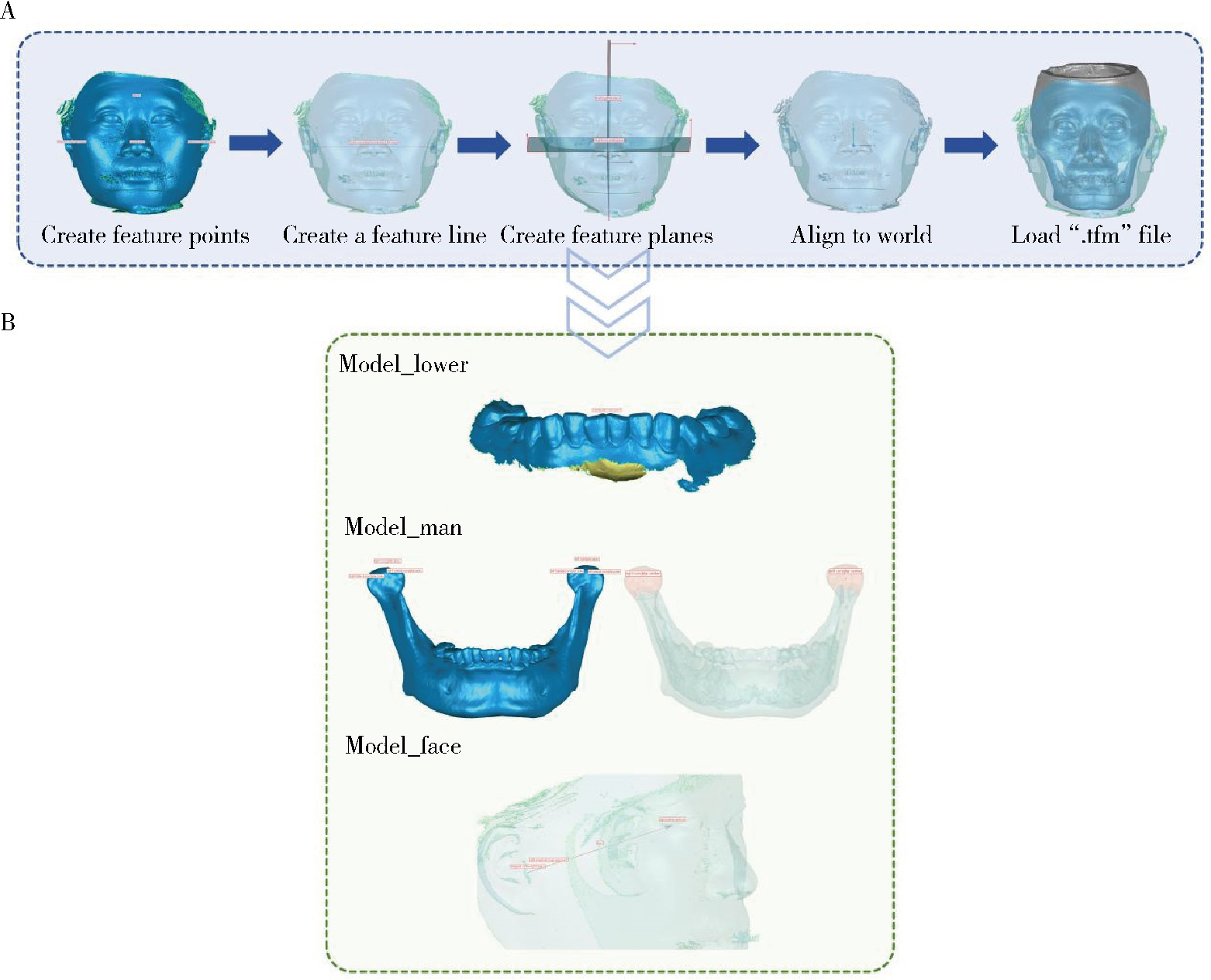
Calculation method of key articulator parameters based on mandibular movement trajectory

Shenyao SHAN1,2, Yongtao YANG2, Wenbo LI2, Aonan WEN1, Zixiang GAO1, Xiangyi SHANG2, Yong WANG1,2,*(), Yijiao ZHAO1,2,*()   

  1. 1. Center for Digital Dentistry, Peking University School and Hospital of Stomatology & National Center for Stomatology & National Clinical Research Center for Oral Diseases & National Engineering Research Center of Oral Biomaterials and Digital Medical Devices & Beijing Key Laboratory of Digital Stomatology & NHC Research Center of Engineering and Technology for Computerized Dentistry, Beijing 100081, China
    2. Institute of Medical Technology, Peking University Health Science Center, Beijing 100191, China
  • Received:2025-10-11 Online:2026-02-18 Published:2025-12-10
  • Contact: Yong WANG, Yijiao ZHAO
  • Supported by:
    the National Natural Science Foundation of China(82271039); Beijing Natural Science Foundation(L232100); Beijing Natural Science Foundation(L242132); National Key Research and Development Plan of China(2022YFC2405401)

RICH HTML


Abstract:

Objective: To explore a mathematical method for calculating key articulator parameters based on mandibular movement trajectory data, and to compare the results of this method with reference values provided by existing foreign mandibular movement recording system, thereby establishing an algorithmic basis for developing a domestic mandibular movement recording system. Methods: Twenty healthy volunteers (7 males, 13 females) meeting inclusion criteria were recruited, with a mean age of (31±8) years. Mandibular movement data during protrusive and left/right lateral movements were recorded using the JMA Optic foreign mandibular movement recording system. A reference plane coordinate system was established using reverse engineering software, the multi-source maxillofacial data were integrated, and the coordinate systems were then unified. The condylar apex, medial condylar pole, lateral condylar pole, condylar center, empirical hinge axis point, and mandibular incisor point were selected as reference points for mandibular movement trajectories. Three-dimensional movement trajectories were generated for each reference point to calculate the sagittal condylar inclination (SCI), transverse condylar inclination (TCI), immediate side shift (ISS), incisal guidance inclination and canine guidance inclination. The calculation results from different reference points served as distinct experimental groups. Reference values provided by the JMA Optic system were used as the control group for comparative analysis. Results: The SCI values of all the experimental groups were significantly higher than that of the control group (P < 0.001), with a systematic positive bias of approximately 3.1°, though the limits of agreement were relatively narrow. The TCI results varied depending on the reference point: Only the condylar apex group (5.7°±6.1°) was significantly lower than the control group (9.2°±6.6°) (t=5.023, P < 0.001). Differences between the remaining groups and the control group were not statistically significant. The empirical hinge axis point group showed the smallest mean bias and the narrowest limits of agreement, indicating optimal consistency with the control group's TCI. The ISS values were 0.0 (0.0) mm in all the groups. The incisal guidance inclination of the mandibular incisor point group (43.1°±8.6°) was significantly lower than that of the control group (50.6°±13.7°) (t=3.749, P=0.001) with poor consistency. However, the canine guidance inclination of the mandibular incisor point group showed no statistically significant difference compared with the control group (t=-1.873, P=0.069), with acceptable consistency. Conclusion: This study proposed a mathematical method for calculating key articulator parameters based on mandibular movement trajectory data, with a clear and traceable computational pathway. The proposed method showed acceptable consistency with the JMA Optic system algorithm in calculating TCI, ISS, and canine guidance inclination, but poor consistency in calculating SCI and incisal guidance inclination. The selection of reference points directly influenced the results of parameter calculation. This mathematical method provided a reliable theoretical foundation for achieving precise, personalized articulator parameter settings.

Key words: Mandibular movement trajectory, Articulator parameters, Sagittal condylar inclination, Transversal condylar inclination, Incisal guidance inclination

CLC Number: 

  • R783.3

Figure 1

Standard maxillary fork three-dimensional model A, the red dots indicate the relationship between three mark points and the standard maxillary fork model; B, the coordinate system for the standard maxillary fork model."

Figure 2

3D multi-source data integration process for the maxillofacial region A, select the labial region of the anterior teeth as the registration area, unify the coordinate systems of Model_fork and Model_upper; B, select the maxillary dental arch region as the registration area, unify the coordinate systems of Model_upper and Model_max, load ".tfm" file to align the coordinate systems of Model_lower and Model_fork; C, select the upper surface region of the maxillary fork as the registration area, unify the coordinate systems of Model_fork and Model_ref, load ".tfm" file to align all 3D models to the coordinate system where Model_ref resides."

Figure 3

Establishment of a reference plane coordinate system and selection of reference points A, procedure for establishing a reference plane coordinate system; B, select reference points based on the reference plane coordinate system, select mandibular incisor point on Model_lower, select bilateral condylar apex, medial condylar pole, lateral condylar pole and condylar center on Model_man, select bilateral empirical hinge axis points on Model_face."

Figure 4

Mandibular movement trajectory based on reference points"

Figure 5

Calculate key articulator parameters based on reference point trajectories Using trajectories of left condylar center and mandibular incisor point as examples to demonstrate the mathematical calculation method of SCI, TCI, ISS, IGI and CGI. SCI, sagittal condylar inclination; TCI, transversal condylar inclination; ISS, immediate side shift; IGI, incisal guidance inclination; CGI, canine guidance inclination."

Table 1

Comparison of left and right SCI, TCI, ISS among experimental groups and the control group"

Group n SCI/(°),${\bar x}$±s TCI/(°),${\bar x}$±s ISS/mm, M (IQR)
Left Right t P Left Right t P Left Right Z P
EG_condylar apex 20 39.7±7.3 40.0±8.5 -0.270 0.790 6.6±5.1 4.8±7.1 1.129 0.273 0.0(0.0) 0.0(0.0) 0.000 >0.999
EG_medial condylar pole 20 39.8±7.1 39.9±8.0 -0.191 0.851 9.0±5.6 6.6±7.5 1.453 0.162 0.0(0.0) 0.0(0.0) 0.000 >0.999
EG_lateral condylar pole 20 40.0±7.2 40.3±8.4 -0.214 0.833 11.0±5.3 9.1±5.8 1.374 0.185 0.0(0.0) 0.0(0.0) 0.000 >0.999
EG_condylar center 20 39.9±7.1 40.1±8.2 -0.240 0.813 10.7±5.5 8.6±6.5 1.367 0.188 0.0(0.0) 0.0(0.0) 0.000 >0.999
EG_empirical hinge axis point 20 39.9±7.4 40.5±8.9 -0.346 0.733 10.2±5.6 8.9±6.3 1.068 0.299 0.0(0.0) 0.0(0.0) 0.000 >0.999
CG 20 37.3±8.6 36.5±9.1 0.639 0.531 9.8±6.5 8.6±6.8 1.019 0.321 0.0(0.0) 0.0(0.0) -0.272 0.785

Table 2

Comparison of left and right canine guidance inclination in EG_mandibular incisor point and the control group"

Group n Canine guidance inclination/(°),${\bar x}$±s
Left Right t P
EG_mandibular incisor point 20 33.9±9.3 32.7±6.5 0.900 0.379
CG 20 29.7±11.5 32.8±8.7 -1.611 0.124

Table 3

Pairwise comparisons of SCI, TCI, ISS between experimental groups and the control group"

Group SCI/(°),${\bar x}$±s t P TCI/(°),${\bar x}$±s t P ISS/mm, M (IQR) Z P
EG_condylar apex 39.8±7.8 -4.635 < 0.001 5.7±6.1 5.023 < 0.001 0.0(0.0) -1.633 0.102
EG_medial condylar pole 39.8±7.5 -4.586 < 0.001 7.8±6.7 1.971 0.056 0.0(0.0) -1.633 0.102
EG_lateral condylar pole 40.1±7.7 -5.206 < 0.001 10.1±5.6 -1.213 0.233 0.0(0.0) -1.633 0.102
EG_condylar center 40.0±7.6 -4.963 < 0.001 9.7±6.0 -0.655 0.517 0.0(0.0) -1.633 0.102
EG_empirical hinge axis point 40.2±8.1 -5.318 < 0.001 9.5±5.9 -0.539 0.593 0.0(0.0) -1.633 0.102
CG 36.9±8.8 9.2±6.6 0.0(0.0)

Figure 6

Bland-Altman plots for consistency of key articulator parameters between experimental and control groups SCI, sagittal condylar inclination; TCI, transversal condylar inclination; IGI, incisal guidance inclination; CGI, canine guidance inclination; EG, experimental group; CG, control group."

1
吴国锋. 𬌗架的发展简史[J]. 实用口腔医学杂志, 2016, 32 (3): 445- 448.
2
Lepidi L , Galli M , Mastrangelo F , et al. Virtual articulators and virtual mounting procedures: Where do we stand?[J]. J Pros-thodont, 2021, 30 (1): 24- 35.
3
Lee VC , Tan MY , Yee SHX , et al. Three-dimensional analysis of the interchangeability of a semiadjustable articulator system in service over time[J]. J Prosthet Dent, 2024, 132 (5): 1028- 1037.

doi: 10.1016/j.prosdent.2023.04.009
4
Lam WYH , Hsung RTC , Choi WWS , et al. A clinical technique for virtual articulator mounting with natural head position by using calibrated stereophotogrammetry[J]. J Prosthet Dent, 2018, 119 (6): 902- 908.

doi: 10.1016/j.prosdent.2017.07.026
5
张良荣. 咬合记录硅橡胶O-BITE在咬合确定中的临床评价[J]. 中外医学研究, 2016, 14 (32): 135- 136.
6
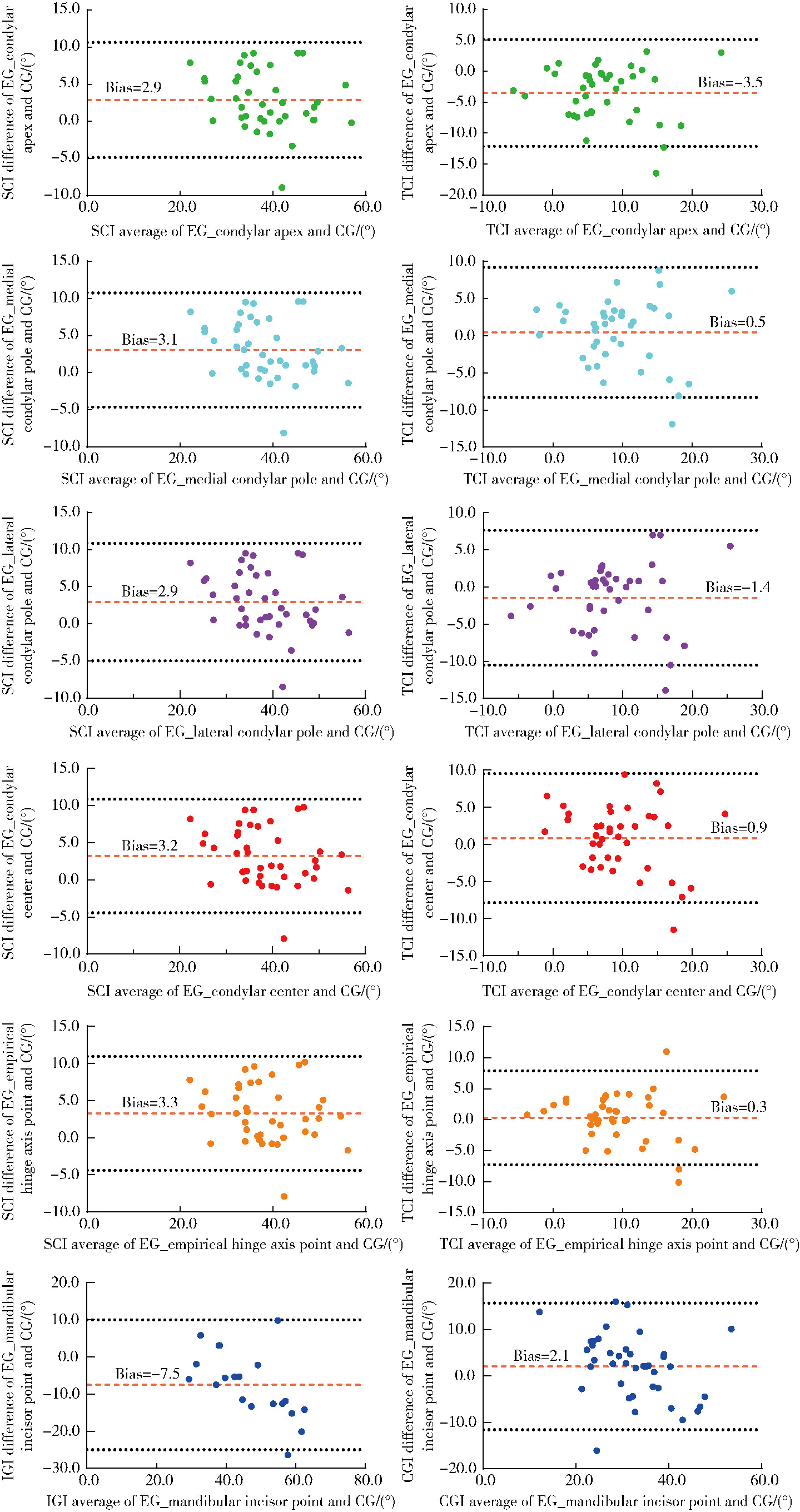
单珅瑶, 朱玉佳, 王俊杰, 等. 基于100名个别正常𬌗成人下颌运动轨迹的平均值𬌗架关键参数初探[J]. 中华口腔医学杂志, 2024, 59 (12): 1228- 1233.
7
徐心雨, 吴灵, 宋凤岐, 等. 基于下颌运动轨迹的正颌外科术中下颌骨髁突定位方法及初步精度验证[J]. 北京大学学报(医学版), 2024, 56 (1): 57- 65.

doi: 10.19723/j.issn.1671-167X.2024.01.010
8
徐欣蕊, 余润平, 袁芸, 等. 基于下颌运动轨迹数据指导的数字化全冠咬合面精度评价[J]. 上海口腔医学, 2023, 32 (1): 58- 62.
9
Li L , Chen H , Li W , et al. Design of wear facets of mandibular first molar crowns by using patient-specific motion with an intraoral scanner: A clinical study[J]. J Prosthet Dent, 2023, 129 (5): 710- 717.

doi: 10.1016/j.prosdent.2021.06.048
10
马丽娅, 巢家瑞, 刘飞, 等. 基于下颌运动轨迹与虚拟𬌗架运动参数模拟调𬌗的对比研究[J]. 华西口腔医学杂志, 2023, 41 (3): 254- 259.
11
Valenti M , Schmitz JH . A reverse digital workflow by using an interim restoration scan and patient-specific motion with an intraoral scanner[J]. J Prosthet Dent, 2021, 126 (1): 19- 23.

doi: 10.1016/j.prosdent.2020.05.011
12
周永胜. 口腔修复学[M]. 3版 北京: 北京大学医学出版社, 2020: 312- 315.
13
王美青. 𬌗学[M]. 4版 北京: 人民卫生出版社, 2020: 93.
14
周哲青, 王思谕, 袁泉, 等. 一种全数字化前伸髁导斜度测量方法的准确性研究[J]. 华西口腔医学杂志, 2024, 42 (1): 67- 74.
15
胡婷姿, 杨海萍, 肖沛, 等. Zebris下颌运动分析系统的应用与研究现状[J]. 口腔医学, 2021, 41 (12): 1129- 1133.
16
Lepidi L , Grande F , Baldassarre G , et al. Preliminary clinical study of the accuracy of a digital axiographic recording system for the assessment of sagittal condylar inclination[J]. J Dent, 2023, 135, 104583.

doi: 10.1016/j.jdent.2023.104583
17
朱家奕, 王俊杰, 王宇轩, 等. 轻咬合和重咬合状态对下颌运动轨迹及虚拟预调𬌗的影响[J]. 中华口腔医学杂志, 2023, 58 (1): 50- 56.
18
Revilla-León M , Zeitler JM , Kois JC . An overview of the different digital facebow methods for transferring the maxillary cast into the virtual articulator[J]. J Esthet Restor Dent, 2024, 36 (12): 1675- 1686.

doi: 10.1111/jerd.13264
19
Lewandowska A , Mańka-Malara K , Kostrzewa-Janicka J . Sagittal condylar inclination and transversal condylar inclination in different skeletal classes[J]. J Clin Med, 2022, 11 (9): 2664.

doi: 10.3390/jcm11092664
20
Tang W , Wu Y , Ma J , et al. 3-Dimensional quantitative analysis of mandibular motion in TMD and healthy subjects: Comparison with clinical observations[J]. J Dent, 2025, 153, 105534.

doi: 10.1016/j.jdent.2024.105534
[1] Kun QIAN, Yihong LIU. Fitness of onlays fabricated with direct and indirect CAD/CAM technology in vitro [J]. Journal of Peking University (Health Sciences), 2025, 57(3): 604-609.
[2] Mengdi LI, Lei LEI, Zhongning LIU, Jian LI, Ting JIANG. Gene silencing of Nemo-like kinase promotes neuralized tissue engineered bone regeneration [J]. Journal of Peking University (Health Sciences), 2025, 57(2): 227-236.
[3] Lingli ZHU, Lin TANG, Bowen LI, Mei WANG, Yuhua LIU. Influence of two methods of smear layer removal on the surface properties of dentin [J]. Journal of Peking University (Health Sciences), 2025, 57(2): 340-346.
[4] Yawen CHENG, Deli LI, Yan ZHAO, Bin XIA, Yunsong LIU. Clinical dilemma and indication selection of restoration for permanent tooth defects in adolescents [J]. Journal of Peking University (Health Sciences), 2025, 57(1): 208-213.
[5] LIU Si-min,ZHAO Yi-jiao,WANG Xiao-yan,WANG Zu-hua. In vitro evaluation of positioning accuracy of trephine bur at different depths by dynamic navigation [J]. Journal of Peking University (Health Sciences), 2022, 54(1): 146-152.
[6] Xiao-xian CHEN,Jie ZHONG,Wen-juan YAN,Hong-mei ZHANG,Xia JIANG,Qian HUANG,Shi-hua XUE,Xing-gang LIU. Clinical performance of rensin-bonded composite strip crowns in primary incisors [J]. Journal of Peking University (Health Sciences), 2020, 52(5): 907-912.
[7] Mei WANG, Bo-wen LI, Si-wen WANG, Yu-hua LIU. Preparation and osteogenic effect study of small intestinal submucosa sponge [J]. Journal of Peking University (Health Sciences), 2020, 52(5): 952-958.
[8] Zhong ZHANG,Huan-xin MENG,Jie HAN,Li ZHANG,Dong SHI. Effect of vertical soft tissue thickness on clinical manifestation of peri-implant tissue in patients with periodontitis [J]. Journal of Peking University (Health Sciences), 2020, 52(2): 332-338.
[9] Wei-ting LI,Peng LI,Mu-zi PIAO,Fang ZHANG,Jie DI. Study on bone volume harvested from the implant sites with different methods [J]. Journal of Peking University(Health Sciences), 2020, 52(1): 103-106.
[10] Qiang LUO,Qian DING,Lei ZHANG,Qiu-fei XIE. Quantitative analysis of occlusal changes in posterior partial fixed implant supported prostheses [J]. Journal of Peking University(Health Sciences), 2019, 51(6): 1119-1123.
[11] Miao ZHENG,Ling-lu ZHAN,Zhi-qiang LIU,He-ping LI,Jian-guo TAN. Effect of different plasma treated zirconia on the adhensive behaviour of human gingival fibroblasts [J]. Journal of Peking University(Health Sciences), 2019, 51(2): 315-320.
[12] Xin-xin LI,Yu-shu LIU,Yu-chun SUN,Hu CHEN,Hong-qiang YE,Yong-sheng ZHOU. Evaluation of one-piece polyetheretherketone removable partial denture fabricated by computer-aided design and computer-aided manufacturing [J]. Journal of Peking University(Health Sciences), 2019, 51(2): 335-339.
[13] Rui-jie WANG,Min LIU,Dan-yang SONG,Sui YANG,Qiao WANG,Lei WANG,Hai-lan FENG. Analysis of edge morphology of partial veneers made by different processing techniques and materials [J]. Journal of Peking University(Health Sciences), 2019, 51(1): 93-99.
[14] ZHOU Tuan-feng, WANG Xin-zhi . Clinical observation of the restoration of computer aided designed and manufactured one-piece zirconia posts and cores: a 5-year prospective follow-up study [J]. Journal of Peking University(Health Sciences), 2018, 50(4): 680-684.
[15] ZHU Meng-meng, WANG Guo-min, SUN Ke, LI Ying-long, PAN Jie. Bonding strength of resin and tooth enamel after teeth bleaching with cold plasma [J]. Journal of Peking University(Health Sciences), 2016, 48(1): 116-120.
Viewed
Full text


Abstract

Cited

  Shared   
  Discussed   
No Suggested Reading articles found!