Journal of Peking University (Health Sciences) ›› 2026, Vol. 58 ›› Issue (1): 220-224. doi: 10.19723/j.issn.1671-167X.2026.01.030
Previous Articles Next Articles
Florid cemento-osseous dysplasia: A case report
Yue WANG1, Yuhong LIANG2,*( )
)
- 1. Department of Stomatology, Peking University International Hospital, Beijing 102206, China
2. Department of Oral Emergency, Peking University School and Hospital of Stomatology & National Center for Stomatology & National Clinical Research Center for Oral Diseases & National Engineering Research Center of Oral Biomaterials and Digital Medical Devices & Beijing Key Laboratory of Digital Stomatology & NHC Key Laboratory of Digital Stomatology & NMPA Key Laboratory for Dental Materials, Beijing 100081, China
CLC Number:
- R782.1
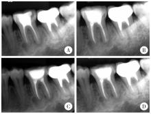
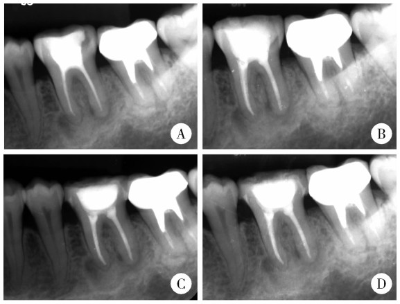
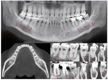
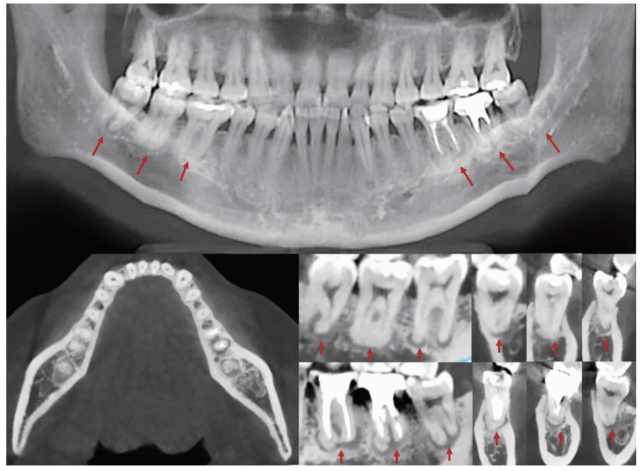
| 1 |
doi: 10.1016/j.oooo.2016.04.013 |
| 2 |
|
| 3 |
史册, 李志民, 孙宏晨. 纤维-骨肿瘤及结构不良的临床病理特征与鉴别诊断[J]. 中华口腔医学杂志, 2023, 58 (2): 124- 130.
|
| 4 |
doi: 10.3390/diagnostics12020238 |
| 5 |
doi: 10.1016/j.cden.2009.12.007 |
| 6 |
doi: 10.1016/j.jds.2021.03.009 |
| 7 |
doi: 10.1007/s10006-025-01394-8 |
| 8 |
doi: 10.1016/j.pathol.2022.10.006 |
| 9 |
|
| 10 |
doi: 10.1016/j.jormas.2020.06.002 |
| 11 |
doi: 10.3390/dj11050138 |
| 12 |
doi: 10.1016/j.oooo.2024.06.011 |
| 13 |
doi: 10.1111/acel.13726 |
| 14 |
doi: 10.1902/jop.2015.140533 |
| 15 |
doi: 10.1007/s10006-012-0314-0 |
| 16 |
|
| 17 |
|
| [1] | Xiaodi XIAO, Youchen XIA, Jianying LIU, Peng FU. Left sided sternocleidomastoid interosseous intravascular papillary endothelial hyperplasia: A case report [J]. Journal of Peking University (Health Sciences), 2025, 57(5): 1002-1004. |
| [2] | Jie LIU, Mingwei MA, Qing'an WANG, Ming SHI, Jinpeng YIN, Zhanping WANG, Jingtao SHEN, Xianshu GAO. Comparison of setup errors between two immobilization methods in prostate cancer radiotherapy based on cone-beam computed tomography [J]. Journal of Peking University (Health Sciences), 2025, 57(4): 692-697. |
| [3] | Yutong SHI, Yiping WEI, Wenjie HU, Tao XU, Haoyun ZHANG. Evaluation of micro crestal flap-alveolar ridge preservation following extraction of mandibular molars with severe periodontitis [J]. Journal of Peking University (Health Sciences), 2025, 57(1): 33-41. |
| [4] | Hua ZHONG, Yuan LI, Liling XU, Mingxin BAI, Yin SU. Application of 18F-FDG PET/CT in rheumatic diseases [J]. Journal of Peking University (Health Sciences), 2024, 56(5): 853-859. |
| [5] | Shishi BO,Chengzhi GAO. Tooth segmentation and identification on cone-beam computed tomography with convolutional neural network based on spatial embedding information [J]. Journal of Peking University (Health Sciences), 2024, 56(4): 735-740. |
| [6] | Xiaotong LING,Liuyang QU,Danni ZHENG,Jing YANG,Xuebing YAN,Denggao LIU,Yan GAO. Three-dimensional radiographic features of calcifying odontogenic cyst and calcifying epithelial odontogenic tumor [J]. Journal of Peking University (Health Sciences), 2024, 56(1): 131-137. |
| [7] | Deng-hui DUAN,Hom-Lay WANG,En-bo WANG. Role of collagen membrane in modified guided bone regeneration surgery using buccal punch flap approach: A retrospective and radiographical cohort study [J]. Journal of Peking University (Health Sciences), 2023, 55(6): 1097-1104. |
| [8] | Jin-hua ZHANG,Jie PAN,Zhi-peng SUN,Xiao WANG. Effect of various intracanal materials on the diagnostic accuracy of cone-beam computed tomography in vertical root fractures [J]. Journal of Peking University (Health Sciences), 2023, 55(2): 333-338. |
| [9] | Xue-mei HA,Yong-zheng YAO,Li-hua SUN,Chun-yang XIN,Yan XIONG. Solid placental transmogrification of the lung: A case report and literature review [J]. Journal of Peking University (Health Sciences), 2023, 55(2): 357-361. |
| [10] | Jia-xue YE,Yu-hong LIANG. A prevalence survey of cone-beam computed tomography use among endodontic practitioners [J]. Journal of Peking University (Health Sciences), 2023, 55(1): 114-119. |
| [11] | Meng-qiao PAN,Jian LIU,Li XU,Xiao XU,Jian-xia HOU,Xiao-tong LI,Xiao-xia WANG. A long-term evaluation of periodontal phenotypes before and after the periodontal-orthodontic-orthognathic combined treatment of lower anterior teeth in patients with skeletal Angle class Ⅲ malocclusion [J]. Journal of Peking University (Health Sciences), 2023, 55(1): 52-61. |
| [12] | Yu FU,Xin-nong HU,Sheng-jie CUI,Jie SHI. Decompensation effectiveness and alveolar bone remodeling analysis of mandibular anterior teeth after preoperative orthodontic treatment in high-angle patients with skeletal class Ⅱ malocclusion [J]. Journal of Peking University (Health Sciences), 2023, 55(1): 62-69. |
| [13] | Juan GAO,Hang-miao LV,Hui-min MA,Yi-jiao ZHAO,Xiao-tong LI. Evaluation of root resorption after surgical orthodontic treatment of skeletal Class Ⅲ malocclusion by three-dimensional volumetric measurement with cone-beam CT [J]. Journal of Peking University (Health Sciences), 2022, 54(4): 719-726. |
| [14] | LIU Wei-tao,WANG Yi-ran,WANG Xue-dong,ZHOU Yan-heng. A cone-beam computed tomography evaluation of three-dimensional changes of circummaxillary sutures following maxillary protraction with alternate rapid palatal expansions and constrictions [J]. Journal of Peking University (Health Sciences), 2022, 54(2): 346-355. |
| [15] | Gang YANG,Wen-jie HU,Jie CAO,Deng-gao LIU. Three-dimensional morphology analysis of the supraosseous gingival profile of periodontally healthy maxillary anterior teeth [J]. Journal of Peking University (Health Sciences), 2021, 53(5): 990-994. |
|
||