Journal of Peking University (Health Sciences) ›› 2025, Vol. 57 ›› Issue (2): 340-346. doi: 10.19723/j.issn.1671-167X.2025.02.019
Previous Articles Next Articles
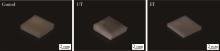
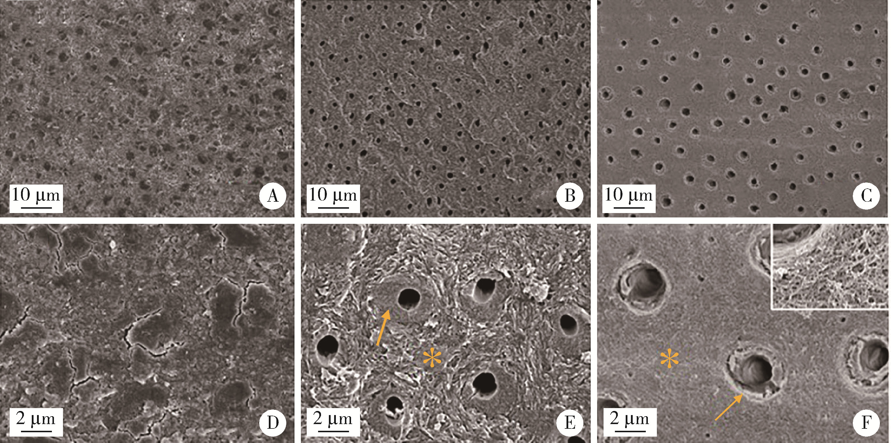
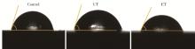
Influence of two methods of smear layer removal on the surface properties of dentin
Lingli ZHU1, Lin TANG1, Bowen LI2, Mei WANG1, Yuhua LIU1,*( )
)
- 1. Department of Prosthodontics, Peking University School and Hospital of Stomatology & National Center for Stomatology & National Clinical Research Center for Oral Diseases & National Engineering Laboratory for Digital and Material Technology of Stomatology & Beijing Key Laboratory of Digital Stomatology, Beijing 100081, China
2. Department of Stomatology, Beijing Hospital; National Center of Gerontology; Institute of Geriatric Medicine, Chinese Academy of Medical Sciences, Beijing 100730, China
CLC Number:
- R783.3


| 1 |
Gu XH , Mao CY , Kern M . Effect of different irrigation on smear layer removal after post space preparation[J]. J Endodont, 2009, 35 (4): 583- 586.
doi: 10.1016/j.joen.2009.01.006 |
| 2 | 刘清, 杨玉琼, 聂蓉蓉, 等. 牙本质玷污层特性对自粘接树脂水门汀粘接强度的影响[J]. 华西口腔医学杂志, 2018, 36 (6): 619- 622. |
| 3 |
Tribst JPM , Dos-Santos AFC , da Cruz-Santos G , et al. Effect of cement layer thickness on the immediate and long-term bond strength and residual stress between lithium disilicate glass-ceramic and human dentin[J]. Materials (Basel), 2021, 14 (18): 5153.
doi: 10.3390/ma14185153 |
| 4 |
李秋菊, 宫玮玉, 董艳梅. 生物活性玻璃预处理对牙本质粘接界面耐久性的影响[J]. 北京大学学报(医学版), 2020, 52 (5): 931- 937.
doi: 10.19723/j.issn.1671-167X.2020.05.023 |
| 5 | 张健, 葛久禹. 微波对化学法根管预备中的玷污层的影响[J]. 口腔医学研究, 2008, 24 (2): 173- 176. |
| 6 | Ghasemi N , Torabi ZS . The effect of photodynamic therapy on the smear layer removal: A scanning electron microscopic study[J]. J Dent (Shiraz), 2021, 22 (3): 162- 168. |
| 7 |
Akter RS , Ahmed Z , Yamauti M , et al. Effects of remaining dentin thickness, smear layer and aging on the bond strengths of selfetch adhesives to dentin[J]. Dent Mater J, 2021, 40 (2): 538- 546.
doi: 10.4012/dmj.2019-436 |
| 8 | 李秋容, 韦小浪, 谢方方. 改性PAMAM促进脱矿牙本质再矿化的体外研究[J]. 牙体牙髓牙周病学杂志, 2014, 24 (3): 151- 154. |
| 9 |
Saikaew P , Matsumoto M , Sattabanasuk V , et al. Ultra-morphological characteristics of dentin surfaces after different preparations and treatments[J]. Eur J Oral Sci, 2020, 128 (3): 246- 254.
doi: 10.1111/eos.12698 |
| 10 |
Dai LL , Mei ML , Chu CH , et al. Remineralizing effect of a new strontium-doped bioactive glass and fluoride on demineralized enamel and dentine[J]. J Dent, 2021, 108, 103633.
doi: 10.1016/j.jdent.2021.103633 |
| 11 |
Feitosa VP , Ogliari FA , van Meerbeek B , et al. Can the hydrophilicity of functional monomers affect chemical interaction?[J]. J Dent Res, 2014, 93 (2): 201- 206.
doi: 10.1177/0022034513514587 |
| 12 |
Karade P , Sharma D , Hoshing UA , et al. Efficiency of different endodontic irrigation and activation systems, self-adjusting file instrumentation/irrigation system, and XP-Endo finisher in removal of the intracanal smear layer: An ex vivo scanning electron microscope study[J]. J Pharm Bioallied Sci, 2021, 13 (Suppl 1): 402- 407.
doi: 10.4103/jpbs.JPBS_775_20 |
| 13 |
Gungormus M , Tulumbaci F . Peptide-assisted pre-bonding remineralization of dentin to improve bonding[J]. J Mech Behav Biomed Mater, 2021, 113, 104119.
doi: 10.1016/j.jmbbm.2020.104119 |
| 14 |
Dai LL , Mei ML , Chu CH , et al. Effect of strontium-doped bioactive glass on preventing formation of demineralized lesion[J]. Materials (Basel), 2021, 14 (16): 4645.
doi: 10.3390/ma14164645 |
| 15 |
Jang JH , Lee MG , Ferracane JL , et al. Effect of bioactive glass-containing resin composite on dentin remineralization[J]. J Dent, 2018, 75, 58- 64.
doi: 10.1016/j.jdent.2018.05.017 |
| 16 |
Anastasiadis K , Verdelis K , Eliades G . The effect of universal adhesives on dentine collagen[J]. Dent Mater, 2021, 37 (8): 1316- 1324.
doi: 10.1016/j.dental.2021.05.004 |
| 17 |
Zhou Z , Ge X , Bian M , et al. Remineralization of dentin slices using casein phosphopeptide-amorphous calcium phosphate combined with sodium tripolyphosphate[J]. Biomed Eng Online, 2020, 19 (1): 18.
doi: 10.1186/s12938-020-0756-9 |
| 18 |
Sereda G , van Laecken A , Turner JA . Monitoring demineralization and remineralization of human dentin by characterization of its structure with resonance-enhanced AFM-IR chemical mapping, nanoindentation, and SEM[J]. Dent Mater, 2019, 35 (4): 617- 626.
doi: 10.1016/j.dental.2019.02.007 |
| 19 |
Ayad MF , Johnston WM , Rosenstiel SF . Influence of dental rotary instruments on the roughness and wettability of human dentin surfaces[J]. J Prosthet Dent, 2009, 102 (2): 81- 88.
doi: 10.1016/S0022-3913(09)60114-1 |
| 20 |
Jing X , Xie B , Li X , et al. Peptide decorated demineralized dentin matrix with enhanced bioactivity, osteogenic differentiation via carboxymethyl chitosan[J]. Dent Mater, 2021, 37 (1): 19- 29.
doi: 10.1016/j.dental.2020.09.019 |
| [1] | Xiaoqiang BAI, Zhiruo YUAN, Yongsheng ZHOU, Longwei LV. Dynamic stretching promotes osteogenic differentiation of human bone marrow mesenchymal stem cells in three-dimensional culture [J]. Journal of Peking University (Health Sciences), 2026, 58(3): 641-649. |
| [2] | Ruo-qing ZHONG,Meng-qian ZHU,Ying-long LI,Jie PAN. Antibacterial effect of low-temperature plasma on Enterococcus faecalis in dentinal tubules in vitro [J]. Journal of Peking University (Health Sciences), 2023, 55(1): 38-43. |
| [3] | Ruo-lan GUO,Gui-bin HUANG,Yun-zi LONG,Yan-mei DONG. Effects of novel bioactive glasses on promoting remineralization of artificial dentin caries [J]. Journal of Peking University (Health Sciences), 2023, 55(1): 82-87. |
| [4] | Wei YONG,Kun QIAN,Wen-hao ZHU,Xiao-yi ZHAO,Chang LIU,Jie PAN. X-ray evaluation of pulp calcification in adult permanent teeth after pulpotomy [J]. Journal of Peking University (Health Sciences), 2023, 55(1): 88-93. |
| [5] | MA Xin-rong,ZHU Xiao-ming,LI Jing,LI De-li,LI He-ping,TAN Jian-guo. Effect of a novel radio-frequency atmospheric-pressure glow discharge plasma jet treatment on crosslinking of dentin collagen [J]. Journal of Peking University (Health Sciences), 2022, 54(1): 83-88. |
| [6] | LUO Chi-yi,PENG Chu-fang,YANG Yuan,QIN Man,WANG Yuan-yuan. Effects of three self-etch adhesives and mild salivary contamination on the bonding durability of deciduous teeth [J]. Journal of Peking University (Health Sciences), 2021, 53(1): 46-53. |
| [7] | Qiu-ju LI,Wei-yu GONG,Yan-mei DONG. Effect of bioactive glass pretreatment on the durability of dentin bonding interface [J]. Journal of Peking University (Health Sciences), 2020, 52(5): 931-937. |
| [8] | Yi-xiang PAN,Xiu-hua LI,Fu-cong TIAN,Xiao-yan WANG. Effect of intrapulpal pressure on the bonding strength of resin cement to dentin [J]. Journal of Peking University(Health Sciences), 2019, 51(2): 321-326. |
| [9] | Xiao-ming ZHU,Xuan QI,De-li LI,Yu-wei ZHANG,He-ping LI,Jian-guo TAN. Effect of a novel cold atmospheric plasma jet treatment with different temperatures on resin-dentin bonding [J]. Journal of Peking University(Health Sciences), 2019, 51(1): 43-48. |
| [10] | LI Fang, LIU Yang, LIU Hao-chen, FENG Hai-lan . Genetic variants analysis and histological observation of teeth in a patient with hereditary opalescent dentin [J]. Journal of Peking University(Health Sciences), 2018, 50(4): 666-671. |
| [11] | LI Shuang, ZHANG Qing. Effect of smear layer on apical sealing ability of mineral trioxide aggregate (MTA) Plus through the sucrose penetration mode [J]. Journal of Peking University(Health Sciences), 2018, 50(3): 560-563. |
| [12] | ZANG Hai-ling, WANG Yue, LIANG Yu-hong. Cleaning efficacy of different solvents on sealer-contaminated dentin surface [J]. Journal of Peking University(Health Sciences), 2018, 50(1): 63-68. |
| [13] | JIAO Yang, WANG Ji-de, DENG Jiu-peng. Effect of different surface treatments on the crystal structure and properties of zirconia [J]. Journal of Peking University(Health Sciences), 2018, 50(1): 49-52. |
| [14] | TANG Lin, ZHANG Yi, LI Hao, LIU Yu-hua, ZHOU Yong-sheng, LI Bo-wen, WU Wei-yi, WANG Si-wen. Influence of EDC ethanol solution on dentin shear bond strength with a self-etch adhesive system [J]. Journal of Peking University(Health Sciences), 2017, 49(6): 1055-1059. |
| [15] | LI Hao, LIU Yu-hua, LUO Zhi-qiang. Effects of bioactive glass on reducing the hypersensitivity after full crown preparation [J]. Journal of Peking University(Health Sciences), 2017, 49(4): 709-713. |
|
||