北京大学学报(医学版) ›› 2022, Vol. 54 ›› Issue (2): 222-226. doi: 10.19723/j.issn.1671-167X.2022.02.004
儿童基底节区生殖细胞瘤30例临床分析
王书磊,高阳旭,张宏武,杨海波,李辉,李宇,沈笠雪,姚红新( )
)
- 北京大学第一医院小儿外科,北京 100034
Clinical analysis of 30 cases of basal ganglia germinoma in children
WANG Shu-lei,GAO Yang-xu,ZHANG Hong-wu,YANG Hai-bo,LI Hui,LI Yu,SHEN Li-xue,YAO Hong-xin()
- Department of Pediatric Surgery, Peking University First Hospital, Beijing 100034, China
摘要:
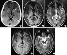
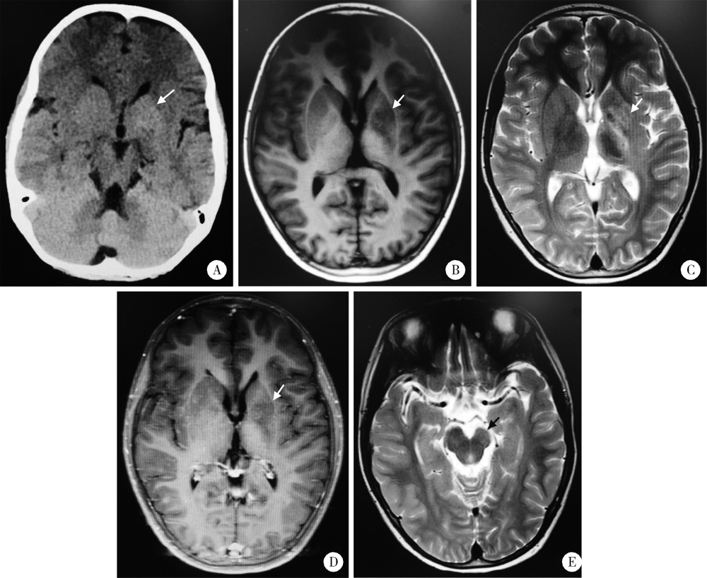
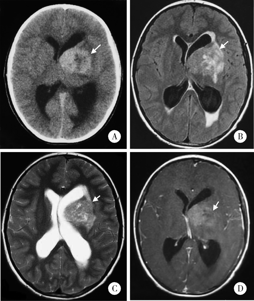
目的: 总结并分析儿童基底节区生殖细胞瘤的临床特征,提高临床早期诊断水平。方法: 选择2013年1月至2020年12月于北京大学第一医院小儿外科病房确诊为基底节区生殖细胞瘤的儿童病例资料进行回顾性分析,利用描述性统计的方法分析儿童基底节区生殖细胞瘤患者的临床资料。结果: 共纳入30例患者,28例为男性,2例为女性,发病平均年龄(9.7±2.2)岁,中位病程7个月,27例单侧发病,3例双侧发病,临床症状表现为偏侧肢体肌力下降、认知功能障碍、多饮多尿、性早熟、颅内高压、发音障碍、吞咽功能障碍。30例患者血清与脑脊液肿瘤标志物甲胎蛋白(alpha-fetoprotein, AFP)均正常, 其中8例患者血清与脑脊液肿瘤标志物β-人绒毛膜促性腺激素(β-human chorionic gonadotropin,β-HCG)均正常,11例患者血清β-HCG正常但脑脊液β-HCG轻度升高,11例患者血清与脑脊液β-HCG均轻度升高。影像学检查共发现33个病灶,病灶形态不规则,其中片状病灶15个(45.5%)、斑片状病灶10个(30.3%)、类圆形高密度病灶8个(24.2%), 计算机断层扫描(computed tomography,CT)肿瘤多呈明显高密度影,磁共振成像(magnetic resonance imaging,MRI)扫描肿瘤T1WI呈低或等信号,T2WI呈等或高信号,瘤周水肿轻,伴有半球萎缩、大脑脚萎缩、钙化、囊变、脑室扩张、华勒氏变性(Wallerian degeneration)。增强扫描肿瘤呈无强化或不均匀强化。结论: 儿童基底节区生殖细胞瘤主要发病年龄为10岁左右,男性占绝对优势,其临床特点与影像学表现有一定的特征,二者结合有利于提高儿童基底节区生殖细胞瘤的早期诊断水平。
中图分类号:
- R725

| [1] |
Woo PYM, Chu ACH, Chan KY, et al. Progressive hemiparesis in a young man: hemicerebral atrophy as the initial manifestation of basal ganglia germinoma[J]. Asian J Neurosurg, 2017, 12(1):65-68.
doi: 10.4103/1793-5482.145083 |
| [2] | Fetcko K, Dey M. Primary central nervous system germ cell tumors: a review and update[J]. Med Res Arch, 2018, 6(3):1719. |
| [3] |
Sonoda Y, Kumabe T, Sugiyama S, et al. Germ cell tumors in the basal ganglia: problems of early diagnosis and treatment[J]. J Neurosurg Pediatr, 2008, 2(2):118-124.
doi: 10.3171/PED/2008/2/8/118 |
| [4] |
Byun HK, Yoon HI, Cho J, et al. Optimization of intracranial germinoma treatment: radiotherapy alone with reduced volume and dose[J]. Int J Radiat Oncol Biol Phys, 2020, 108(3):657-666.
doi: 10.1016/j.ijrobp.2020.05.019 |
| [5] |
Wei XH, Shen HC, Tang SX, et al. Radiologic features of primary intracranial ectopic germinomas: case reports and literature review[J]. Medicine (Baltimore), 2016, 95(52):e5543.
doi: 10.1097/MD.0000000000005543 |
| [6] | Tso WW, Yung AW, Lau HY, et al. Basal ganglia germinoma: MRI classification correlates well with neurological and cognitive outcome[J]. J Pediatr Hematol Oncol, 2014, 36(7):443-447. |
| [7] |
Ogino H, Shibamoto Y, Takanaka T, et al. CNS germinoma with elevated serum human chorionic gonadotropin level: clinical characteristics and treatment outcome[J]. Int J Radiat Oncol Biol Phys, 2005, 62(1):803-808.
doi: 10.1016/j.ijrobp.2004.10.026 |
| [8] | Reddy MP, Saad AF, Doughty KE, et al. Intracranial germinoma[J]. Proc (Bayl Univ Med Cent), 2015, 28(1):43-45. |
| [9] | 程龙飞, 吴嘉铭, 张茂营, 等. 儿童基底节生殖细胞瘤的早期诊断和治疗[J]. 中国临床解剖学杂志, 2019, 37(2):190-195. |
| [10] | Takeda N, Fujita K, Katayama S, et al. Germinoma of the basal ganglia. An 8 year asymptomatic history after detection of abnormality on CT[J]. Pediatr Neurosurg, 2004, 40(6):306311. |
| [11] | Vialatte D, Bielle F, Mokhtari K, et al. Basal ganglia germinoma in an adult[J]. World Neurosurg, 2016, 92: 584.e11-584.e14. |
| [12] |
Kang YM, Lin SC, Lee YY, et al. A single-center study of treatment outcomes of pediatric basal ganglia germinoma in Taiwan[J]. Child’s Nerv Syst, 2020, 36(8):1745-1753.
doi: 10.1007/s00381-020-04543-4 |
| [13] |
Wong TT, Chen YW, Guo WY, et al. Germinoma involving the basal ganglia in children[J]. Child’s Nerv Syst, 2008, 24(1):71-78.
doi: 10.1007/s00381-007-0495-2 |
| [14] |
Phi JH, Cho BK, Kim SK, et al. Germinomas in the basal gang-lia: magnetic resonance imaging classification and the prognosis[J]. J Neurooncol, 2010, 99(2):227-236.
doi: 10.1007/s11060-010-0119-7 |
| [15] | Konovalov AN, Kadyrov SU, Tarasova EM, et al. Basal ganglia germinomas in children. Four clinical cases and a literature review[J]. Zh Vopr Neirokhir Im N N Burdenko, 2016, 80(1):71-82. |
| [16] |
Louis DN, Perry A, Reifenberger G, et al. The 2016 world health organization classification of tumors of the central nervous system: a summary[J]. Acta Neuropathol, 2016, 131(6):803-820.
doi: 10.1007/s00401-016-1545-1 |
| [17] |
Fujimaki T, Mishima K, Asai A, et al. Levels of beta-human chorionic gonadotropin in cerebrospinal fluid of patients with malignant germ cell tumor can be used to detect early recurrence and monitor the response to treatment[J]. Jpn J Clin Oncol, 2000, 30(7):291-294.
pmid: 11007160 |
| [18] | 张大千, 李桥, 何慧瑾, 等. 基底节区生殖细胞瘤的MRI 和MRS征象分析[J]. 中国医学计算机成像杂志, 2017, 23(2):101-106. |
| [19] | 夏正荣, 刘明, 曹雯君, 等. 儿童及青少年颅内生殖细胞瘤的临床和影像学特点[J]. 中国临床医学影像杂志, 2017, 28(8):542-545. |
| [20] | Ozelame RV, Shroff M, Wood B, et al. Basal ganglia germinoma in children with associated ipsilateral cerebral and brain stem hemiatrophy[J]. Pediatr Radiol, 2006, 36(4):325330. |
| [21] | Wong ST, Yuen SC, Fong D. Pathophysiological mechanism of ipsilateral cerebral and brainstem hemiatrophy in basal ganglia germ cell tumors: case report[J]. Child’s Nerv Syst, 2009, 25(6):693699. |
| [22] | Murray MJ, Bartels U, Nishikawa R, et al. Consensus on the management of intracranial germ cell tumors[J]. Lancet Oncol, 2015, 16(9):470-477. |
| [23] | 黄立敏, 雷竹, 曹雪, 等. 低剂量诊断性放疗联合化疗在诊治颅内生殖细胞肿瘤中的价值[J]. 中国癌症杂志, 2018, 28(4):270-275. |
| [24] |
Höftberger R, Lassmann H. Inflammatory demyelinating diseases of the central nervous system[J]. Handb Clin Neurol, 2017, 145:263-283.
doi: B978-0-12-802395-2.00019-5 pmid: 28987175 |
| [25] |
Fu W, Ju Y, Zhang S, et al. Pediatric basal ganglia region tumors: clinical and radiologic features correlated with histopathologic fin-dings[J]. World Neurosurg, 2017, 103:504-516.
doi: 10.1016/j.wneu.2017.04.004 |
| [26] | 阮胜, 魏根霞. 基底节区胶质瘤与生殖细胞肿瘤的 MRI 诊断和鉴别诊断[J]. 功能与分子医学影像学杂志, 2016, 5(2):926-929. |
| [1] | 邢念增,王明帅,杨飞亚,尹路,韩苏军. 前列腺免活检创新理念的临床实践及其应用前景[J]. 北京大学学报(医学版), 2024, 56(4): 565-566. |
| [2] | 田宇轩,阮明健,刘毅,李德润,吴静云,沈棋,范宇,金杰. 双参数MRI改良PI-RADS评分4分和5分病灶的最大径对临床有意义前列腺癌的预测效果[J]. 北京大学学报(医学版), 2024, 56(4): 567-574. |
| [3] | 刘毅,袁昌巍,吴静云,沈棋,肖江喜,赵峥,王霄英,李学松,何志嵩,周利群. 靶向穿刺+6针系统穿刺对PI-RADS 5分患者的前列腺癌诊断效能[J]. 北京大学学报(医学版), 2023, 55(5): 812-817. |
| [4] | 袁昌巍,李德润,李志华,刘毅,山刚志,李学松,周利群. 多参数磁共振成像中动态对比增强状态在诊断PI-RADS 4分前列腺癌中的应用[J]. 北京大学学报(医学版), 2023, 55(5): 838-842. |
| [5] | 刘颖,霍然,徐慧敏,王筝,王涛,袁慧书. 磁共振血管壁成像评估颈动脉中重度狭窄患者斑块特征与脑血流灌注的相关性[J]. 北京大学学报(医学版), 2023, 55(4): 646-651. |
| [6] | 傅强,高冠英,徐雁,林卓华,孙由静,崔立刚. 无症状髋关节前上盂唇撕裂超声与磁共振检查的对比研究[J]. 北京大学学报(医学版), 2023, 55(4): 665-669. |
| [7] | 叶珊,金萍萍,张楠,邬海博,石林,赵强,杨坤,袁慧书,樊东升. 肌萎缩侧索硬化患者认知功能改变与脑皮层厚度分析[J]. 北京大学学报(医学版), 2022, 54(6): 1158-1162. |
| [8] | 蔡颖,万巧琴,蔡宪杰,高亚娟,韩鸿宾. 光生物调节加速脑组织间液引流及其机制[J]. 北京大学学报(医学版), 2022, 54(5): 1000-1005. |
| [9] | 张帆,陈曲,郝一昌,颜野,刘承,黄毅,马潞林. 术前及术后膜性尿道长度与腹腔镜根治性前列腺切除术后控尿功能恢复的相关性[J]. 北京大学学报(医学版), 2022, 54(2): 299-303. |
| [10] | 吴一凡,张晓圆,任爽,玉应香,常翠青. 基于磁共振的青年男性股四头肌的测量和评估[J]. 北京大学学报(医学版), 2021, 53(5): 843-849. |
| [11] | 李蓬,朴牧子,胡洪成,王勇,赵一姣,申晓婧. 经嵴顶上颌窦底提升术后不植骨同期种植的影像研究[J]. 北京大学学报(医学版), 2021, 53(1): 95-101. |
| [12] | 盛荟,梁磊,周童亮,贾彦兴,王彤,袁兰,韩鸿宾. 光磁双模态探针钆-[4,7-双-羧甲基-10-(2-荧光素硫脲乙基)-1,4,7,10-四氮杂环十二烷-1-基]-乙酸络合物合成方法的改进[J]. 北京大学学报(医学版), 2020, 52(5): 959-963. |
| [13] | 赵世明,杨铁军,许春苗,郭孝峰,马永康,陈学军,李祥,何朝宏. 3.0T磁共振成像在接受过经尿道膀胱肿瘤切除术膀胱癌中诊断肌层浸润的应用[J]. 北京大学学报(医学版), 2020, 52(4): 701-704. |
| [14] | 宋宇,韩鸿宾,杨军,王艾博,和清源,李媛媛,赵国梅,高亚娟,王睿,韩易兴,刘爱连,宋清伟. 脑对流增强给药对老年大鼠脑细胞外间隙微观结构的影响[J]. 北京大学学报(医学版), 2020, 52(2): 362-367. |
| [15] | 吴静云,米悦,刘水,姚林,唐琦,何志嵩,王霄英. MRI对肾细胞癌静脉瘤栓侵犯下腔静脉壁的术前评估[J]. 北京大学学报(医学版), 2019, 51(4): 673-677. |
| Viewed | ||||||||||||||||||||||||||||||||||||||||||||||||||
|
Full text 267
|
|
|||||||||||||||||||||||||||||||||||||||||||||||||
|
Abstract 959
|
|
|||||||||||||||||||||||||||||||||||||||||||||||||
Cited |
|
|||||||||||||||||||||||||||||||||||||||||||||||||
| Shared | ||||||||||||||||||||||||||||||||||||||||||||||||||
| Discussed | ||||||||||||||||||||||||||||||||||||||||||||||||||
|
||
