北京大学学报(医学版) ›› 2024, Vol. 56 ›› Issue (1): 99-105. doi: 10.19723/j.issn.1671-167X.2024.01.016
常温流动牙胶封闭剂GuttaFlow2单尖充填弯曲根管的封闭效果
- 北京大学口腔医学院·口腔医院牙体牙髓科,国家口腔医学中心,国家口腔疾病临床医学研究中心,口腔生物材料和数字诊疗装备国家工程研究中心,口腔数字医学北京市重点实验室,北京 100081
Sealing effect of GuttaFlow2 in curved root canals
- Department of Cariology and Endodoontology, Peking University School and Hospital of Stomatology & National Center for Stomatology & National Clinical Research Center for Oral Diseases & National Engineering Research Center of Oral Biomaterials and Digital Medical Devices & Beijing Key Laboratory of Digital Stomatology, Beijing 100081, China
摘要:
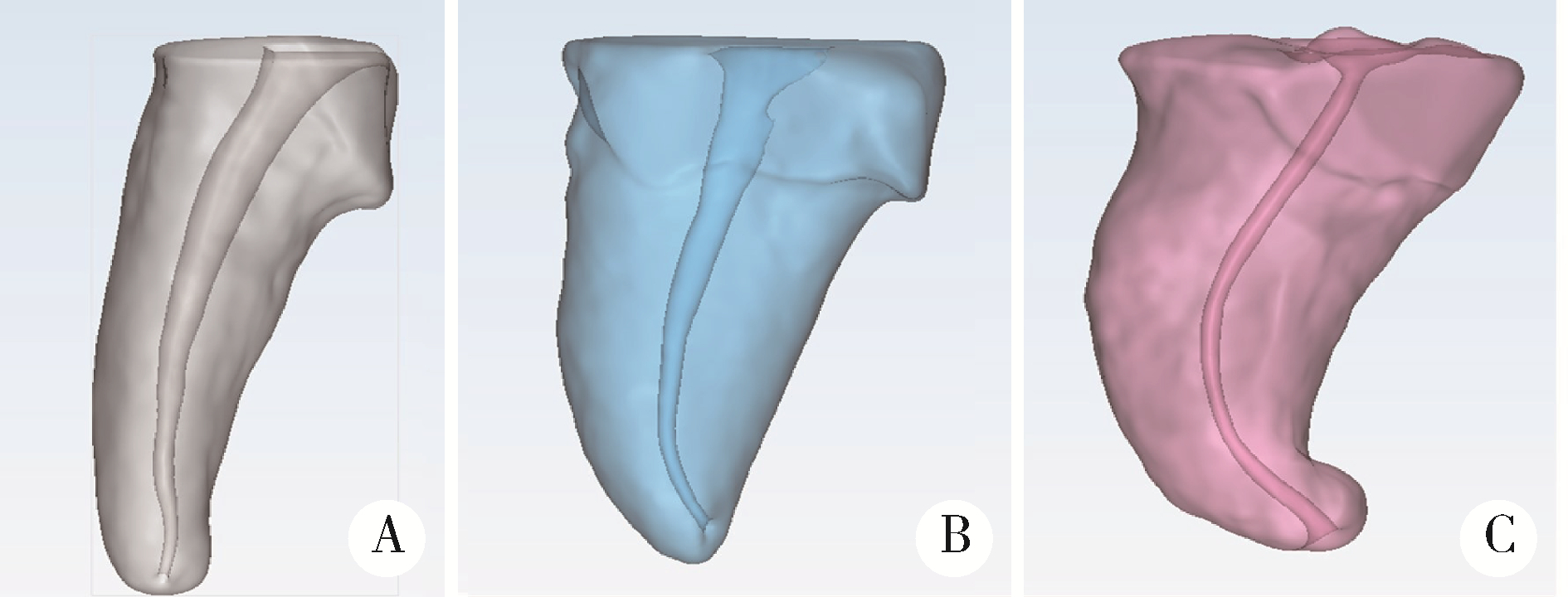
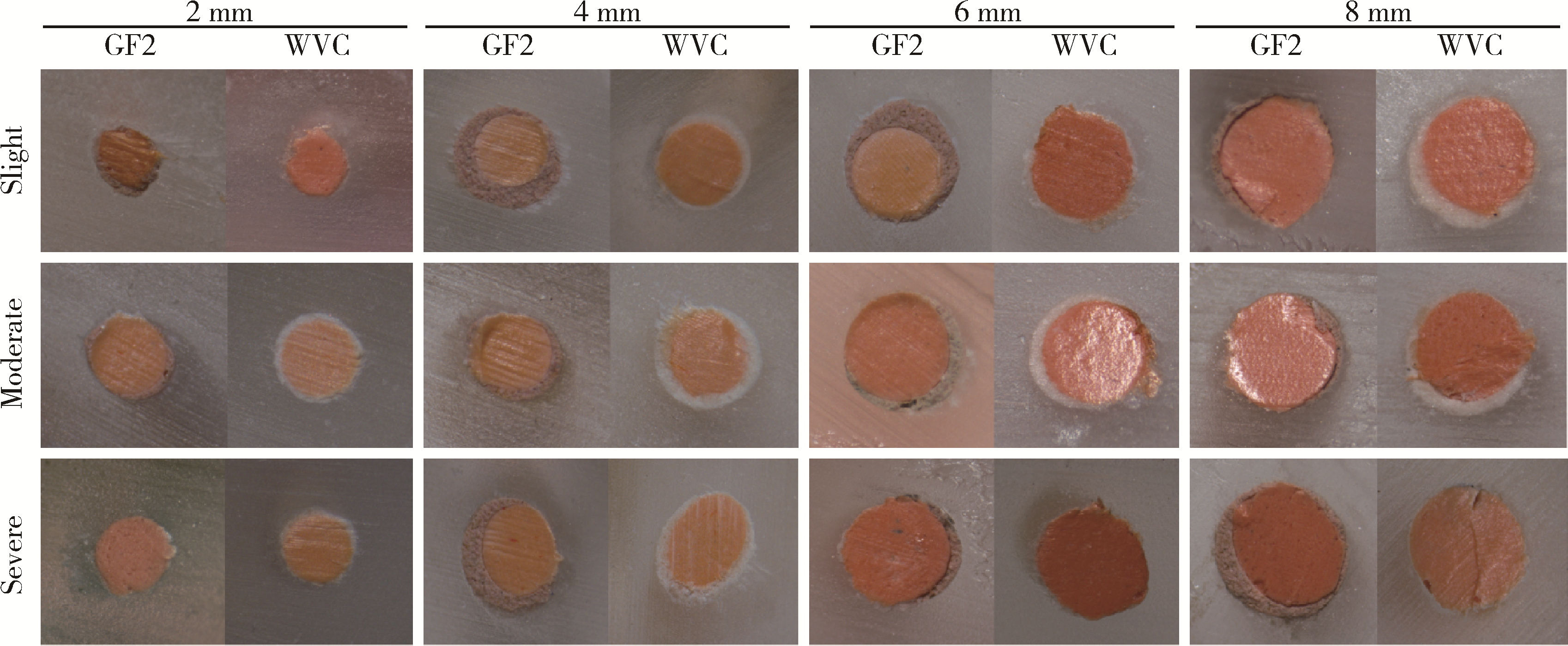
目的: 评估常温流动牙胶封闭剂单尖充填法对弯曲根管的封闭效果。方法: (1) 3D打印轻、中、重度弯曲根管模型各20例, 分别采用热垂直加压技术(warm vertical compaction, 以AH-Plus为封闭剂, 以下称WVC组, n = 10)和单尖充填技术(以GuttaFlow2为封闭剂, 以下称GF2组, n = 10)充填根管, 在距根尖2 mm(根尖部)、4 mm和6 mm(根中部), 以及距根尖8 mm(根冠部)处切片, 使用体视显微镜观察计算根管截面的空隙面积百分比。(2)选取离体恒磨牙轻、中、重度弯曲根管各16例, 同样分别采用热垂直加压技术(WVC组, n = 8)和GuttaFlow2单尖充填技术(GF2组, n = 8)充填根管, 使用激光共聚焦显微镜观察计算距根尖2 mm(根尖部)、5 mm(根中部)和8 mm(根冠部)处各个截面上封闭剂渗入牙本质小管区域的渗透百分比。结果: (1) 根管空隙面积百分比: GF2组和WVC组轻、中、重度弯曲根管根尖部均无明显空隙, 两组间差异无统计学意义(P>0.05);根中部两种充填方法产生的空隙面积百分比差异也没有统计学意义; 但在根冠部GF2组相较于WVC组在轻度弯曲根管中出现了更多的空隙, 差异有统计学意义(P = 0.009)。(2)封闭剂对牙本质小管渗透百分比:GF2组在轻、中、重度弯曲根管根尖部, 封闭剂对牙本质小管渗透百分比分别为36.10%、55.80%、65.08%, 重度弯曲根管显著高于轻度弯曲根管(P = 0.001);组间比较提示, 根尖部GF2组轻、中、重度弯曲根管牙本质小管渗透百分比均明显高于WVC组(分别为15.78%、20.70%、15.61%, P = 0.001), 根中部GF2组轻、中度弯曲根管牙本质小管渗透百分比也明显高于WVC组(P = 0.001), 但根冠部两组间差异没有统计学意义(P>0.05)。结论: 常温流动牙胶封闭剂单尖法充填弯曲根管在根尖和根中段对根管的封闭效果和热垂直加压技术相同, 而对牙本质小管则具有更好的渗透封闭效果, 尤其对于重度弯曲根管。
中图分类号:
- R781.3
| 1 |
Clinton K , Himel VT . Comparison of a warm gutta-percha obturation technique and lateral condensation[J]. J Endod, 2001, 27 (11): 692- 695.
doi: 10.1097/00004770-200111000-00010 |
| 2 |
Libonati A , Montemurro E , Nardi R , et al. Percentage of gutta-percha-filled areas in canals obturated by 3 different techniques with and without the use of endodontic sealer[J]. J Endod, 2018, 44 (3): 506- 509.
doi: 10.1016/j.joen.2017.09.019 |
| 3 |
Jafarzadeh H , Abbott PV . Dilaceration: Review of an endodontic challenge[J]. J Endod, 2007, 33 (9): 1025- 1030.
doi: 10.1016/j.joen.2007.04.013 |
| 4 |
Iglecias EF , Freire LG , de Miranda Candeiro GT , et al. Presence of voids after continuous wave of condensation and single-cone obturation in mandibular molars: A micro-computed tomography analysis[J]. J Endod, 2017, 43 (4): 638- 642.
doi: 10.1016/j.joen.2016.11.027 |
| 5 |
Collado-Gonzalez M , Tomas-Catala CJ , Onate-Sanchez RE , et al. Cytotoxicity of GuttaFlow Bioseal, GuttaFlow2, MTA Fillapex, and AH Plus on human periodontal ligament stem cells[J]. J Endod, 2017, 43 (5): 816- 822.
doi: 10.1016/j.joen.2017.01.001 |
| 6 |
Pedullà E , Abiad RS , Conte G , et al. Root fillings with a matched-taper single cone and two calcium silicate-based sealers: An analysis of voids using micro-computed tomography[J]. Clin Oral Investig, 2020, 24 (12): 4487- 4492.
doi: 10.1007/s00784-020-03313-5 |
| 7 |
Akcay M , Arslan H , Durmus , et al. Dentinal tubule penetration of AH Plus, iRoot SP, MTA fillapex, and GuttaFlow bioseal root canal sealers after different final irrigation procedures: A confocal microscopic study[J]. Lasers Surg Med, 2016, 48 (1): 70- 76.
doi: 10.1002/lsm.22446 |
| 8 |
Zheng QH , Zhou XD , Jiang Y , et al. Radiographic investigation of frequency and degree of canal curvatures in Chinese mandibular permanent incisors[J]. J Endod, 2009, 35 (2): 175- 178.
doi: 10.1016/j.joen.2008.10.028 |
| 9 |
Schneider SW . A comparison of canal preparations in straight and curved root canals[J]. Oral Surg Oral Med Oral Pathol, 1971, 32 (2): 271- 275.
doi: 10.1016/0030-4220(71)90230-1 |
| 10 |
Wu MK , Wesselink PR . A primary observation on the preparation and obturation of oval canals[J]. Int Endod J, 2001, 34 (2): 137- 141.
doi: 10.1046/j.1365-2591.2001.00361.x |
| 11 |
Eymirli A , Sungur DD , Uyanik O , et al. Dentinal tubule penetration and retreatability of a calcium silicate-based sealer tested in bulk or with different main core material[J]. J Endod, 2019, 45 (8): 1036- 1040.
doi: 10.1016/j.joen.2019.04.010 |
| 12 | Kumar NS , Prabu PS , Prabu N , et al. Sealing ability of lateral condensation, thermoplasticized gutta-percha and flowable gutta-percha obturation techniques: A comparative in vitro study[J]. J Pharm Bioallied Sci, 2012, 4 (Suppl 2): S131- S135. |
| 13 | 张琛. 根管充填的难点和误区[J]. 华西口腔医学杂志, 2017, 35 (3): 232- 238. |
| 14 |
Elyassi Y , Moinzadeh AT , Kleverlaan CJ . Characterization of leachates from 6 root canal sealers[J]. J Endod, 2019, 45 (5): 623- 627.
doi: 10.1016/j.joen.2019.01.011 |
| 15 |
Zhong XY , Shen Y , Ma JZ , et al. Quality of root filling after obturation with gutta-percha and 3 different sealers of minimally instrumented root canals of the maxillary first molar[J]. J Endod, 2019, 45 (8): 1030- 1035.
doi: 10.1016/j.joen.2019.04.012 |
| 16 |
Oguntebi BR . Dentine tubule infection and endodontic therapy implications[J]. Int Endod J, 1994, 27 (4): 218- 222.
doi: 10.1111/j.1365-2591.1994.tb00257.x |
| 17 |
Tuncer AK , Tuncer S . Effect of different final irrigation solutions on dentinal tubule penetration depth and percentage of root canal sealer[J]. J Endod, 2012, 38 (6): 860- 863.
doi: 10.1016/j.joen.2012.03.008 |
| [1] | 臧海玲,王月,梁宇红. 有机溶剂对牙本质表面残留根管封闭剂的清除效果[J]. 北京大学学报(医学版), 2018, 50(1): 63-68. |
| [2] | 范聪,袁重阳,张继川,王晓燕. 牙胶类根管充填材料导热性对根尖封闭的影响[J]. 北京大学学报(医学版), 2017, 49(1): 110-114. |
| [3] | 乔迪, 董艳梅, 高学军. 体外评价新型根尖倒充填材料iRoot的生物学性能[J]. 北京大学学报(医学版), 2016, 48(2): 324-329. |
| [4] | 渠薇,白伟,梁宇红,高学军. 热牙胶充填系统携热头工作状态下的实际温度[J]. 北京大学学报(医学版), 2015, 47(5): 834-837. |
| [5] | 陈小贤, 林碧琛, 钟洁, 葛立宏. 改良乳牙根管充填材料体内降解匹配性与临床效果[J]. 北京大学学报(医学版), 2015, 47(3): 529-535. |
| [6] | 卫彦, 严红, 马琦. 四种根管封闭剂的根尖封闭性研究[J]. 北京大学学报(医学版), 2008, 40(1): 71-73. |
|
||


