北京大学学报(医学版) ›› 2025, Vol. 57 ›› Issue (2): 354-359. doi: 10.19723/j.issn.1671-167X.2025.02.021
基于CBCT图像构建牙颌面虚拟患者的两种配准方法比较
叶嘉慧1, 王时敏2, 王子轩2, 刘云松1, 孙玉春3, 叶红强1,*( ), 周永胜1,*()
- 1. 北京大学口腔医学院·口腔医院修复科,北京 100081
2. 北京大学口腔医学院·口腔医院义齿加工中心,北京 100081
3. 北京大学口腔医学院·口腔医院口腔医学数字化中心,国家口腔医学中心,国家口腔疾病临床医学研究中心,口腔生物材料和数字诊疗装备国家工程研究中心,口腔数字医学北京市重点实验室,国家卫生健康委员会口腔医学计算机应用工程技术研究中心,国家药品监督管理局口腔材料重点实验室,北京 100081
Comparison of two registration methods for constructing virtual craniodentofacial patients based on cone beam computed tomography images
Jiahui YE1, Shimin WANG2, Zixuan WANG2, Yunsong LIU1, Yuchun SUN3, Hongqiang YE1,*(), Yongsheng ZHOU1,*()
- 1. Department of Prosthodontics, Peking University School and Hospital of Stomatology, Beijing 100081, China
2. Center of Dental Laboratory, Peking University School and Hospital of Stomatology, Beijing 100081, China
3. Center of Digital Dentistry, Peking University School and Hospital of Stomatology & National Center for Stomatology & National Clinical Research Center for Oral Diseases & National Engineering Research Center of Oral Biomaterials and Digital Medical Devices & Beijing Key Laboratory of Digital Stomatology & NHC Research Center of Engineering and Technology for Computerized Dentistry & NMPA Key Laboratory for Dental Materials, Beijing 100081, China
摘要:
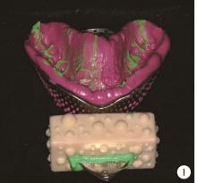
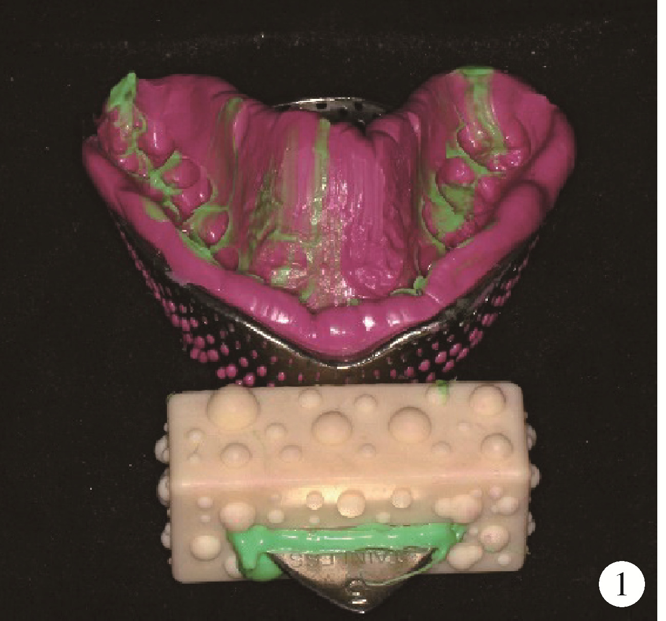
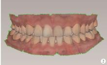
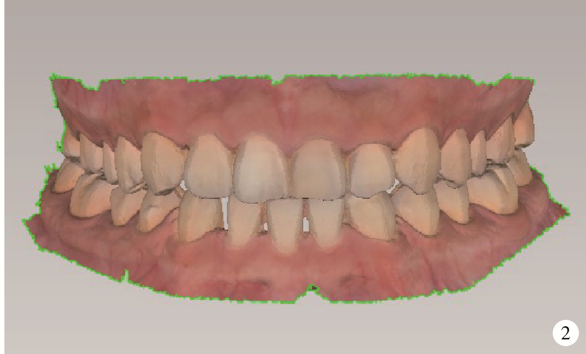
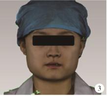
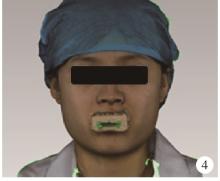
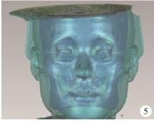
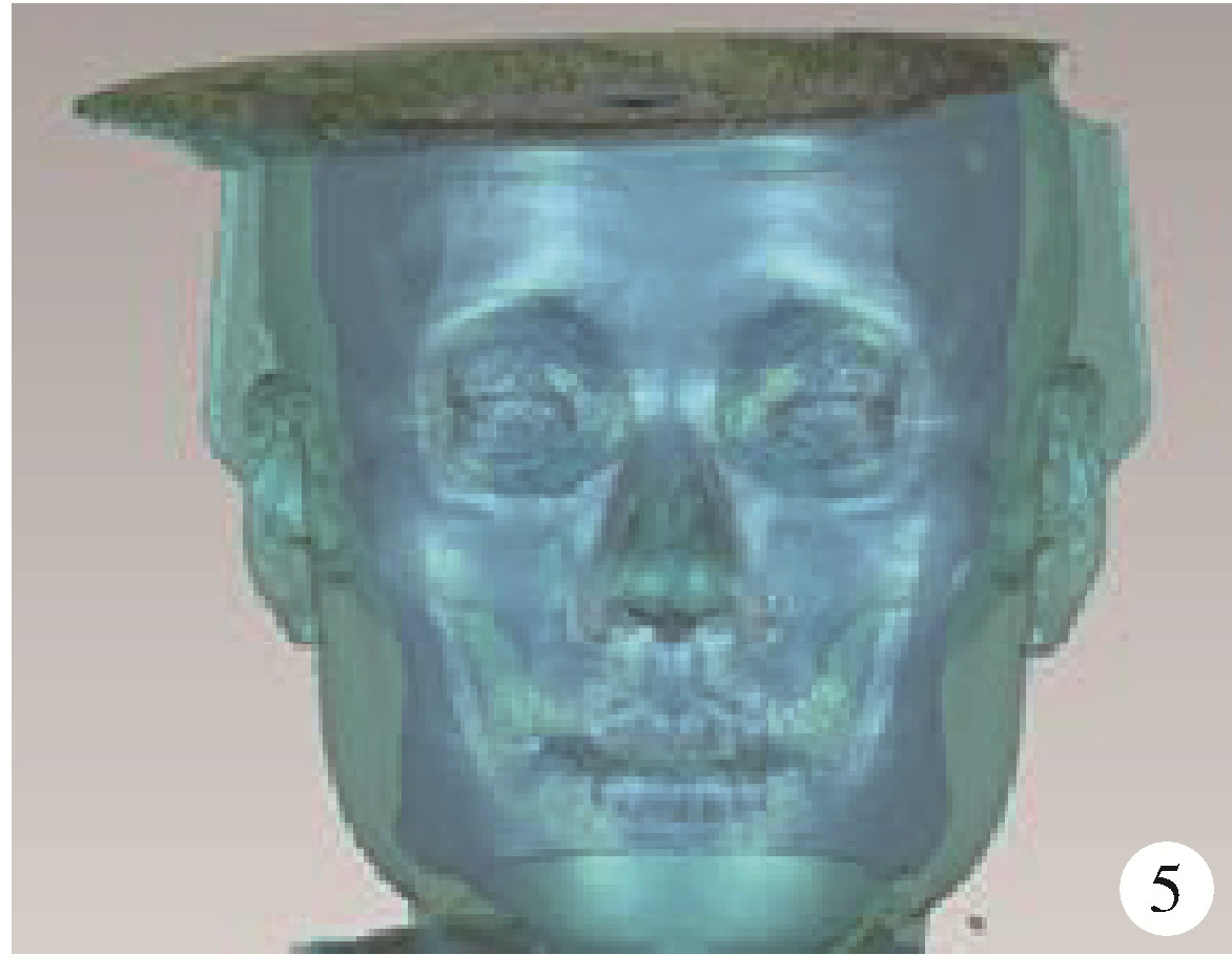
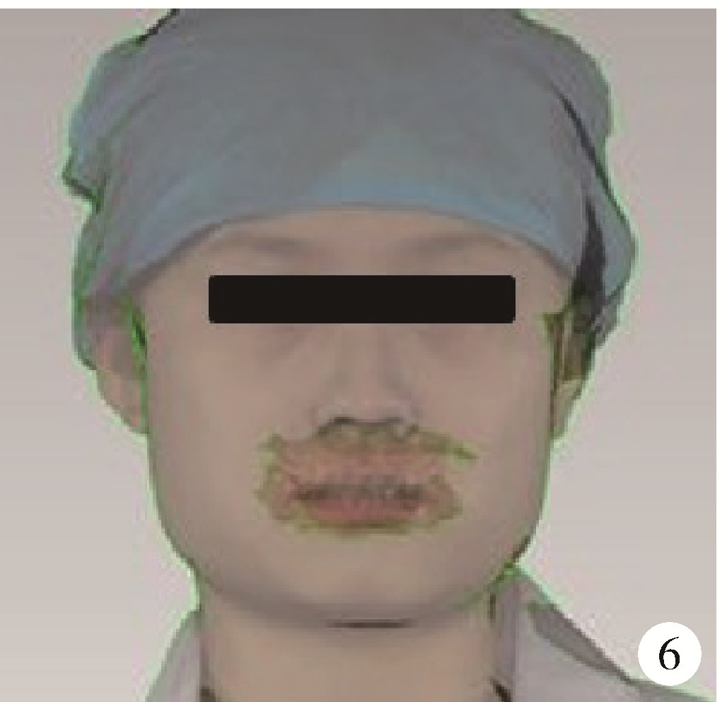
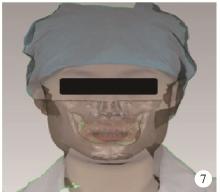
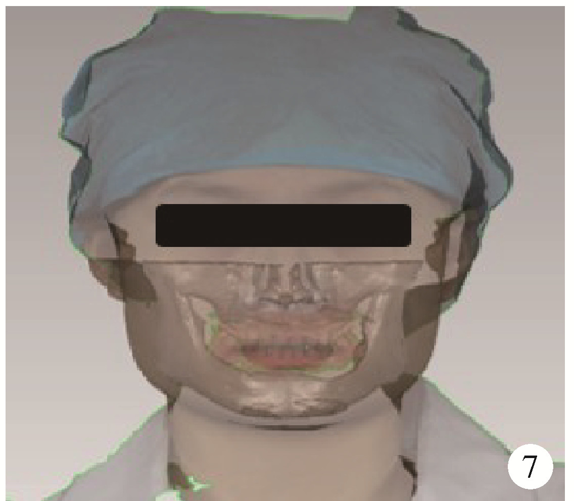
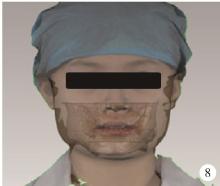
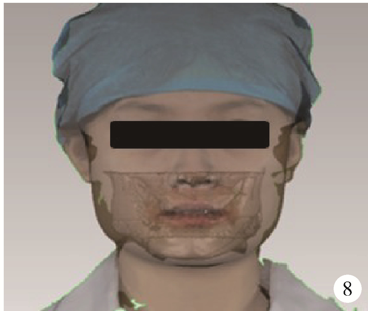
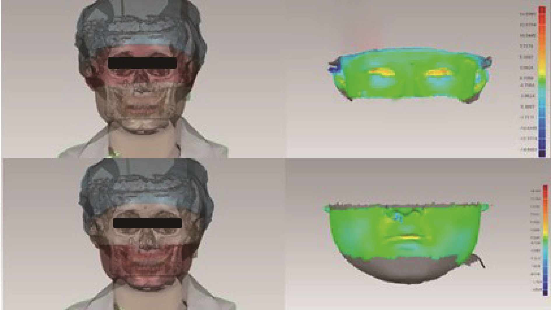
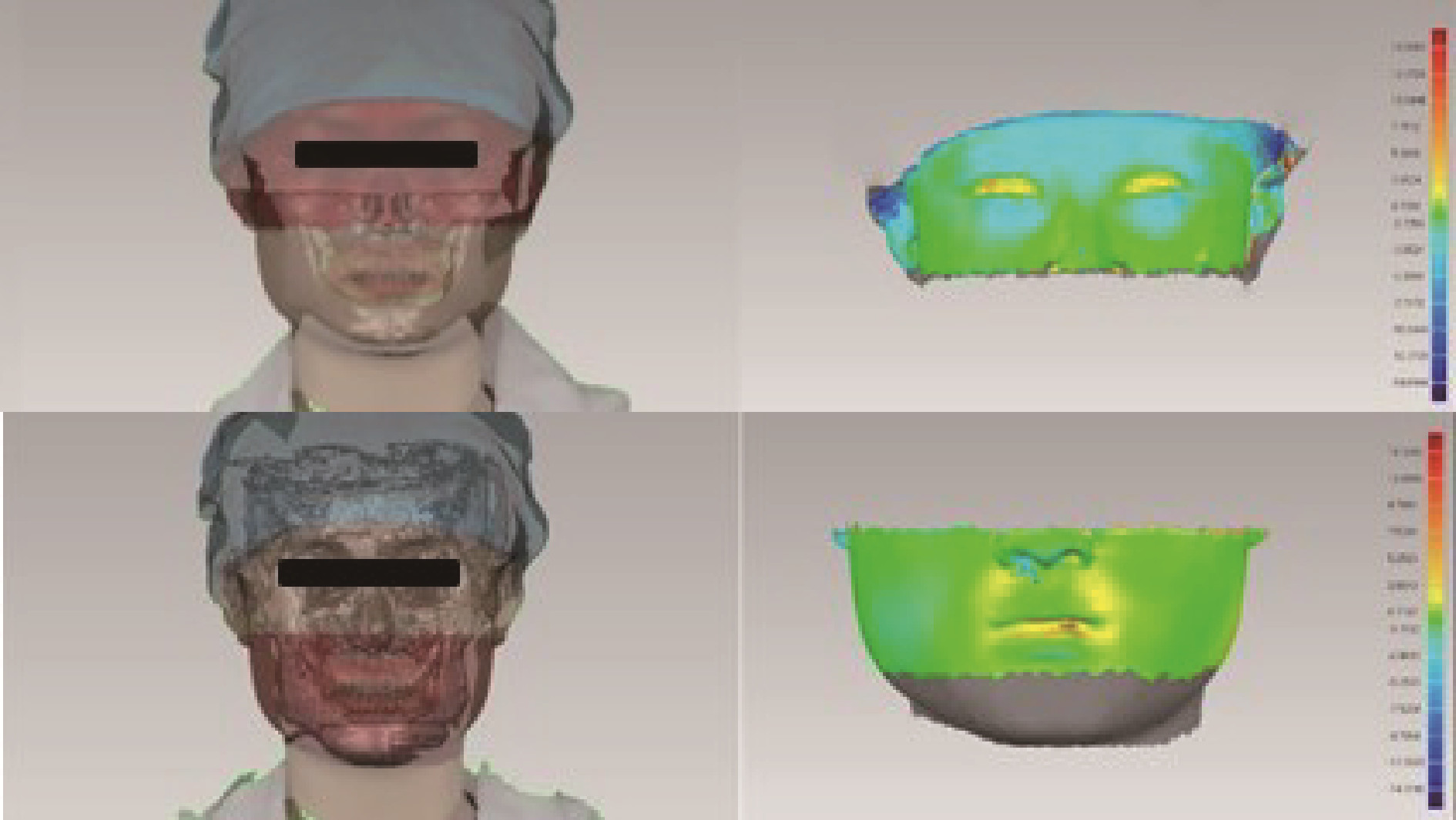
目的: 比较锥形束计算机体层摄影术(cone beam computed tomography,CBCT)数据参与构建牙颌面虚拟患者时,软组织配准法和牙列配准法的准确度。方法: 选择13名需要行CBCT检查的患者,分别获取其数字牙列图像、配准印模三维图像、面部三维图像和颌面部数据。以配准印模三维图像为媒介建立包含牙列和面部软组织的牙面虚拟患者,将CBCT数据三维重建并形成包含面部软组织、颌骨和牙列的牙颌面虚拟患者,分别通过面中下部软组织标志点(软组织配准法)及牙列标志点(牙列配准法)将CBCT来源的牙颌面虚拟患者和配准印模法建立的牙面虚拟患者进行配准,选择口内扫描来源的数字牙列图像、面部扫描来源的面部三维图像和CBCT来源的颌骨图像建立2种牙颌面虚拟患者(隐藏CBCT来源的面部图像和牙列图像)。以配准印模法建立的牙面虚拟患者面部软组织为参考,对2种牙颌面虚拟患者中CBCT来源面部图像的面下1/2区域及面上1/2区域分别进行三维偏差分析,记录各组偏差的均方根误差(root mean square error,RMSE)。用配对t检验分别比较2种牙颌面虚拟患者中CBCT面部图像的面上1/2与面下1/2区域之间,以及面上1/2区域之间的RMSE差异是否有统计学意义。结果: 配对t检验结果表明,2种牙颌面虚拟患者中CBCT来源面部图像的面上1/2与面下1/2区域之间的RMSE值差异无统计学意义(P>0.05),面上1/2区域软组织配准法的RMSE值小于牙列配准法[(1.696±0.420) mm vs. (1.752±0.424) mm,P < 0.01]。结论: 基于数字牙列图像、面部三维图像和CBCT来源颌骨图像建立牙颌面虚拟患者时,用软组织配准法将CBCT数据配准到牙面虚拟患者中的准确度较高。
中图分类号:
- R783.4


| 1 |
Joda T , Gallucci GO . The virtual patient in dental medicine[J]. Clin Oral Implants Res, 2015, 26 (6): 725- 726.
doi: 10.1111/clr.12379 |
| 2 |
Su TS , Sun J . Comparison of repeatability between intraoral digital scanner and extraoral digital scanner: An in-vitro study[J]. J Prosthodont Res, 2015, 59 (4): 236- 242.
doi: 10.1016/j.jpor.2015.06.002 |
| 3 | 刘静. 口内扫描仪全牙列扫描精度及不同操作者间扫描精度差异的研究[D]. 济南: 山东大学, 2017. |
| 4 |
Ye H , Lv L , Liu Y , et al. Evaluation of the accuracy, reliability, and reproducibility of two different 3D face-scanning systems[J]. Int J Prosthodont, 2016, 29 (3): 213- 218.
doi: 10.11607/ijp.4397 |
| 5 |
Ye H , Wang KP , Liu Y , et al. Four-dimensional digital prediction of the esthetic outcome and digital implementation for rehabilitation in the esthetic zone[J]. J Prosthet Dent, 2020, 123 (4): 557- 563.
doi: 10.1016/j.prosdent.2019.04.007 |
| 6 |
Becker K , Schmücker U , Schwarz F , et al. Accuracy and eligibi-lity of CBCT to digitize dental plaster casts[J]. Clin Oral Investig, 2018, 22 (4): 1817- 1823.
doi: 10.1007/s00784-017-2277-x |
| 7 |
Jayaratne YS , McGrath CP , Zwahlen RA . How accurate are the fusion of cone-beam CT and 3-D stereophotographic images[J]. PLoS One, 2012, 7 (11): e49585.
doi: 10.1371/journal.pone.0049585 |
| 8 | Xiao Z , Liu Z , Gu Y . Integration of digital maxillary dental casts with 3D facial images in orthodontic patients[J]. Angle Orthod, 2019, 90 (3): 397- 404. |
| 9 |
Bechtold TE , Göz TG , Schaupp E , et al. Integration of a maxillary model into facial surface stereophotogrammetry[J]. J Orofac Orthop, 2012, 73 (2): 126- 137.
doi: 10.1007/s00056-011-0060-1 |
| 10 |
Codari M , Pucciarelli V , Tommasi DG , et al. Validation of a technique for integration of a digital dental model into stereophotogrammetric images of the face using cone-beam computed tomographic data[J]. Br J Oral Maxillofac Surg, 2016, 54 (5): 584- 586.
doi: 10.1016/j.bjoms.2016.01.019 |
| 11 |
Granata S , Giberti L , Vigolo P , et al. Incorporating a facial scanner into the digital workflow: A dental technique[J]. J Prosthet Dent, 2020, 123 (6): 781- 785.
doi: 10.1016/j.prosdent.2019.05.021 |
| 12 |
Bohner L , Gamba DD , Hanisch M , et al. Accuracy of digital technologies for the scanning of facial, skeletal, and intraoral tissues: A systematic review[J]. J Prosthet Dent, 2019, 121 (2): 246- 251.
doi: 10.1016/j.prosdent.2018.01.015 |
| 13 |
Kim J , Heo G , Lagravère MO . Accuracy of laser-scanned models compared to plaster models and cone-beam computed tomography[J]. Angle Orthod, 2014, 84 (3): 443- 450.
doi: 10.2319/051213-365.1 |
| 14 |
Akyalcin S , Dyer DJ , English JD , et al. Comparison of 3-dimensional dental models from different sources: Diagnostic accuracy and surface registration analysis[J]. Am J Orthod Dentofacial Orthop, 2013, 144 (6): 831- 837.
doi: 10.1016/j.ajodo.2013.08.014 |
| 15 | 吕燕, 严斌, 王林, 等. 利用锥形束CT和激光快速成型技术制作牙颌模型的可靠性分析[J]. 上海口腔医学, 2012, 21 (2): 175- 179. |
| 16 |
Lim MY , Lim SH . Comparison of model analysis measurements among plaster model, laser scan digital model, and cone beam CT image[J]. Korean J Orthod, 2009, 39 (1): 6- 17.
doi: 10.4041/kjod.2009.39.1.6 |
| 17 |
Nahm KY , Kim Y , Choi YS , et al. Accurate registration of cone-beam computed tomography scans to 3-dimensional facial photographs[J]. Am J Orthod Dentofacial Orthop, 2014, 145 (2): 256- 264.
doi: 10.1016/j.ajodo.2013.10.018 |
| 18 | Pérez-Giugovaz MG , Park SH , Revilla-León M . Three-dimensional virtual representation by superimposing facial and intraoral digital scans with an additively manufactured intraoral scan body[J]. J Prosthet Dent, 2020, 126 (4): 459- 463. |
| 19 |
国丹妮, 潘韶霞, 衡墨笛, 等. 应用于无牙颌种植修复设计的三维面部扫描配准方法的对比[J]. 北京大学学报(医学版), 2021, 53 (1): 83- 87.
doi: 10.19723/j.issn.1671-167X.2021.01.013 |
| [1] | 吴灵, 方嘉琨, 刘筱菁, 李自力, 李阳, 王晓霞. 基于牙颌面畸形患者三维颅面特征相似性度量模型的建立及评估[J]. 北京大学学报(医学版), 2025, 57(1): 128-135. |
| [2] | 付玉,胡鑫浓,崔圣洁,施捷. 骨性Ⅱ类高角错 |
| [3] | 国丹妮,潘韶霞,衡墨笛,屈健,魏秀霞,周永胜. 应用于无牙颌种植修复设计的三维面部扫描配准方法的对比[J]. 北京大学学报(医学版), 2021, 53(1): 83-87. |
| [4] | 周境,刘怡. 不同垂直骨面型骨性Ⅱ类青少年女性颞下颌关节锥形束CT测量分析[J]. 北京大学学报(医学版), 2021, 53(1): 109-119. |
| [5] | 王怡然,周彦恒,王雪东,魏松,刘伟涛. 上颌反复扩缩前方牵引三维变化的锥形束CT分析[J]. 北京大学学报(医学版), 2018, 50(4): 685-693. |
| [6] | 王兴, 林野, 伊彪, 周彦恒, 梁成, 王晓霞, 李自力, 张震康. 内置式颌骨牵引成骨在疑难牙颌面畸形矫治中的应用--附88例报告[J]. 北京大学学报(医学版), 2001, 33(3): 258-262. |
|
||
