北京大学学报(医学版) ›› 2025, Vol. 57 ›› Issue (2): 369-375. doi: 10.19723/j.issn.1671-167X.2025.02.023
单尖根管充填峡部模拟根管的体外研究
马雨琪, 梁宇红*( )
)
- 北京大学口腔医学院·口腔医院牙体牙髓科,国家口腔医学中心,国家口腔疾病临床医学研究中心,口腔生物材料和数字诊疗装备国家工程研究中心,口腔数字医学北京市重点实验室,国家卫生健康委员会口腔医学计算机应用工程技术研究中心,北京 100081
In vitro study of using single cone obturation technique in artificial canals with an isthmus
Yuqi MA, Yuhong LIANG*()
- Department of Cariology and Endodontology, Peking University School and Hospital of Stomatology & National Center for Stomatology & National Clinical Research Center for Oral Diseases & National Engineering Research Center of Oral Biomaterials and Digital Medical Devices & Beijing Key Laboratory of Digital Stomatology & NHC Key Laboratory of Digital Stomatology, Beijing 100081, China
摘要:
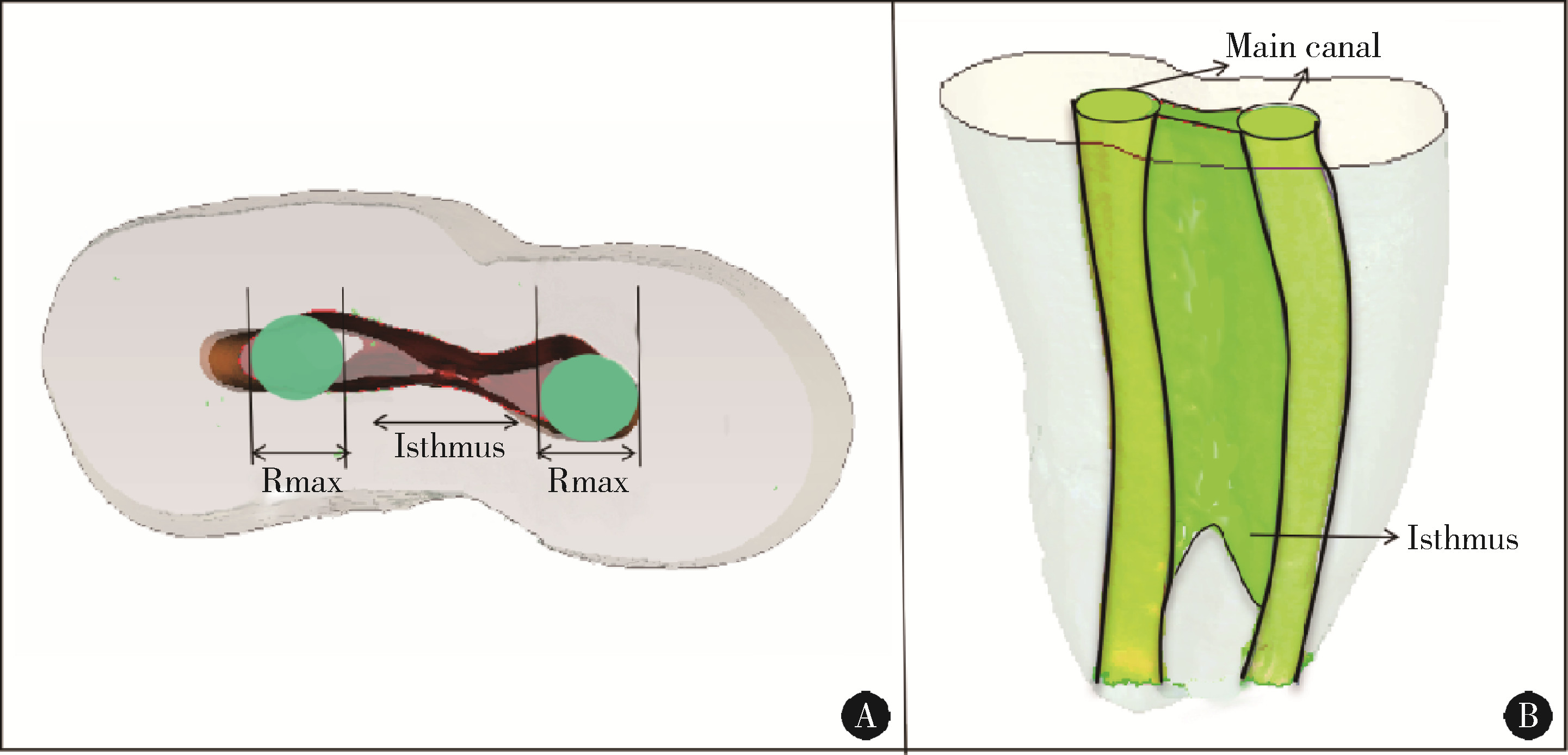
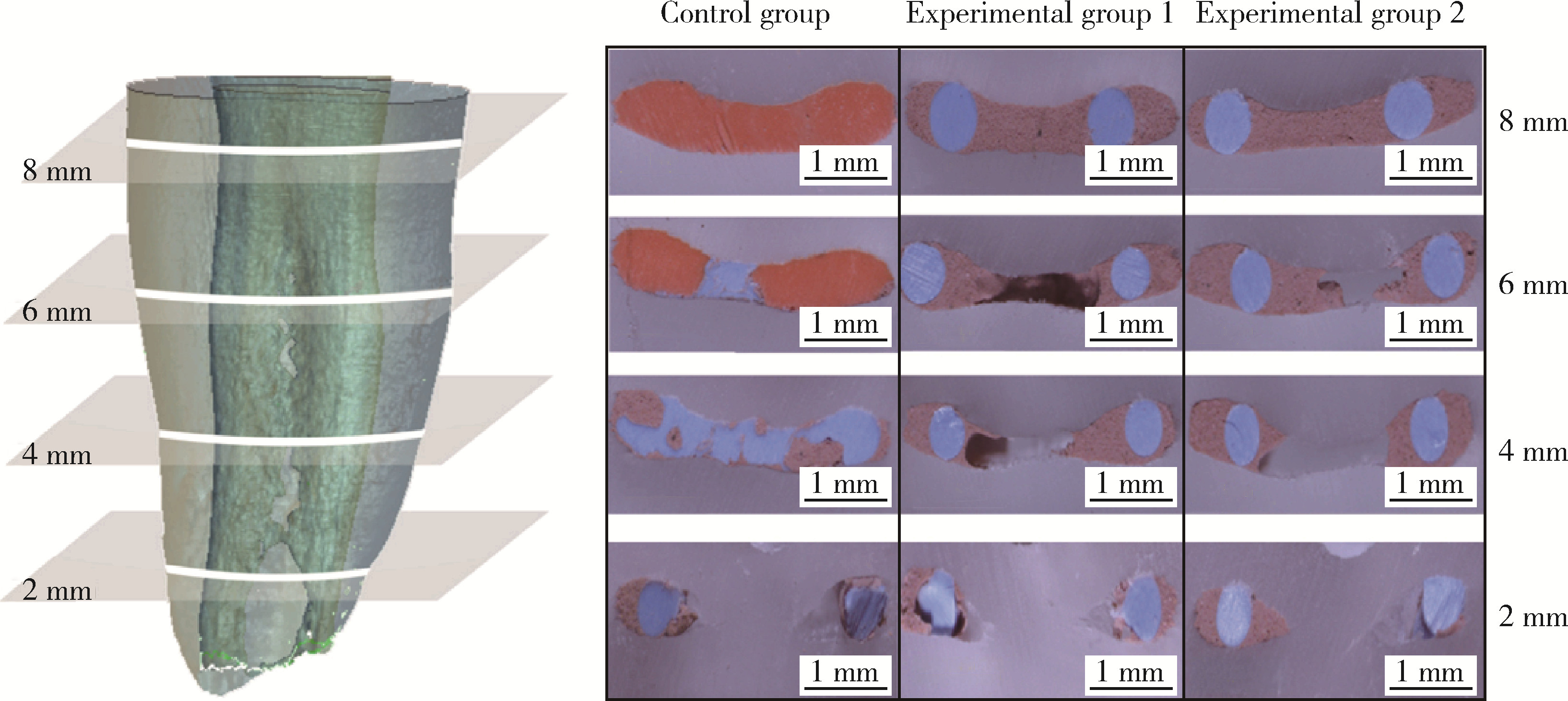
目的: 通过3D打印技术建立带有不规则结构(Hus&Kim Ⅴ型、Yin Ⅱ型峡部)的根管模型,并利用切片和影像学手段评价单尖根管充填的效果,以期对临床应用单尖根管充填技术提供参考。方法: (1) 纳入具备Hus&Kim Ⅴ型、Yin Ⅱ型峡部根管结构的人离体前磨牙,使用锥形束计算机断层扫描(cone-beam computed tomography, CBCT)并重建根管系统,设计打印标准化3D根管模型,并用罗丹明B染色和偏差拟合验证模型可用性。(2)将30个模型随机分为3组(n=10),使用不同充填方法进行根管充填,分别为对照组:热垂直加压充填组;实验组1:0.06锥度(30#)单尖充填组;实验组2:0.04锥度(35#)单尖充填组,均采用多组分混合根管封闭剂(GuttaFlow 2)。样本储存1周后,在距根尖2、4、6、8 mm处进行横切片,体式显微镜下观察,计算充填率和充填物构成比。(3)在上述结果基础上,选择充填率较高的实验组1(n=4)与对照组(n=4)通过微计算机断层扫描(micro-computed tomography,Micro-CT)进一步分析充填效果,测量根管系统、主根管及峡部内充填物体积,并计算体积充填率。采用两因素多元方差分析评估差异,并通过Tukey’ s检验对各组数据进行组间和组内两两比较。结果: (1) 用罗丹明B染色后可见染色液从模型根尖孔溢出,染色范围达主根管系统及峡部区域,切片显示无残余支撑材料,打印模型偏差为0.03 mm,拟合平均距离为0.02 mm,证实模型可用。(2)实验组1和2在距根尖4、6、8 mm层面上的充填率均低于对照组,差异具有统计学意义(F=45.04,P<0.01)。两实验组在根尖2、4 mm处的充填率差异无统计学意义(P>0.01),但在根管中上段6、8 mm处,实验组1的充填率高于实验组2(P<0.01)。同一实验组内,根尖区2、4 mm处的充填率均低于根管中上段6、8 mm处(P<0.01)。两实验组在根尖段2、4 mm的牙胶及封闭剂占比差异无统计学意义(F=2.383,P>0.01),根尖2 mm处牙胶占比较大,根尖4 mm处封闭剂占比较大(P<0.01)。(3)Micro-CT扫描结果显示,实验组1与对照组的体积充填率之间的差异具有统计学意义(F=47.33,P<0.01),其中在峡部结构中,实验组1的体积充填率为54.33%±4.35%,在主根管内为78.31%±4.21%,均低于对照组的峡部体积充填率76.48%±4.89%和主根管体积充填率86.90%±3.29%(P<0.01),实验组1的体积充填率在主根管高于峡部(P<0.01),而对照组在峡部与主根管之间的差异无统计学意义(P>0.01)。结论: 在带有峡部的不规则根管结构中,使用大锥度牙胶尖可改善单尖根管充填在根管中上段的充填效果,但峡部的体积充填率仍低于主根管,需进一步改进技术。
中图分类号:
- R781.3


| 1 | Marshall FS , Massler M . The sealing of pulpless teeth evaluated with radioisotopes[J]. J Dent Med, 1961, 16 (4): 172- 184. |
| 2 | Wu MK , Ozok AR , Wesselink PR . Sealer distribution in root canals obturated by three techniques[J]. Int Endod J, 2010, 33 (4): 340- 345. |
| 3 | Weis MV , Parashos P , Messer HH . Effect of obturation technique on sealer cement thickness and dentinal tubule penetration[J]. Int Endod J, 2010, 37 (10): 653- 663. |
| 4 |
Kazemi RB , Safavi KE , Spångberg LS . Dimensional changes of endodontic sealers[J]. Oral Surg Oral Med Oral Pathol, 1993, 76 (6): 766- 771.
doi: 10.1016/0030-4220(93)90050-E |
| 5 |
Schäfer E , Zandbiglari T . Solubility of root-canal sealers in water and artificial saliva[J]. Int Endod J, 2003, 36 (10): 660- 669.
doi: 10.1046/j.1365-2591.2003.00705.x |
| 6 |
Beatty R . The effect of standard or serial preparation on single cone obturations[J]. Int Endod J, 1987, 20 (6): 276- 281.
doi: 10.1111/j.1365-2591.1987.tb00627.x |
| 7 | Smith CS , Setchell DJ , Harty FJ . Factors influencing the success of conventional root canal therapy: A five-year retrospective study[J]. Int Endod J, 2010, 26 (6): 321- 333. |
| 8 |
Kim SY , Jang YE , Kim BS , et al. Effects of ultrasonic activation on root canal filling quality of single-cone obturation with calcium silicate-based sealer[J]. Materials, 2021, 14 (5): 1292- 1300.
doi: 10.3390/ma14051292 |
| 9 |
Kim S , Kim S , Park JW , et al. Comparison of the percentage of voids in the canal filling of a calcium silicate-based sealer and gutta percha cones using two obturation techniques[J]. Materials, 2017, 10 (10): 1170- 1179.
doi: 10.3390/ma10101170 |
| 10 |
Iglecias EF , Freire LG , de Miranda Candeiro GT , et al. Presence of voids after continuous wave of condensation and single-cone obturation in mandibular molars: A micro-computed tomography analysis[J]. J Endod, 2017, 43 (4): 638- 642.
doi: 10.1016/j.joen.2016.11.027 |
| 11 |
Zhang P , Yuan K , Jin Q , et al. Presence of voids after three obturation techniques in band: Haped isthmuses: A micro computed tomography study[J]. BMC Oral Health, 2021, 21 (1): 227- 235.
doi: 10.1186/s12903-021-01584-2 |
| 12 | Koch KA, Brave GD, Nasseh AA. Bioceramic technology: Closing the endo-restorative circle, Part 2[J]. Dent Today, 2010, 29(3), 98: 102-105. |
| 13 |
Alcalde MP , Bramante CM , Vivan RR , et al. Intradentinal antimicrobial action and filling quality promoted by ultrasonic agitation of epoxy resin-based sealer in endodontic obturation[J]. J Appl Oral Sci, 2017, 25 (6): 641- 649.
doi: 10.1590/1678-7757-2017-0090 |
| 14 |
Villalta-Briones N , Baca P , Bravo M , et al. A laboratory study of root canal and isthmus disinfection in extracted teeth using various activation methods with a mixture of sodium hypochlorite and etidronic acid[J]. Int Endod J, 2021, 54 (2): 268- 278.
doi: 10.1111/iej.13417 |
| 15 | 顾永春, 皮昕, 周培刚, 等. 3 803个恒牙侧副根管的解剖形态研究[J]. 临床口腔医学杂志, 2004, 20 (6): 334- 338. |
| 16 |
Yin X , Chang JWW , Wang Q , et al. Three-dimensional morphologic classifications and analysis of canal isthmuses in permanent molars[J]. Surg Radiol Anat, 2021, 43 (11): 1793- 1799.
doi: 10.1007/s00276-021-02796-5 |
| 17 |
Teixeira FB , Sano CL , Gomes BP , et al. A preliminary in vitro study of the incidence and position of the root canal isthmus in maxillary and mandibular first molars[J]. Int Endod J, 2003, 36 (4): 276- 280.
doi: 10.1046/j.1365-2591.2003.00638.x |
| 18 |
Marceliano-Alves MF , Lima CO , Bastos L , et al. Mandibular mesial root canal morphology using micro-computed tomography in a Brazilian population[J]. Aust Endod J, 2019, 45 (1): 51- 56.
doi: 10.1111/aej.12265 |
| 19 |
Yin X , Cheung GS , Zhang C , et al. Micro-computed tomographic comparison of nickel-titanium rotary versus traditional instruments in C-shaped root canal system[J]. J Endod, 2010, 36 (4): 708- 712.
doi: 10.1016/j.joen.2010.01.003 |
| 20 |
Keleş A , Keskin C . Presence of voids after warm vertical compaction and single-cone obturation in band-shaped isthmuses using micro-computed tomography: A phantom study[J]. Microsc Res Tech, 2020, 83 (4): 370- 374.
doi: 10.1002/jemt.23423 |
| 21 |
Chybowski EA , Glickman GN , Patel Y , et al. Clinical outcome of non-surgical root canal treatment using a single-cone technique with endosequence bioceramic sealer: A retrospective analysis[J]. J Endod, 2018, 44 (6): 941- 945.
doi: 10.1016/j.joen.2018.02.019 |
| 22 |
Figueiredo F , Lima LF , Oliveira LS , et al. Effectiveness of a reciprocating single file, single cone endodontic treatment approach: A randomized controlled pragmatic clinical trial[J]. Clin Oral Investig, 2020, 24 (7): 2247- 2257.
doi: 10.1007/s00784-019-03077-7 |
| [1] | 张燕, 韩志慧, 钟延丰, 王盛兰, 李玲玲, 郑丹枫. 骨骼肌活组织检查病理诊断技术的改进及应用[J]. 北京大学学报(医学版), 2009, 41(4): 459-462. |
| [2] | 武登诚, 李盛林. 软硬组织切磨技术在口腔医学研究中的应用[J]. 北京大学学报(医学版), 2007, 39(1): 94-95. |
|
||
