北京大学学报(医学版) ›› 2025, Vol. 57 ›› Issue (5): 903-910. doi: 10.19723/j.issn.1671-167X.2025.05.014

• 论著 • 上一篇    下一篇

丝氨酸蛋白酶23在系统性硬化病皮肤纤维化中的作用和机制

袁显墩, 李照华, 徐丹, 李婷, 方丹, 穆荣*()   

  1. 北京大学第三医院风湿免疫科,北京 100191
  • 收稿日期:2023-02-28 出版日期:2025-10-18 发布日期:2024-04-10
  • 通讯作者: 穆荣
  • 基金资助:
    国家自然科学基金(81771706)

Pathogenesis and mechanism of serine protease 23 in skin fibrosis of systemic sclerosis

Xiandun YUAN, Zhaohua LI, Dan XU, Ting LI, Dan FANG, Rong MU*()   

  1. Department of Rheumatology and Immunology, Peking University Third Hospital, Beijing 100191, China
  • Received:2023-02-28 Online:2025-10-18 Published:2024-04-10
  • Contact: Rong MU
  • Supported by:
    the National Natural Science Foundation of China(81771706)

RICH HTML


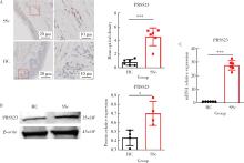
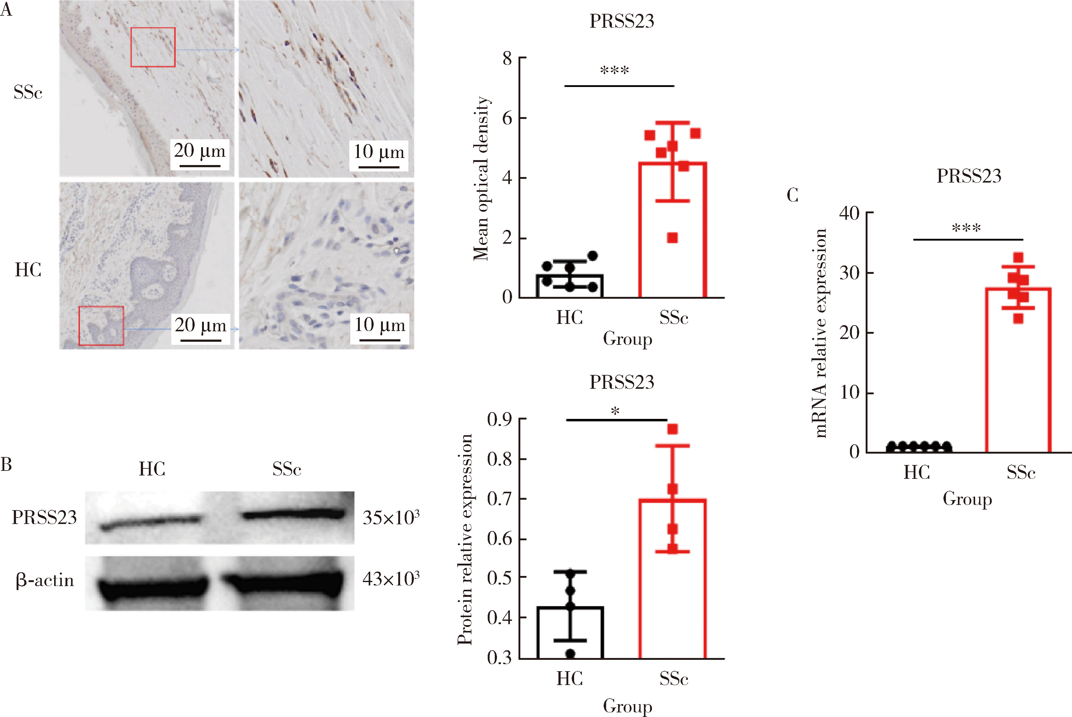
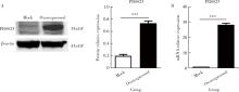
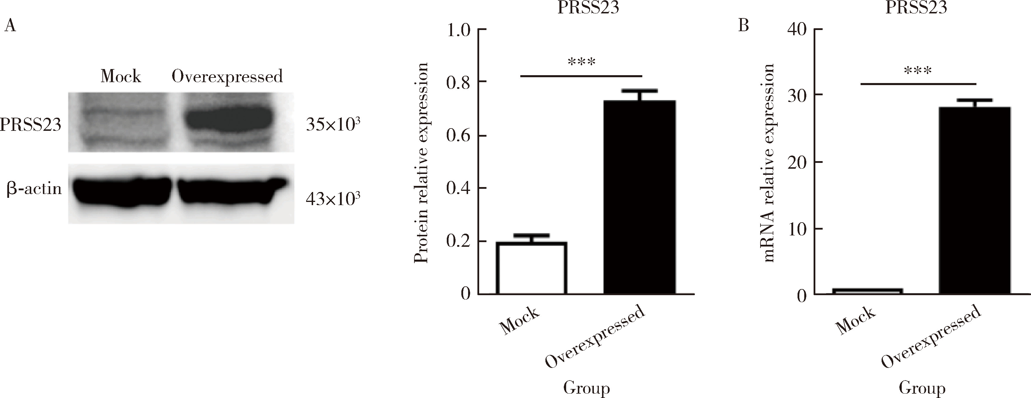
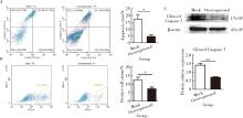
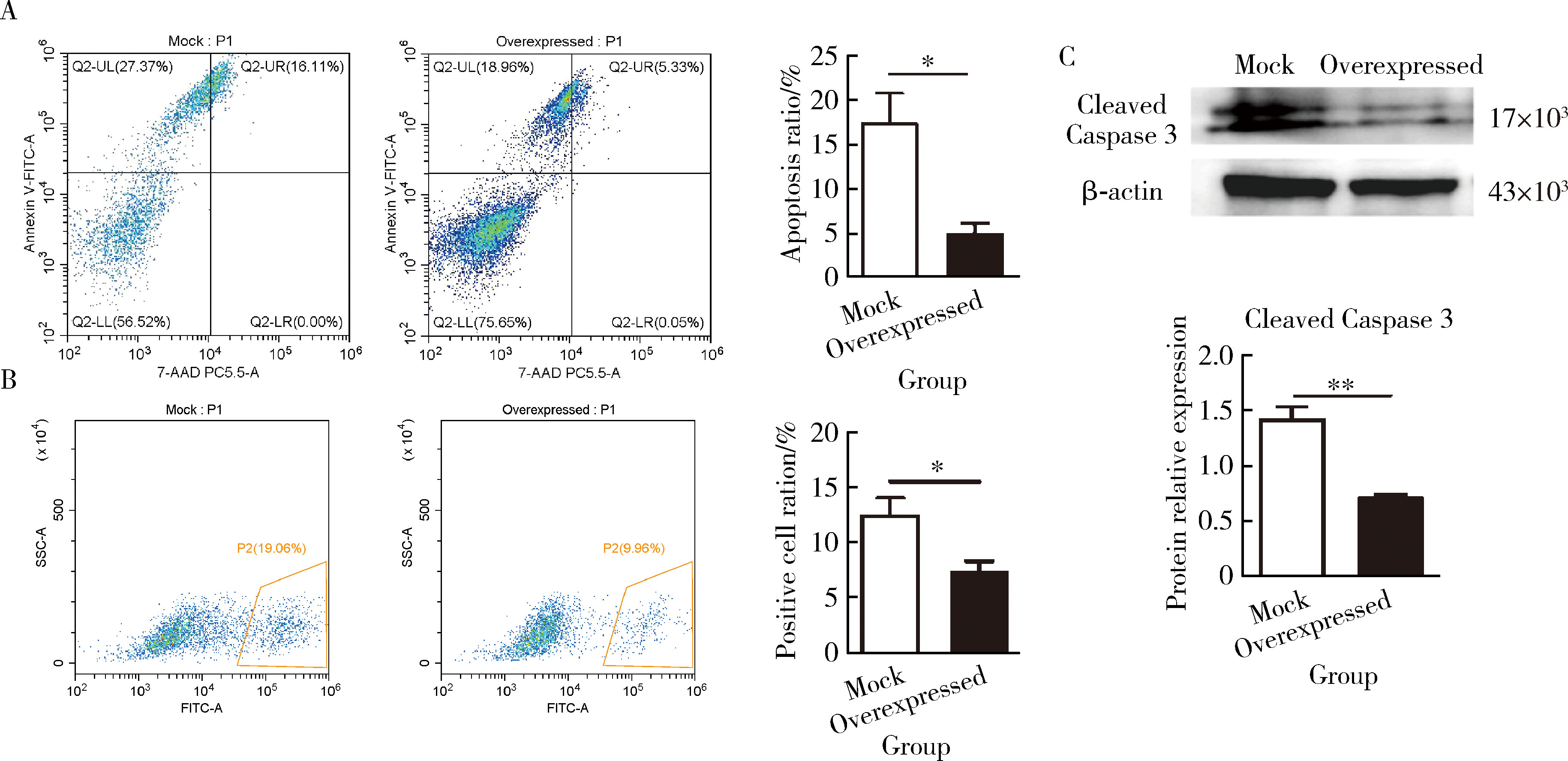
摘要: 目的: 系统性硬化病(systemic sclerosis, SSc)患者的皮肤成纤维细胞中丝氨酸蛋白酶23(serine protease 23,PRSS23)mRNA水平升高,本研究旨在探索PRSS23表达上调对SSc患者皮肤纤维化的作用及机制。方法: 采用免疫组织化学方法检测SSc患者和健康对照者皮肤组织石蜡切片中PRSS23蛋白的表达水平。分离新鲜皮肤组织的成纤维细胞,采用实时荧光定量PCR(real-time quantitative PCR,RT-qPCR)和蛋白免疫印迹法检测PRSS23在皮肤成纤维细胞中的mRNA和蛋白表达。采用慢病毒感染的方式构建过表达PRSS23的皮肤成纤维细胞BJ细胞系,以400 μmol/L过氧化氢刺激12 h后,采用膜联蛋白V/7-AAD染色检测凋亡成纤维细胞的比例,采用流式细胞仪和蛋白免疫印迹法检测凋亡相关蛋白激活型半胱氨酸-天冬氨酸蛋白酶-3(Caspase-3)的表达。采用RT-qPCR和酶联免疫吸附试验(enzyme linked immunosorbent assay, ELISA)法检测成纤维细胞分泌的白细胞介素-6(interleukin 6,IL-6)和肿瘤坏死因子-α(tumor necrosis factor alpha,TNF-α)水平。结果: 与健康对照组比较,SSc患者皮肤组织中的PRSS23蛋白表达显著升高[4.952 (3.806~5.439) vs. 0.806 (0.395~1.173),P<0.001]。PRSS23主要表达在成纤维细胞中,SSc患者分离的皮肤成纤维细胞中PRSS23的mRNA[27.59 (25.02~30.00) vs. 1.00,P<0.001]和蛋白表达水平[0.675 (0.587~0.837) vs. 0.451 (0.342~0.502),P=0.029]均显著上调。相较于对照组,过表达PRSS23的皮肤成纤维细胞抗凋亡能力增强,过氧化氢诱导后凋亡细胞的比例显著减少[(5.043±1.097)% vs. (17.480±3.212)%,P=0.022],凋亡相关蛋白激活型Caspase-3表达水平也显著降低[(0.718±0.022) vs. (1.422±0.105),P=0.003],而炎症因子IL-6的mRNA水平[(99.780±1.796) vs. (1.000±0.004),P<0.001]和蛋白分泌水平[(211.600±2.431) ng/L vs. (65.930±1.768) ng/L,P<0.001]显著上调,同时,TNF-α的mRNA水平[(3.555±0.555) vs. (1.000±0.004),P<0.001]和蛋白分泌水平[(41.190±0.949) ng/L vs. (31.150±0.360) ng/L,P<0.001]显著上调。结论: PRSS23在SSc患者皮肤成纤维细胞中的表达增加,可能通过抑制细胞凋亡,并促进炎症因子IL-6和TNF-α的分泌,调控皮肤成纤维细胞向促炎型转化,参与皮肤纤维化的发展。

关键词: 系统性硬化病, 成纤维细胞, 丝氨酸蛋白酶类, 细胞凋亡

Abstract: Objective: It has been reported that the mRNA expression of serine protease 23 (PRSS23) was increased in skin fibroblasts from systemic sclerosis patients (SSc). The purpose of this study is to explore the pathogenetic effect and mechanism of PRSS23 in skin fibrosis of SSc. Methods: The expression of PRSS23 in skin tissues from the SSc patients and healthy controls was detected by immunohisto-chemistry. Fibroblasts isolated from fresh skin tissue were used to detect the expression of PRSS23 by real-time quantitative PCR (RT-qPCR) and Western blot. Overexprssion of PRSS23 in BJ, the fibroblasts cell line of skin, was constructed by lentivirus. After stimulation with 400 μmol/L hydrogen peroxide for 12 h, Annexin V/7-AAD staining was used to detect apoptosis of fibroblasts; flow cytometry and Western blot were used to detect the expression of apoptosis-related protein cleaved Caspase-3. The expression of interleukin 6 (IL-6) and tumor necrosis factor alpha (TNF-α) in fibroblasts was detected by RT-qPCR and enzyme linked immunosorbent assay (ELISA). Results: Compared with the healthy controls, the expression of PRSS23 in skin tissues of the SSc patients was significantly increased [4.952 (3.806-5.439) vs. 0.806 (0.395-1.173), P < 0.001], and fibroblast was the main cell that expressed PRSS23. The mRNA [27.59 (25.02-30.00) vs. 1.00, P < 0.001] and protein [0.675 (0.587-0.837) vs. 0.451 (0.342-0.502), P=0.029] of PRSS23 in skin fibroblasts isolated from the SSc patients were significantly up-regulated. Compared with the control group, the anti-apoptotic ability of skin fibroblasts overexpressing PRSS23 was enhanced, and the proportion of apoptotic cells was significantly reduced after hydrogen peroxide induction [(5.043±1.097)% vs. (17.480±3.212)%, P=0.022], the expression of apoptosis-related protein cleaved Caspase-3 was also markedly reduced [(0.718±0.022) vs. (1.422±0.105), P=0.003]. In addition, the mRNA [(99.780±1.796) vs. (1.000±0.004), P < 0.001] and protein [(211.600±2.431) ng/L vs. (65.930±1.768) ng/L, P < 0.001] of IL-6 in the fibroblasts overexpressing PRSS23 were significantly up-regulated; the mRNA[(3.555±0.555) vs. (1.000±0.004), P < 0.001] and protein levels [(41.190±0.949) ng/L vs. (31.150±0.360) ng/L, P < 0.001] of TNF-α in the fibroblasts overexpressing PRSS23 were also significantly up-regulated. Conclusion: The expression of PRSS23 is increased in skin fibroblasts of SSc patients. PRSS23 can inhibit cell apoptosis, promote the secretion of inflammatory factors such as IL-6 and TNF-α, and regulate the process that skin fibroblasts transform into pro-inflammatory type. So, PRSS23 is associated with the development of skin fibrosis.

Key words: Systemic sclerosis, Fibroblasts, Serine proteases, Apoptosis

中图分类号: 

  • R593.2

表1

IL-6、TNF-α、PRSS23引物序列"

Gene GenBank ID Primer sequences (5′ to 3′) Length/bp
IL-6 NM_000600 Forward: 5′-ACTCACCTCAGAACGAATTG-3′
Reversed: 5′-CCATCTTGGAAGGTTCAGGTTG-3′
149
TNF-α NM_000594 Forward: 5′-CCTCTAATCAGCCTCTG-3′
Reversed: 5′-GAGGACCTGGAGTAGAG-3′
220
PRSS23 NM_009104 Forward: 5′-TGTGCTGGGCAAGTGAG-3′
Reversed: 5′-AGTTCCCTTATGACTGGGG-3′
174

图1

SSc患者皮肤组织成纤维细胞中PRSS23的表达"

表2

PRSS23在SSc患者皮肤组织成纤维细胞中的表达"

Items Sample size (n) Value, M (P25-P75)
Mean optical density
  HC 6 0.806 (0.395-1.173)
  SSc 6 4.952 (3.806-5.439)***
Protein relative expression
  HC 4 0.451 (0.342-0.502)
  SSc 4 0.675 (0.587-0.837)*
mRNA relative expression
  HC 6 1.000
  SSc 6 27.590 (25.020-30.000)***

图2

转染PRSS23质粒的皮肤成纤维细胞过表达PRSS23"

表3

PRSS23在转染PRSS23质粒的皮肤成纤维细胞中的表达"

Items Sample size (n) Value, ${\bar x}$±SE
Protein relative expression (PRSS23)
  Mock 3 0.199±0.023
  Overexpressed 3 0.730±0.037***
mRNA relative expression
  Mock 4 1.000
  Overexpressed 4 28.160±0.507***

图3

过表达PRSS23抑制皮肤成纤维细胞凋亡"

表4

PRSS23抑制皮肤成纤维细胞凋亡率和相关凋亡蛋白的测定"

Items Sample size (n) Value, ${\bar x}$±SE
Apoptosis ratio
  Mock 3 (17.480±3.212)%
  Overexpressed 3 (5.043±1.097)%*
Positive cell ratio
  Mock 4 (12.510±1.472)%
  Overexpressed 4 (7.460±0.866)%*
Protein relative expression (cleaved Caspase 3)
  Mock 4 1.422±0.105
  Overexpressed 4 0.718±0.022**

图4

过表达PRSS23促进皮肤成纤维细胞分泌促炎因子IL-6和TNF-α"

表5

PRSS23对皮肤成纤维细胞炎症因子分泌的影响"

Items Sample size (n) Value, ${\bar x}$±SE
IL-6 mRNA relative expression
  Mock 3 1.000
  Overexpressed 3 99.780±1.796***
TNF-α mRNA relative expression
  Mock 4 1.000
  Overexpressed 4 3.555±0.555**
IL-6 protein content (cell supernatant)/(ng/L)
  Mock 4 65.930±1.768
  Overexpressed 4 211.600±2.431***
TNF-α protein content (cell supernatant)/(ng/L)
  Mock 4 31.150±0.360
  Overexpressed 4 41.190±0.949***
1
Domsic RT, Rodriguez-Reyna T, Lucas M, et al. Skin thickness progression rate: A predictor of mortality and early internal organ involvement in diffuse scleroderma[J]. Ann Rheum Dis, 2011, 70(1): 104- 109.

doi: 10.1136/ard.2009.127621
2
Hinz B, Lagares D. Evasion of apoptosis by myofibroblasts: A hallmark of fibrotic diseases[J]. Nat Rev Rheumatol, 2020, 16(1): 11- 31.

doi: 10.1038/s41584-019-0324-5
3
Dodi AE, Ajayi IO, Chang C, et al. Regulation of fibroblast Fas expression by soluble and mechanical pro-fibrotic stimuli[J]. Respir Res, 2018, 19(1): 91.

doi: 10.1186/s12931-018-0801-4
4
Xu C, Meng LB, Duan YC, et al. Screening and identification of biomarkers for systemic sclerosis via microarray technology[J]. Int J Mol Med, 2019, 44(5): 1753- 1770.
5
Tabib T, Huang M, Morse N, et al. Myofibroblast transcriptome indicates SFRP2(hi) fibroblast progenitors in systemic sclerosis skin[J]. Nat Commun, 2021, 12(1): 4384.

doi: 10.1038/s41467-021-24607-6
6
Kanno Y. The role of fibrinolytic regulators in vascular dysfunction of systemic sclerosis[J]. Int J Mol Sci, 2019, 20(3): 619.

doi: 10.3390/ijms20030619
7
Menou A, Duitman J, Crestani B. The impaired proteases and anti-proteases balance in idiopathic pulmonary fibrosis[J]. Matrix Biol, 2018(68/69): 382- 403.
8
Page MJ, Di Cera E. Serine peptidases: Classification, structure and function[J]. Cell Mol Life Sci, 2008, 65(7/8): 1220- 1236.
9
Han B, Yang Y, Chen J, et al. PRSS23 knockdown inhibits gastric tumorigenesis through EIF2 signaling[J]. Pharmacol Res, 2019, 142, 50- 57.

doi: 10.1016/j.phrs.2019.02.008
10
Rawlings ND, Barrett AJ, Bateman A. MEROPS: The peptidase database[J]. Nucleic Acids Res, 2010, 38(Database issue): D227- D233.
11
Deng CC, Hu YF, Zhu DH, et al. Single-cell RNA-seq reveals fibroblast heterogeneity and increased mesenchymal fibroblasts in human fibrotic skin diseases[J]. Nat Commun, 2021, 12(1): 3709.

doi: 10.1038/s41467-021-24110-y
12
Xiong J, Li Y, Tan X, et al. Targeting PRSS23 with tipranavir induces gastric cancer stem cell apoptosis and inhibits growth of gastric cancer via the MKK3/p38 MAPK-IL24 pathway[J]. Acta Pharmacol Sin, 2024, 45(2): 405- 421.

doi: 10.1038/s41401-023-01165-9
13
Fan L, Wang Q, Liu R, et al. Citrullinated fibronectin inhibits apoptosis and promotes the secretion of pro-inflammatory cytokines in fibroblast-like synoviocytes in rheumatoid arthritis[J]. Arthritis Res Ther, 2012, 14(6): R266.

doi: 10.1186/ar4112
14
Chaudhuri V, Zhou L, Karasek M. Inflammatory cytokines induce the transformation of human dermal microvascular endothelial cells into myofibroblasts: A potential role in skin fibrogenesis[J]. J Cutan Pathol, 2007, 34(2): 146- 153.

doi: 10.1111/j.1600-0560.2006.00584.x
15
Johnson BZ, Stevenson AW, Prêle CM, et al. The role of IL-6 in skin fibrosis and cutaneous wound healing[J]. Biomedicines, 2020, 8(5): 101.

doi: 10.3390/biomedicines8050101
16
Borthwick LA, Wynn TA, Fisher AJ. Cytokine mediated tissue fibrosis[J]. Biochim Biophys Acta, 2013, 1832(7): 1049- 1060.

doi: 10.1016/j.bbadis.2012.09.014
17
Li Y, Zhao J, Yin Y, et al. The role of IL-6 in fibrotic diseases: Molecular and cellular mechanisms[J]. Int J Biol Sci, 2022, 18(14): 5405- 5414.

doi: 10.7150/ijbs.75876
18
Khan K, Xu S, Nihtyanova S, et al. Clinical and pathological significance of interleukin 6 overexpression in systemic sclerosis[J]. Ann Rheum Dis, 2012, 71(7): 1235- 1242.

doi: 10.1136/annrheumdis-2011-200955
19
Khanna D, Denton CP, Jahreis A, et al. Safety and efficacy of subcutaneous tocilizumab in adults with systemic sclerosis (faSScinate): A phase 2, randomised, controlled trial[J]. Lancet, 2016, 387(10038): 2630- 2640.

doi: 10.1016/S0140-6736(16)00232-4
20
Bahcecioglu IH, Koca SS, Poyrazoglu OK, et al. Hepatoprotective effect of infliximab, an anti-TNF-alpha agent, on carbon tetrachloride-induced hepatic fibrosis[J]. Inflammation, 2008, 31(4): 215- 221.

doi: 10.1007/s10753-008-9067-1
21
Messadi DV, Doung HS, Zhang Q, et al. Activation of NFkappaB signal pathways in keloid fibroblasts[J]. Arch Dermatol Res, 2004, 296(3): 125- 133.
22
Zhu G, Cai J, Zhang J, et al. Abnormal nuclear factor (NF)-kappaB signal pathway and aspirin inhibits tumor necrosis factor alpha-induced NF-kappaB activation in keloid fibroblasts[J]. Dermatol Surg, 2007, 33(6): 697- 708.
23
Li X, Sun Z, Peng G, et al. Single-cell RNA sequencing reveals a pro-invasive cancer-associated fibroblast subgroup associated with poor clinical outcomes in patients with gastric cancer[J]. Theranostics, 2022, 12(2): 620- 638.

doi: 10.7150/thno.60540
24
Solé-Boldo L, Raddatz G, Schütz S, et al. Single-cell transcriptomes of the human skin reveal age-related loss of fibroblast priming[J]. Commun Biol, 2020, 3(1): 188.

doi: 10.1038/s42003-020-0922-4
25
Griffin MF, desJardins-Park HE, Mascharak S, et al. Understanding the impact of fibroblast heterogeneity on skin fibrosis[J]. Dis Model Mech, 2020, 13(6): dmm044164.

doi: 10.1242/dmm.044164
26
Sato Y, Yanagita M. Resident fibroblasts in the kidney: A major driver of fibrosis and inflammation[J]. Inflamm Regen, 2017, 37, 17.

doi: 10.1186/s41232-017-0048-3
[1] 赵柯林, 夏雪, 史乃旭, 周韩, 盖婧雯, 李萍. 铁死亡标志物4-HNE在系统性硬化症细胞模型中的表达及意义[J]. 北京大学学报(医学版), 2024, 56(6): 950-955.
[2] 何珊,陈炘,程琦,朱灵江,张培玉,童淑婷,薛静,杜燕. 托法替布通过JAK/STAT3通路抑制肺成纤维细胞向肌成纤维细胞转化[J]. 北京大学学报(医学版), 2024, 56(3): 505-511.
[3] 刘耘充,吴宗龙,葛力源,杜坦,吴雅倩,宋一萌,刘承,马潞林. 肾透明细胞癌中核蛋白1对阿昔替尼耐药的作用及机制[J]. 北京大学学报(医学版), 2023, 55(5): 781-792.
[4] 蔡天玉,朱振鹏,徐纯如,吉星,吕同德,郭振可,林健. 成纤维细胞生长因子受体2在肾透明细胞癌中的表达及意义[J]. 北京大学学报(医学版), 2022, 54(4): 628-635.
[5] 郜洪宇,孟焕新,侯建霞,黄宝鑫,李玮. 钙结合蛋白在健康牙周组织和实验性牙周炎组织的表达分布[J]. 北京大学学报(医学版), 2021, 53(4): 744-749.
[6] 耿良,吕静,范敬. 肺瘤平膏联合环磷酰胺化疗对肺癌的抑瘤作用和酸性微环境的影响[J]. 北京大学学报(医学版), 2020, 52(2): 247-253.
[7] 石冰清,袁晓静,赵玉鸣. 比较矿物三氧化物凝聚体及山东蜂胶乙醇提取物对牙髓成纤维细胞生物学性能的影响[J]. 北京大学学报(医学版), 2019, 51(6): 1108-1114.
[8] 郑苗,詹凌璐,刘志强,李和平,谭建国. 不同等离子体处理氧化锆对人牙龈成纤维细胞黏附能力的影响[J]. 北京大学学报(医学版), 2019, 51(2): 315-320.
[9] 王昊,陈亮,叶小云. 雷公藤甲素对TM4细胞氧化应激及PI3K/AKT通路的影响[J]. 北京大学学报(医学版), 2018, 50(4): 607-612.
[10] 李乾,常亮,苏冬梅,马旭. 粉防己碱对心肌成纤维细胞增殖、活化的影响[J]. 北京大学学报(医学版), 2018, 50(2): 331-334.
[11] 王玉洁,郭向阳,王军. 重复异丙酚麻醉对新生大鼠海马细胞凋亡及远期学习记忆能力的影响[J]. 北京大学学报(医学版), 2017, 49(2): 310-314.
[12] 杨光,程庆砾,李春霖,贾雅丽,岳文,裴雪涛,刘洋,赵佳慧,杜婧,敖强国. 高糖减弱肾组织干细胞条件培养液对缺氧损伤肾小管上皮细胞的修复作用[J]. 北京大学学报(医学版), 2017, 49(1): 125-130.
[13] 曹珮,姜学军,席志军. 舒尼替尼通过抑制Akt/mTOR信号通路诱导肾癌细胞自噬[J]. 北京大学学报(医学版), 2016, 48(4): 584-589.
[14] 贾双双,李伟阳,刘欣,李丽英. 转化生长因子-β1 通过产生活性氧诱导骨髓间充质干细胞分化为肌成纤维细胞[J]. 北京大学学报(医学版), 2015, 47(5): 737-742.
[15] 李刚,张洪宪,王云鹏,张径,洪锴,田晓军,马潞林. 间苯三酚对大鼠肾缺血再灌注损伤的保护作用[J]. 北京大学学报(医学版), 2015, 47(5): 743-748.
Viewed
Full text


Abstract

Cited

  Shared   
  Discussed   
No Suggested Reading articles found!