北京大学学报(医学版) ›› 2026, Vol. 58 ›› Issue (1): 22-29. doi: 10.19723/j.issn.1671-167X.2026.01.003

• 论著 • 上一篇    下一篇

细胞膜囊泡递送靶向肿瘤坏死因子-α的小干扰RNA对牙髓干细胞的抗炎作用

高若凡, 马天宇, 王润楷, 殷雨辰, 李芮迪, 王丹丹*(), 夏斌*()   

  1. 北京大学口腔医学院·口腔医院儿童口腔科, 国家口腔医学中心, 国家口腔疾病临床医学研究中心, 口腔生物材料和数字诊疗装备国家工程研究中心, 北京 100081
  • 收稿日期:2025-09-19 出版日期:2026-02-18 发布日期:2025-11-27
  • 通讯作者: 王丹丹, 夏斌
  • 基金资助:
    国家自然科学基金(82401182); 北京市自然科学基金-海淀原始创新联合基金(L232110)

Anti-inflammatory effects of cell membrane vesicle-mediated delivery of small interfering RNA targeting tumor necrosis factor-α on dental pulp stem cells

Ruofan GAO, Tianyu MA, Runkai WANG, Yuchen YIN, Ruidi LI, Dandan WANG*(), Bin XIA*()   

  1. Department of Pediatric Dentistry, Peking University School and Hospital of Stomatology & National Center of Stomatology & National Clinical Research Center for Oral Diseases & National Engineering Laboratory for Digital and Material Technology of Stomatology, Beijing 100081, China
  • Received:2025-09-19 Online:2026-02-18 Published:2025-11-27
  • Contact: Dandan WANG, Bin XIA
  • Supported by:
    the National Natural Science Foundation of China(82401182); the Natural Science Foundation of Beijing-Haidian Original Innovation Joint Fund(L232110)

RICH HTML


摘要:

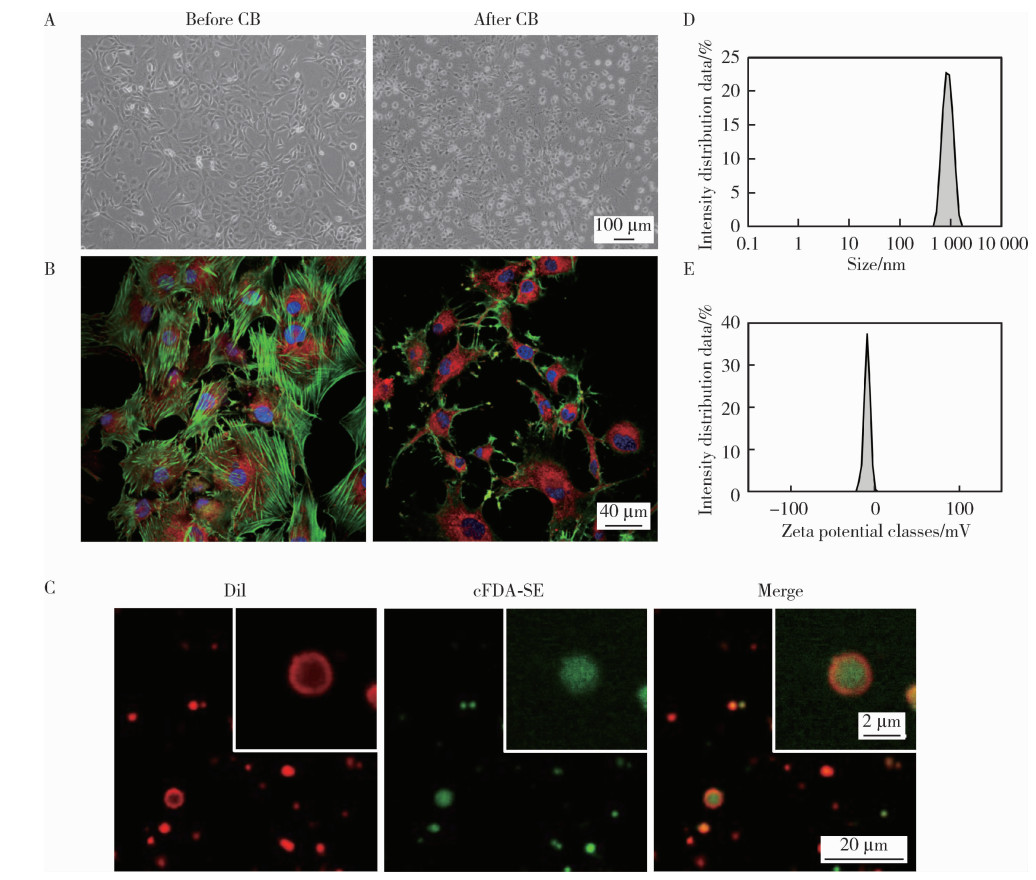
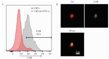
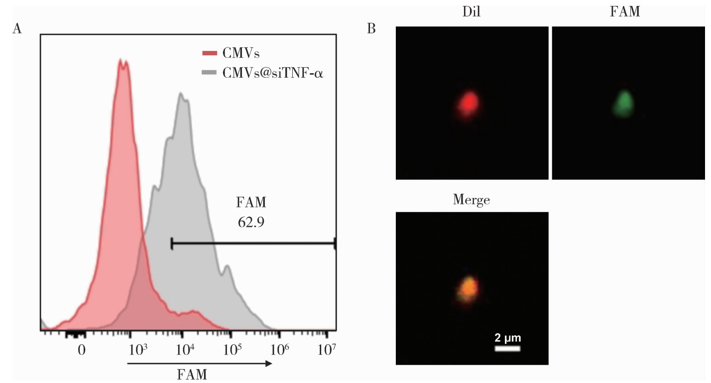
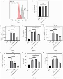
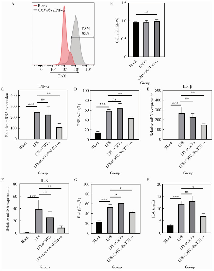
目的: 探索细胞膜囊泡(cell membrane vesicles, CMVs)作为广泛适用的基因沉默工具小干扰RNA(small interfering RNA, siRNA)递送平台的可行性, 并以脂多糖(lipopolysaccharide, LPS)诱导的人牙髓干细胞(dental pulp stem cells, DPSCs)炎症模型验证其应用效果。方法: 采用细胞松弛素B处理3T3细胞制备CMVs, 并通过共聚焦显微镜、动态光散射检测CMVs理化性质。利用洋地黄皂苷透化CMVs将靶向肿瘤坏死因子-α(tumor necrosis factor-α, TNF-α)的siRNA(siTNF-α)负载至CMVs中, 构建CMVs@siTNF-α。通过流式细胞术和共聚焦显微镜检测siRNA负载与胞内转运效率。以LPS(1 mg/L)刺激DPSCs建立炎症模型, 并与CMVs或CMVs@siTNF-α共培养。采用细胞活力检测试剂盒(cell counting kit-8, CCK-8)检测细胞毒性, 实时荧光定量聚合酶链反应(quantitative real-time polymerase chain reaction, qRT-PCR)和酶联免疫吸附试验(enzyme linked immunosorbent assay, ELISA)检测TNF-α、白介素(interleukin, IL)-1β和IL-6的表达与分泌。结果: CMVs呈球形, 由细胞膜及细胞质构成, 平均粒径为903 nm, 表面电位为-9.39 mV。流式细胞术结果显示, 负载荧光素酰胺(fluorescein amidite, FAM)标记siRNA(FAM-siRNA)后CMVs荧光强度升高; DPSCs加入CMVs@FAM-siTNF-α培养24 h后, 荧光信号增强。CCK-8检测结果显示CMVs及CMVs@siTNF-α对DPSCs无明显毒性。LPS处理显著上调DPSCs中TNF-α、IL-1β和IL-6的表达, 与LPS组相比, CMVs@siTNF-α下调TNF-α mRNA及其蛋白表达水平, 并抑制IL-1β和IL-6的转录与分泌。单独CMVs处理无明显差异。结论: CMVs可作为一种低毒性的siRNA递送平台, 在体外炎症模型中实现TNF-α沉默及下游炎症反应抑制, 具有进一步研究和应用的潜力。

关键词: 小干扰RNA, 细胞膜囊泡, 牙髓干细胞, 肿瘤坏死因子

Abstract:

Objective: To evaluate the feasibility of using cell membrane vesicles (CMVs) as a delivery system for small interfering RNA (siRNA) and to assess their performance in a lipopolysaccharide (LPS)-induced inflammatory model of human dental pulp stem cells (DPSCs). Methods: CMVs were generated from cytochalasin B-treated 3T3 cells and characterized for their physicochemical properties, including morphology, size distribution, and zeta potential, using confocal microscopy and dynamic light scattering. To construct CMVs@siTNF-α, saponin-mediated transient permeabilization was used to facilitate siRNA loading, after which encapsulation efficiency was evaluated by flow cytometry and confocal imaging. Intracellular uptake behaviors were examined using flow cytometry in DPSCs. LPS (1 mg/L) was employed to establish a robust in vitro inflammatory microenvironment. DPSCs were subsequently treated with CMVs or CMVs@siTNF-α, and cell viability was assessed via CCK-8 (cell counting kit-8). The expression and secretion of tumor necrosis factor-α (TNF-α), interleukin-1β (IL-1β), and interleukin-6 (IL-6) were analyzed using quantitative real-time polymerase chain reaction (qRT-PCR) and enzyme linked immunosorbent assay (ELISA) to evaluate both gene-silencing efficiency and downstream anti-inflammatory effects. Results: CMVs exhibited uniform spherical morphology with an average diameter of approximately 903 nm and a zeta potential of -9.39 mV, confirming successful vesicle formation. CMVs efficiently encapsulated FAM-labeled siRNA, as indicated by a pronounced fluorescence shift in flow cytometry and clear colocalization signals in confocal imaging. DPSCs cultured with CMVs@FAM-siTNF-α demonstrated increased intracellular fluorescence, reflecting efficient vesicle uptake and cytoplasmic siRNA release. Importantly, both CMVs and CMVs@siTNF-α displayed negligible cytotoxicity. LPS stimulation significantly elevated TNF-α, IL-1β, and IL-6 expression, validating the inflammatory model. CMVs alone did not affect cytokine levels, indicating biological inertness. In contrast, CMVs@siTNF-α significantly suppressed the LPS-induced upregulation of TNF-α at both mRNA and protein levels., demonstrating potent gene-silencing activity. Furthermore, suppression of TNF-α led to downstream attenuation of IL-1β and IL-6, with both transcription and secretion significantly decreased compared with the LPS group. These findings collectively confirmed that CMVs enabled efficient intracellular siRNA transport and effectively mitigate inflammatory responses in DPSCs. Conclusion: CMVs represent a biocompatible and effective siRNA delivery platform capable of achieving robust TNF-α knockdown and ameliorating inflammatory cytokine production in vitro, highlighting their potential for future nucleic acid-based anti-inflammatory therapies.

Key words: Small interfering RNA, Cell membrane vesicles, Dental pulp stem cells, Tumor necrosis factor

中图分类号: 

  • R78

表1

实时荧光定量PCR引物序列"

Gene Forward primer (5′ to 3′) Reverse primer (5′ to 3′)
TNF-α AGCAAGGACAGCAGAGGA GGGGAGAGAGGGTGGAG
IL-1β TGTGCTGAATGTGGACTCA ACAAAAGGGCTGGGGAT
IL-6 TGTGTGAAAGCAGCAAAGA ACCAGGCAAGTCTCCTCA
GAPDH GCCAACGTGTCAGTGGTG AAGGTGGAGGAGTGGGTGT

图1

CMVs的制备及表征"

图2

CMVs对siTNF-α的加载"

图3

CMVs@siTNF-α在DPSCs中的转运与抗炎效果"

1
Jadhav V, Vaishnaw A, Fitzgerald K, et al. RNA interference in the era of nucleic acid therapeutics[J]. Nat Biotechnol, 2024, 42(3): 394- 405.

doi: 10.1038/s41587-023-02105-y
2
Alshaer W, Zureigat H, Al Karaki A, et al. siRNA: Mechanism of action, challenges, and therapeutic approaches[J]. Eur J Pharmacol, 2021, 905, 174178.

doi: 10.1016/j.ejphar.2021.174178
3
Moazzam M, Zhang M, Hussain A, et al. The landscape of nanoparticle-based siRNA delivery and therapeutic development[J]. Mol Ther, 2024, 32(2): 284- 312.

doi: 10.1016/j.ymthe.2024.01.005
4
Kulkarni JA, Witzigmann D, Chen S, et al. Lipid nanoparticle technology for clinical translation of siRNA therapeutics[J]. Acc Chem Res, 2019, 52(9): 2435- 2444.

doi: 10.1021/acs.accounts.9b00368
5
Tang Q, Khvorova A. RNAi-based drug design: Considerations and future directions[J]. Nat Rev Drug Discov, 2024, 23(5): 341- 364.

doi: 10.1038/s41573-024-00912-9
6
Biscans A, Coles A, Haraszti R, et al. Diverse lipid conjugates for functional extra-hepatic siRNA delivery in vivo[J]. Nucleic Acids Res, 2019, 47(3): 1082- 1096.

doi: 10.1093/nar/gky1239
7
Tanowitz M, Hettrick L, Revenko A, et al. Asialoglycoprotein receptor 1 mediates productive uptake of N-acetylgalactosamine-conjugated and unconjugated phosphorothioate antisense oligonucleotides into liver hepatocytes[J]. Nucleic Acids Res, 2017, 45(21): 12388- 12400.

doi: 10.1093/nar/gkx960
8
Schlegel MK, Janas MM, Jiang Y, et al. From bench to bedside: Improving the clinical safety of GalNAc-siRNA conjugates using seed-pairing destabilization[J]. Nucleic Acids Res, 2022, 50(12): 6656- 6670.

doi: 10.1093/nar/gkac539
9
Liu YJ, Wang C. A review of the regulatory mechanisms of extracellular vesicles-mediated intercellular communication[J]. Cell Commun Signal, 2023, 21(1): 77.

doi: 10.1186/s12964-023-01103-6
10
Schulz-Siegmund M, Aigner A. Nucleic acid delivery with extracellular vesicles[J]. Adv Drug Deliv Rev, 2021, 173, 89- 111.

doi: 10.1016/j.addr.2021.03.005
11
Wang D, Jiang S, Zhang F, et al. Cell membrane vesicles with enriched CXCR4 display enhances their targeted delivery as drug carriers to inflammatory sites[J]. Adv Sci, 2021, 8(23): 2101562.

doi: 10.1002/advs.202101562
12
Sama S, Jerz G, Schmieder P, et al. Sapofectosid-ensuring non-toxic and effective DNA and RNA delivery[J]. Int J Pharm, 2017, 534(1/2): 195- 205.
13
Sama S, Woith E, Walther W, et al. Targeted suicide gene transfections reveal promising results in nu/nu mice with aggressive neuroblastoma[J]. J Control Release, 2018, 275, 208- 216.

doi: 10.1016/j.jconrel.2018.02.031
14
Oshchepkova A, Neumestova A, Matveeva V, et al. Cytochalasin-B-inducible nanovesicle mimics of natural extracellular vesicles that are capable of nucleic acid transfer[J]. Micromachines, 2019, 10(11): 750.

doi: 10.3390/mi10110750
15
Ni Z, Cai L, Tsai IC, et al. NAT10 regulates LPS-induced inflammation via stabilization of N4-acetylated PTX3 mRNA in human dental pulp stem cells[J]. Int J Mol Sci, 2025, 26(9): 4325.

doi: 10.3390/ijms26094325
16
Yu S, Liu XM, Liu Y, et al. Inflammatory microenvironment of moderate pulpitis enhances the osteo-/odontogenic potential of dental pulp stem cells by autophagy[J]. Int Endodontic J, 2024, 57(10): 1465- 1477.

doi: 10.1111/iej.14108
17
Lan C, Chen S, Jiang S, et al. Different expression patterns of inflammatory cytokines induced by lipopolysaccharides from Escherichia coli or Porphyromonas gingivalis in human dental pulp stem cells[J]. BMC Oral Health, 2022, 22(1): 121.

doi: 10.1186/s12903-022-02161-x
18
Chang J, Zhang C, Tani-Ishii N, et al. NF-κB activation in human dental pulp stem cells by TNF and LPS[J]. J Dent Res, 2005, 84(11): 994- 998.

doi: 10.1177/154405910508401105
19
Christ R, Siemes D, Zhao S, et al. Inhibition of tumour necrosis factor alpha by Etanercept attenuates Shiga toxin-induced brain pathology[J]. J Neuroinflammation, 2025, 22(1): 33.

doi: 10.1186/s12974-025-03356-z
20
Chen P, Wu L, Zhang S, et al. Combining TNF-α silencing with Wnt3a overexpression: A promising gene therapy for particle-induced periprosthetic osteolysis[J]. Front Cell Dev Biol, 2025, 13, 1511577.

doi: 10.3389/fcell.2025.1511577
21
Pick H, Schmid EL, Tairi AP, et al. Investigating cellular signaling reactions in single attoliter vesicles[J]. J Am Chem Soc, 2005, 127(9): 2908- 2912.

doi: 10.1021/ja044605x
22
Brown CR, Gupta S, Qin J, et al. Investigating the pharmacodynamic durability of GalNAc-siRNA conjugates[J]. Nucleic Acids Res, 2020, 48(21): 11827- 11844.

doi: 10.1093/nar/gkaa670
23
Hosseini-Kharat M, Bremmell KE, Prestidge CA. Why do lipid nanoparticles target the liver? Understanding of biodistribution and liver-specific tropism[J]. Mol Ther Meth Clin Dev, 2025, 33(1): 101436.

doi: 10.1016/j.omtm.2025.101436
24
Peng LH, Zhang YH, Han LJ, et al. Cell membrane capsules for encapsulation of chemotherapeutic and cancer cell targeting in vivo[J]. ACS Appl Mater Interfaces, 2015, 7(33): 18628- 18637.

doi: 10.1021/acsami.5b05065
25
Salloum G, Bresnick AR, Backer JM. Macropinocytosis: Mechanisms and regulation[J]. Biochem J, 2023, 480(5): 335- 362.

doi: 10.1042/BCJ20210584
26
Wang D, Guo Y, Heng BC, et al. Cell membrane vesicles derived from hBMSCs and hUVECs enhance bone regeneration[J]. Bone Res, 2024, 12, 23.

doi: 10.1038/s41413-024-00325-9
27
Yuan Z, Kolluri KK, Gowers KHC, et al. TRAIL delivery by MSC-derived extracellular vesicles is an effective anticancer therapy[J]. J Extracellular Vesicle, 2017, 6, 1265291.

doi: 10.1080/20013078.2017.1265291
28
Smyth T, Petrova K, Payton NM, et al. Surface functionalization of exosomes using click chemistry[J]. Bioconjugate Chem, 2014, 25(10): 1777- 1784.

doi: 10.1021/bc500291r
29
Nawal RR, Yadav S, Duncan HF, et al. Discriminatory performance of the pulpal inflammatory biomarkers; Interleukin-8 and TNF-α in patients with symptoms indicative of reversible and irreversible pulpitis: A diagnostic accuracy study[J]. Int Endodontic J, 2024, 57(9): 1200- 1211.

doi: 10.1111/iej.14078
30
Wang RP, Huang J, Chan KWY, et al. IL-1β and TNF-α play an important role in modulating the risk of periodontitis and Alzheimer's disease[J]. J Neuroinflammation, 2023, 20(1): 71.

doi: 10.1186/s12974-023-02747-4
31
Qin Y, Cai ML, Jin HZ, et al. Age-associated B cells contribute to the pathogenesis of rheumatoid arthritis by inducing activation of fibroblast-like synoviocytes via TNF-α-mediated ERK1/2 and JAK-STAT1 pathways[J]. Ann Rheum Dis, 2022, 81(11): 1504- 1514.

doi: 10.1136/ard-2022-222605
32
Neurath MF. Strategies for targeting cytokines in inflammatory bowel disease[J]. Nat Rev Immunol, 2024, 24(8): 559- 576.

doi: 10.1038/s41577-024-01008-6
33
Chugh V, Vijaya Krishna K, Pandit A. Cell membrane-coated mimics: A methodological approach for fabrication, characterization for therapeutic applications, and challenges for clinical translation[J]. ACS Nano, 2021, 15(11): 17080- 17123.

doi: 10.1021/acsnano.1c03800
[1] 李梦迪, 雷蕾, 刘中宁, 李健, 姜婷. siRNA沉默NLK基因促进神经化组织工程骨再生[J]. 北京大学学报(医学版), 2025, 57(2): 227-236.
[2] 叶雨阳,岳林,邹晓英,王晓燕. 成牙本质方向分化牙髓干细胞外泌体形态及微小RNA表达谱特征[J]. 北京大学学报(医学版), 2023, 55(4): 689-696.
[3] 娄雪,廖莉,李兴珺,王楠,刘爽,崔若玫,徐健. 类风湿关节炎患者外周血TWEAK基因启动子区甲基化状态及其表达[J]. 北京大学学报(医学版), 2021, 53(6): 1020-1025.
[4] 胡永玮,刘蕊,罗莉. 慢性多灶性骨髓炎1例及文献回顾[J]. 北京大学学报(医学版), 2020, 52(6): 1140-1145.
[5] 高晓敏,邹晓英,岳林. 根尖牙乳头干细胞摄取外泌体的介导途径[J]. 北京大学学报(医学版), 2020, 52(1): 43-50.
[6] 谢静,赵玉鸣,饶南荃,汪晓彤,方滕姣子,李晓霞,翟越,李静芝,葛立宏,王媛媛. 3种口腔颌面部来源的间充质干细胞成血管内皮分化潜能的比较研究[J]. 北京大学学报(医学版), 2019, 51(5): 900-906.
[7] 汪晓彤,饶南荃,方腾姣子,赵玉鸣,葛立宏. 乳牙牙髓干细胞CD146阳性/阴性细胞亚群生物学特性的比较[J]. 北京大学学报(医学版), 2018, 50(2): 284-292.
[8] 贾维茜,赵玉鸣,葛立宏. 人重组转化生长因子β1促进牙髓干细胞的增殖和矿化[J]. 北京大学学报(医学版), 2017, 49(4): 680-681.
[9] 王艳飞, 贾新未, 赵文萍, 王凤娟, 刘亚宁, 张芳, 张丽敏, 王鸿超. 辛伐他汀后适应对缺血再灌注损伤大鼠TNF-α及NF-κB的影响[J]. 北京大学学报(医学版), 2014, 46(6): 990-992.
[10] 余日月*, 曾百进. 重组人肿瘤坏死因子α对人脂肪基质细胞体外成骨向分化的影响[J]. 北京大学学报(医学版), 2012, 44(3): 475-480.
[11] 王秀茹*, 苏茵, 安媛, 周云杉, 张晓盈, 段天骄, 朱佳鑫, 李小峰, 王彩虹, 王莉枝, 王永福, 杨荣, 王国春, 卢昕, 朱平, 陈丽娜, 王轶, . 我国类风湿关节炎患者应用肿瘤坏死因子抑制剂现况调查[J]. 北京大学学报(医学版), 2012, 44(2): 182-187.
[12] 梁建涛, 王振宇. 脑动脉狭窄患者血清中炎性因子干扰素-γ、白细胞介素-6和肿瘤坏死因子-α的水平分析[J]. 北京大学学报(医学版), 2011, 43(6): 837-840.
[13] 刘洋, 金建秋, 袁振芳, 刘晓松, 曹婕, 郭晓蕙, 刘宏伟. 2型糖尿病伴口腔扁平苔藓患者唾液白细胞介素-6和肿瘤坏死因子-α水平[J]. 北京大学学报(医学版), 2011, 43(4): 596-599.
[14] 曾百进, 余日月, 周永胜, 徐军, 倪永伟, 刘云松, 许永伟. rhTNF-α对成骨向分化前后的人脂肪基质细胞分泌血管生成相关生长因子的影响[J]. 北京大学学报(医学版), 2009, 41(5): 565-570.
[15] 孙晓军, 孟焕新, 陈智滨, 徐莉, 张立, 释栋, 冯向辉. 侵袭性牙周炎患者血浆白细胞介素-1β和肿瘤坏死因子-α的检测[J]. 北京大学学报(医学版), 2008, 40(1): 24-27.
Viewed
Full text


Abstract

Cited

  Shared   
  Discussed   
No Suggested Reading articles found!