北京大学学报(医学版) ›› 2026, Vol. 58 ›› Issue (1): 43-49. doi: 10.19723/j.issn.1671-167X.2026.01.006

• 论著 • 上一篇    下一篇

无窦型与有窦型根管治疗后慢性根尖周炎根管外菌群的组成及差异

唐仁韬*, 杨流畅*, 聂杰*(), 王晓燕   

  1. 北京大学口腔医学院·口腔医院牙体牙髓科, 国家口腔医学中心, 国家口腔疾病临床医学研究中心, 口腔生物材料和数字化诊疗装备国家工程研究中心, 北京 100081
  • 收稿日期:2025-10-10 出版日期:2026-02-18 发布日期:2025-12-09
  • 通讯作者: 聂杰
  • 作者简介:

    * These authors contributed equally to this work

  • 基金资助:
    北京大学口腔医学院青年科研基金(PKUSS20180101)

Microbial communities in extraradicular infections of post-treatment apical periodontitis without or with sinus tracts

Rentao TANG, Liuchang YANG, Jie NIE*(), Xiaoyan WANG   

  1. Department of Cariology and Endodontology, Peking University School and Hospital of Stomatology & National Center for Stomatology & National Clinical Research Center for Oral Diseases & National Engineering Research Center of Oral Biomaterials and Digital Medical Devices, Beijing 100081, China
  • Received:2025-10-10 Online:2026-02-18 Published:2025-12-09
  • Contact: Jie NIE
  • Supported by:
    the Foundation of Peking University School and Hospital of Stomatology(PKUSS20180101)

RICH HTML


摘要:

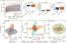
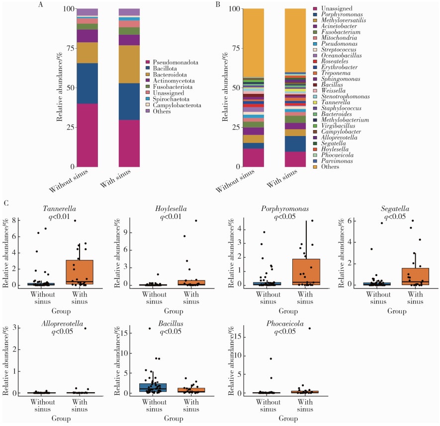
目的: 比较无窦型与有窦型根管治疗后慢性根尖周炎(post-treatment apical periodontitis, PoAP)患牙根管外菌群的组成与结构差异。方法: 纳入需要进行显微根尖手术治疗的PoAP患牙。手术中收集患牙根尖周病变组织, 并对细菌进行16S rRNA高通量测序, 测序区域为V3~V4。利用QIIME2软件进行生物信息学分析, 计算样本的α多样性, 采用Wilcoxon秩和检验进行组间差异分析。采用基于Weighted Unifrac距离的主坐标分析评估样本的β多样性, 通过置换多元方差分析(permutational multivariate analysis of variance, PERMANOVA)比较组间差异。采用Wilcoxon秩和检验比较组间物种相对丰度的差异, 并通过Benjamini-Hochberg法进行错误发现率(false discovery rate, FDR)校正, 将P值校正为q值。结果: 共纳入66例受试者, 男性21例, 女性45例, 平均年龄(33.91±9.16)岁。66例受试者中3例各有2颗患牙被纳入, 共纳入69颗患牙, 其中无窦型47颗, 有窦型22颗, 无窦型和有窦型两组在根充质量及病理类型上的分布差异均无统计学意义(P>0.05)。两组样本的α多样性指数差异无统计学意义(P>0.05), β多样性分析显示两组样本整体菌群结构差异有统计学意义(P < 0.01)。在门水平上, 有窦型组中拟杆菌门(Bacteroidota)的相对丰度显著高于无窦型组(16.98% vs. 9.22%, q < 0.01), 而假单胞菌门(Pseudomonadota)的相对丰度显著低于无窦型组(30.70% vs. 42.19%, q < 0.05);在属水平上, 卟啉单胞菌属(Porphyromonas)、坦纳菌属(Tannerella)、SegatellaPhocaeicolaHoylesella在有窦型组中丰度更高(q < 0.05), 而芽孢杆菌属(Bacillus)在无窦型组中丰度更高(q < 0.05)。结论: 无窦型与有窦型PoAP的根管外菌群结构存在显著差异, 卟啉单胞菌属、坦纳菌属、SegatellaPhocaeicolaHoylesella可能与窦道的形成和持续存在相关。

关键词: 慢性根尖周炎, 显微根尖手术, 窦道, 16S rRNA, 高通量测序

Abstract:

Objective: To compare the composition and structure of extraradicular bacterial communities in periapical lesions of teeth with post-treatment apical periodontitis (PoAP) between cases with and without sinus tracts. Methods: Patients requiring apical surgery were recruited from the Department of Cariology and Endodontology, Peking University School and Hospital of Stomatology. Periapical lesion samples were collected from teeth with sinus tracts and without sinus tracts during apical microsurgery. The remaining lesion samples were submitted for pathological examination. Following the extraction of the bacterial DNA, the V3-V4 region of the bacterial 16S rRNA gene was subjected to high-throughput sequencing using Illumina NovaSeq 6000. Bioinformatics analysis was performed using QIIME2 software. α diversity indices (Shannon, Simpson, Chao1) were calculated, and intergroup differences were analyzed using Wilcoxon test. β diversity was assessed by principal coordinate analysis based on Weighted Unifrac test, and community composition differences were analyzed with permutational multivariate analysis of variance (PERMANOVA). Differences in relative abundance between the groups were compared using the Wilcoxon test, followed by Benjamini-Hochberg false discovery rate (FDR) correction to convert P value to q value. Results: A total of 66 subjects were enrolled, including 21 male and 45 female patients, with a mean age of (33.91±9.16) years. Three subjects each had two teeth enrolled. In total, 69 teeth were included. These teeth included 47 cases without sinus tracts and 22 cases with sinus tracts. No significant differences were observed between the two groups in the distribution of obturation quality or pathological types (P>0.05). No statistically significant differences were found in α diversity indices between the two groups (P>0.05). However, β diversity analysis revealed a significant difference between the two groups (P=0.008). At the phylum level, the relative abundance of Bacteroidota was significantly higher in the group with sinus tracts (16.98% vs. 9.22%, q < 0.01), while the relative abundance of Pseudomonadota was significantly lower in the group with sinus tracts (30.70% vs. 42.19%, q < 0.05). At the genus level, Porphyromonas, Tannerella, Segatella, Phocaeicola, and Hoylesella were significantly enriched in the group with sinus tracts (q < 0.05), whereas Bacillus was more abundant in the group without sinus tracts (q < 0.05). Conclusion: The extraradicular bacterial community structure significantly differs between PoAP with and without sinus tracts. Porphyromonas, Tannerella, Segatella, Phocaeicola, and Hoylesella may be associated with the formation and persistence of sinus tracts.

Key words: Apical periodontitis, Apical microsurgery, Sinus tract, 16S rRNA, High-throughput sequencing

中图分类号: 

  • R781.3

图1

菌群的多样性分析"

图2

菌群物种组成及差异"

1
Nair PNR, Henry S, Cano V, et al. Microbial status of apical root canal system of human mandibular first molars with primary apical periodontitis after "one-visit" endodontic treatment[J]. Oral Surg Oral Med Oral Pathol Oral Radiol Endod, 2005, 99(2): 231- 252.

doi: 10.1016/j.tripleo.2004.10.005
2
Nair PNR. On the causes of persistent apical periodontitis: A review[J]. Int Endod J, 2006, 39(4): 249- 281.

doi: 10.1111/j.1365-2591.2006.01099.x
3
Bronzato JD, Davidian MES, de Castro M, et al. Bacteria and virulence factors in periapical lesions associated with teeth following primary and secondary root canal treatment[J]. Int Endod J, 2021, 54(5): 660- 671.

doi: 10.1111/iej.13457
4
Sun X, Yang Z, Nie Y, et al. Microbial communities in the extraradicular and intraradicular infections associated with persistent apical periodontitis[J]. Front Cell Infect Microbiol, 2022, 11, 798367.

doi: 10.3389/fcimb.2021.798367
5
Zhang JL, Yun J, Yue L, et al. Distinctive microbiota distribution from healthy oral to post-treatment apical periodontitis[J]. Front Cell Infect Microbiol, 2022, 12, 980157.

doi: 10.3389/fcimb.2022.980157
6
Gupta R, Hasselgren G. Prevalence of odontogenic sinus tracts in patients referred for endodontic therapy[J]. J Endod, 2003, 29(12): 798- 800.

doi: 10.1097/00004770-200312000-00003
7
Ng YL, Mann V, Gulabivala K. A prospective study of the factors affecting outcomes of nonsurgical root canal treatment: Part 1: Periapical health[J]. Int Endod J, 2011, 44(7): 583- 609.

doi: 10.1111/j.1365-2591.2011.01872.x
8
Wang W, Qu Y, Chen H, et al. The microbial co-infection interaction network in apical periodontitis with sinus tracts[J]. J Dent, 2025, 153, 105496.

doi: 10.1016/j.jdent.2024.105496
9
漆正楠, 尹君, 唐子圣, 等. 有窦型与无窦型根尖周炎微生物群落比较[J]. 牙体牙髓牙周病学杂志, 2016, 26(1): 1- 6.
10
Griffee MB, Patterson SS, Miller CH, et al. The relationship of Bacteroides melaninogenicus to symptoms associated with pulpal necrosis[J]. Oral Surg Oral Med Oral Pathol, 1980, 50(5): 457- 461.

doi: 10.1016/S0030-4220(80)80015-6
11
Sassone LM, Fidel R, Faveri M, et al. Microbiological evaluation of primary endodontic infections in teeth with and without sinus tract[J]. Int Endod J, 2008, 41(6): 508- 515.

doi: 10.1111/j.1365-2591.2008.01397.x
12
Qi Z, Cao H, Jiang H, et al. Combinations of bacterial species associated with symptomatic endodontic infections in a Chinese population[J]. Int Endod J, 2016, 49(1): 17- 25.

doi: 10.1111/iej.12428
13
Zhang C, Yang Z, Hou B. Diverse bacterial profile in extraradicular biofilms and periradicular lesions associated with persistent apical periodontitis[J]. Int Endod J, 2021, 54(9): 1425- 1433.

doi: 10.1111/iej.13512
14
Bolyen E, Rideout JR, Dillon MR, et al. Reproducible, interactive, scalable and extensible microbiome data science using QIIME 2[J]. Nat Biotechnol, 2019, 37(8): 852- 857.

doi: 10.1038/s41587-019-0209-9
15
Alquria TA, Acharya A, Kabir B, et al. Clinical investigation of bacteriome in primary endodontic infections with apical periodontitis using high-throughput sequencing analysis[J]. J Endod, 2024, 50(10): 1393- 1402.

doi: 10.1016/j.joen.2024.07.016
16
Buonavoglia A, Zamparini F, Lanave G, et al. Endodontic microbial communities in apical periodontitis[J]. J Endod, 2023, 49(2): 178- 189.

doi: 10.1016/j.joen.2022.11.015
17
李昱志, 苏旭, 陈晓涛, 等. 基于16S rDNA测序的慢性牙髓炎及根尖周炎感染根管内菌群多样性研究[J]. 安徽医科大学学报, 2024, 59(9): 1669- 1674.
18
Mominkhan D, Brito LCN, Yakubu AR, et al. Exploring relationships within the microbiome of root canal infections and the influence of associated clinical parameters[J]. Int Endod J, 2025, 58(11): 1751- 1763.

doi: 10.1111/iej.70011
19
Lyu P, Song Y, Bi R, et al. Protective actions in apical periodontitis: The regenerative bioactivities led by mesenchymal stem cells[J]. Biomolecules, 2022, 12(12): 1737.

doi: 10.3390/biom12121737
20
Barbosa-Ribeiro M, Arruda-Vasconcelos R, Louzada LM, et al. Microbiological analysis of endodontically treated teeth with apical periodontitis before and after endodontic retreatment[J]. Clin Oral Investig, 2021, 25(4): 2017- 2027.

doi: 10.1007/s00784-020-03510-2
21
Ordinola-Zapata R, Costalonga M, Dietz M, et al. The root canal microbiome diversity and function: A whole-metagenome shotgun analysis[J]. Int Endod J, 2024, 57(7): 872- 884.

doi: 10.1111/iej.13911
22
Darveau RP. Periodontitis: A polymicrobial disruption of host homeostasis[J]. Nat Rev Microbiol, 2010, 8(7): 481- 490.

doi: 10.1038/nrmicro2337
23
Tak EJ, Park OJ, Lee JS, et al. Microbiota associated with caries and apical periodontitis: A next-generation sequencing study[J]. Int Endod J, 2025, 58(6): 890- 901.

doi: 10.1111/iej.14218
24
Zhao W, Ji L, Li J, et al. Mesaconate from bacillus subtilis R0179 supernatant attenuates periodontitis by inhibiting porphyromonas gingivalis in mice[J]. J Periodontal Res, 2025, 60(6): 617- 627.

doi: 10.1111/jre.13363
25
Tsubura S, Mizunuma H, Ishikawa S, et al. The effect of Bacillus subtilis mouth rinsing in patients with periodontitis[J]. Eur J Clin Microbiol Infect Dis, 2009, 28(11): 1353- 1356.

doi: 10.1007/s10096-009-0790-9
26
Turan Gökduman C, Çanakçi BC, Arili Öztürk E, et al. Factors affecting long-term success and survival in non-surgical root canal retreatment: A retrospective cohort study[J]. Clin Oral Investig, 2025, 29(11): 491.

doi: 10.1007/s00784-025-06592-y
[1] 谢京城,陈晓东,杨军. 成人先天性皮窦道脊髓拴系综合征的诊断和治疗[J]. 北京大学学报(医学版), 2022, 54(6): 1163-1166.
[2] 张家赫, 史佳琪, 陈章健, 贾光. 基于人消化道微生态体外模拟系统观察纳米二氧化钛对肠道菌群的影响[J]. 北京大学学报(医学版), 2022, 54(3): 468-476.
[3] 朱诗玮, 刘作静, 李默, 朱怀球, 段丽萍. 不同软件平台分析肠易激综合征患者肠道菌群结构的比较[J]. 北京大学学报(医学版), 2018, 50(2): 231-238.
Viewed
Full text


Abstract

Cited

  Shared   
  Discussed   
No Suggested Reading articles found!