北京大学学报(医学版) ›› 2021, Vol. 53 ›› Issue (6): 1144-1151. doi: 10.19723/j.issn.1671-167X.2021.06.023

• 论著 • 上一篇    下一篇

不同转流标准对颈动脉内膜切除术后脑梗死的影响

白鹏1,王涛2,周阳1,陶立元3,李刚1,李正迁1,郭向阳1,()   

  1. 1.北京大学第三医院 麻醉科, 北京 100191
    2.北京大学第三医院 神经外科, 北京 100191
    3.北京大学第三医院 临床流行病研究中心,北京 100191
  • 收稿日期:2020-06-04 出版日期:2021-12-18 发布日期:2021-12-13
  • 通讯作者: 郭向阳 E-mail:puthmzk@hsc.pku.edu.cn
  • 基金资助:
    北京市自然科学基金(7172230)

Effect of different shunt strategies on cerebral infarction after carotid endarterectomy

BAI Peng1,WANG Tao2,ZHOU Yang1,TAO Li-yuan3,LI Gang1,LI Zheng-qian1,GUO Xiang-yang1,()   

  1. 1. Department of Anesthesiology, Peking University Third Hospital, Beijing 100191, China
    2. Department of Neurosurgery, Peking University Third Hospital, Beijing 100191, China
    3. Clinical Epidemiology Research Center, Peking University Third Hospital, Beijing 100191, China
  • Received:2020-06-04 Online:2021-12-18 Published:2021-12-13
  • Contact: Xiang-yang GUO E-mail:puthmzk@hsc.pku.edu.cn
  • Supported by:
    Beijing Natural Science Foundation(7172230)

摘要:

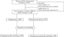
目的:颈动脉内膜切除术 (carotid endarterectomy, CEA)的麻醉管理要点是颈动脉阻断期间维持足够的颅脑灌注,放置转流管是外科常用方法之一,分析CEA手术不同转流标准对术后脑梗死的影响。方法:回顾收集2年内全身麻醉下接受CEA手术的443例患者,根据不同转流标准分为影像组 (以术前影像学资料作为是否转流的依据)和残端压组 (以术中残端压作为是否转流的依据)。分析患者术前人口学资料、既往病史、颈部血管狭窄程度、围术期各时间点血压情况、血管阻断时间、是否放置转流管、术后住院时间、住院期间脑梗死和其他不良事件,进行组间对比,在此基础上对组间差异有统计学意义的术前、术中情况进行倾向性评分匹配,分析不同转流标准对术后发生脑梗死的影响。结果:影像组患者268例、残端压组患者175例均接受全身麻醉下CEA手术,两组间基本情况和各时间点血压差异有统计学意义。经倾向性评分匹配后,匹配出影像组和残端压组各105例患者,两组患者的术前基本情况、各时间点血压差异无统计学意义。两组患者术后脑梗死发生率差异无统计学意义 (1.9% vs. 1.0%,P>0.999), 影像组术中转流率明显低于残端压组 (0 vs. 22.9%, P<0.001),影像组患者术后住院时间8 (7, 8) d明显长于残端压组5 (4, 6) d (P<0.001)。结论:依据术前影像学检查决定是否转流的患者组中,术中转流管使用率低于依据术中残端压决定是否转流的患者组,两组患者术后住院期间脑梗死的发生率差异无统计学意义,不同转流标准对患者脑梗死的影响有待于进一步深入研究。

关键词: 颈动脉内膜切除术, 脑梗死, 脑血管循环, 颈动脉狭窄

Abstract:

Objective: The key point of anesthesia management in carotid endarterectomy (CEA) is to maintain adequate cerebral perfusion during carotid artery occlusion. Placement of shunt is one of the common surgical methods. This study analyzed the effects of different shunt strategies on cerebral infarction after carotid endarterectomy. Methods: A total of 443 patients who underwent CEA under general anesthesia within 2 years were divided into imaging group (based on preoperative imaging data as the basis for shunt) and stump pressure group (based on intraoperative stump pressure as the basis for shunt). The preoperative demographic data, past medical history, degree of cervical vascular stenosis, blood pressure at each time point during the perioperative period, vascular blocking time, whether to place the shunt, postoperative hospital stay, cerebral infarction during hospitalization, and other adverse events were collected and compared between the two groups. On this basis, the preoperative and intraoperative conditions with significant differences were matched with propensity scores, and the influence of different shunt strategies on postoperative cerebral infarction was analyzed. Results: In the study, 268 patients in the imaging group and 175 patients in the stump pressure group underwent CEA under general anesthesia. There were statistically significant differences in basic conditions and blood pressure at each time point between the two groups. After matching the propensity scores, 105 patients in each of the two groups were matched. The basic conditions of the patients before surgery and the difference in blood pressure of the two groups at each time point were not statistically significant. There was no significant diffe-rence in the incidence of postoperative cerebral infarction between the two groups (1.9% vs. 1.0%, P>0.999). The intraoperative shunt rate in the imaging group was lower than that in the stump pressure group (0 vs. 22.9%, P<0.001). The postoperative hospital stay in the imaging group was 8 (7, 8) days, which was longer than the stump pressure group 5 (4, 6) days (P<0.001). Conclusion: The rate of the shunt was lower according to preoperative imaging examination than that according to the residual pressure in our hospital. There is no significant difference in the incidence of cerebral infarction during the postoperative hospital stay. The effect of different shunt strategies on cerebral infarction needs further study.

Key words: Carotid endarterectomy, Cerebral infarction, Cerebrovascular circulation, Carotid stenosis

中图分类号: 

  • R651.12

图1

不同转流策略流程图"

表1

两组术前的基本资料"

Items Imaging group (n=268) Stump pressure group (n=175) t/χ2 P value
Gender (male/female) 219 (81.7%)/49 (18.3%) 132 (75.4%)/43 (24.6%) 2.544 0.111
Age/years 65.0 (60.0, 71.0) 68.0 (62.0, 75.0) 6.814 0.009
BMI/(kg/m2) 24.5 (22.6, 26.9) 25.1 (22.9, 27.4) 1.738 0.187
Hypertension 198 (73.9%) 128 (73.1%) 0.030 0.863
Diabetes 89 (33.2%) 68 (38.9%) 1.476 0.224
Coronary heart disease 76 (28.4%) 57 (32.6%) 0.895 0.344
Preoperative neurological symptoms 0.699 0.403
Yes (cerebral infarction/TIA) 138 (51.5%)
[82 (30.6%)/56 (20.9%)]
83 (47.4%)
[66 (37.7%)/17 (9.7%)]
No 130 (48.5%) 92 (52.6%)
Carotid artery stenosis on the operative side 47.810 <0.001
Light 0 (0) 5 (2.9%)
Moderate 10 (3.7%) 40 (22.9%)
Severe 258 (96.3%) 130 (74.3%)
Carotid artery stenosis on the other side 7.147 0.028
Light 156 (58.2%) 119 (68.0%)
Moderate 52 (19.4%) 34 (19.4%)
Severe 60 (22.4%) 22 (12.6%)
Vertebral artery stenosis on the operative side 2.346 0.309
Light 220 (82.1%) 153 (87.4%)
Moderate 17 (6.3%) 7 (4.0%)
Severe 31 (11.6%) 15 (8.8%)
Vertebral artery stenosis on the operative side 1.150 0.563
Light 222 (82.8%) 150 (85.7%)
Moderate 18 (6.7%) 12 (6.9%)
Severe 28 (10.4%) 13 (7.4%)

表2

两组患者血压、转流管使用及血管阻断时间"

Items Imaging group (n=268) Stump pressure group (n=175) t/χ2 P value
T1 SBP/mmHg 130 (130, 140) 133 (127, 142) 6.523 0.011
T1 DBP/mmHg 75 (70, 80) 74 (68, 80) 6.219 0.013
T1 MBP/mmHg 93 (90, 97) 93 (89, 99) 0.164 0.685
T2 SBP/mmHg 160 (145, 165) 156 (145, 169) 0.694 0.405
T2 DBP/mmHg 80 (70, 85) 79 (71, 86) 0.045 0.831
T2 MBP/mmHg 105 (97, 112) 105 (98, 113) 0.344 0.563
T3 SBP/mmHg 130 (120, 149) 138 (126, 147) 5.645 0.018
T3 DBP/mmHg 65 (60, 70) 65 (58, 73) 0.005 0.946
T3 MBP/mmHg 87 (82, 97) 90 (83, 98) 1.187 0.276
T4 SBP/mmHg 157±14 156±18 0.900 0.369
T4 standard deviation of SBP/mmHg 5 (3, 8) 5 (2, 7) 3.113 0.078
T4 DBP/mmHg 72±10 70±11 1.850 0.065
T4 standard deviation of DBP/mmHg 3 (2, 4) 3 (1, 5) 0.080 0.777
T4 MBP/mmHg 101±9 99±11 1.787 0.075
T5 SBP/mmHg 130 (120, 140) 130 (121, 139) 3.378 0.066
T5 DBP/mmHg 65 (60, 70) 60 (55, 67) 9.289 0.002
T5 MBP/mmHg 87 (80, 93) 84 (78, 90) 9.557 0.002
T6 SBP/mmHg 140 (130, 140) 133 (122, 142) 11.762 0.001
T6 DBP/mmHg 75 (70, 80) 70 (65, 76) 51.968 <0.001
T6 MBP/mmHg 97 (93, 100) 92 (86, 97) 45.535 <0.001
Blocking time/min 25.0 (25.0, 29.0) 35.0 (23.0, 42.0) 16.197 <0.001
Shunt 14 (5.2%) 32 (18.3%) 19.410 <0.001

表3

两组患者术后不良事件及住院时间"

Items Imaging group (n=268) Stump pressure group (n=175) t/χ2 P value
Cerebral infarction 5 (1.9%) 1 (0.6%) 1.327 0.410
Cerebral hemorrhage 3 (1.1%) 0 (0) 1.972 0.281
Myocardial ischemia and myocardial infarction 6 (2.2%) 1 (0.6%) 1.893 0.253
Atrial fibrillation (new onset) 2 (0.7%) 1 (0.6%) 0.048 >0.999
Pulmonary infection and respiratory failure 1 (0.4%) 0 (0) 0.654 >0.999
Wound infection 0 (0) 1 (0.6%) 1.535 0.395
Postoperative hospital stay/d 8 (7, 8) 5 (5, 6) 67.918 <0.001

表4

倾向性评分匹配后两组患者的术前基本资料"

Items Imaging group (n=105) Stump pressure group (n=105) t/χ2 P value
Gender (male/female) 77 (73.3%)/28 (26.7%) 78 (74.3%)/27 (25.7%) 0.025 0.875
Age/years 66.0 (62.0, 72.0) 66.0 (61.0, 74.5) 0.011 0.918
BMI/(kg/m2) 24.4 (22.5, 27.1) 24.9 (22.3, 27.1) 0.284 0.594
Hypertension 81 (77.1%) 82 (78.1%) 0.027 0.868
Diabetes 41 (39.0%) 43 (41.0%) 0.079 0.778
Coronary heart disease 26 (24.8%) 35 (33.3%) 1.871 0.171
Preoperative neurological symptoms 0.019 0.890
Yes (cerebral infarction/TIA) 52 (49.5%)
[29 (27.6%)/23 (21.9%)]
53 (50.5%)
[44 (41.9%)/9 (8.6%)]
No 53 (50.5%) 52 (49.5%)
Carotid artery stenosis on the operative side 0.000 >0.999
Light 0 (0) 0 (0)
Moderate 8 (7.6%) 8 (7.6%)
Severe 97 (92.4%) 97 (92.4%)
Carotid artery stenosis on the other side 0.029 0.986
Light 68 (64.8%) 67 (63.8%)
Moderate 23 (21.9%) 24 (22.9%)
Severe 14 (13.3%) 14 (13.3%)
Vertebral artery stenosis on the operative side 1.593 0.451
Light 220 (82.1%) 153 (87.4%)
Moderate 17 (6.3%) 7 (4.0%)
Severe 31 (11.6%) 15 (8.8%)
Vertebral artery stenosis on the operative side 2.339 0.311
Light 87 (82.9%) 93 (88.6%)
Moderate 9 (8.6%) 5 (4.8%)
Severe 9 (8.6%) 7 (6.7%)

表5

倾向性评分匹配后两组患者血压、转流管使用及血管阻断时间"

Items Imaging group (n=105) Stump pressure group (n=105) t/χ2 P value
T1 SBP/mmHg 130 (130, 140) 133 (128, 142) 0.150 0.699
T1 DBP/mmHg 75 (70, 80) 75 (70, 80) 0.320 0.571
T1 MBP/mmHg 93 (90, 98) 93 (90, 100) 0.805 0.370
T2 SBP/mmHg 155 (140, 170) 157 (145, 170) 0.690 0.406
T2 DBP/mmHg 80 (70, 85) 80 (71, 86) 0.366 0.545
T2 MBP/mmHg 103 (96, 113) 106 (98, 112) 0.977 0.323
T3 SBP/mmHg 135 (130, 150) 138 (127, 146) 0.276 0.606
T3 DBP/mmHg 65 (60, 70) 66 (58, 74) 0.023 0.879
T3 MBP/mmHg 90 (83, 97) 90 (82, 98) 0.009 0.925
T4 SBP/mmHg 156±13 156±17 0.150 0.881
T4 standard deviation of SBP/mmHg 5 (4, 8) 5 (2, 7) 8.584 0.003
T4 DBP/mmHg 70±11 70±11 0.160 0.873
T4 standard deviation of DBP/mmHg 3 (2, 4) 2 (1, 4) 6.424 0.011
T4 MBP/mmHg 99±9 99±11 0.187 0.852
T5 SBP/mmHg 130 (120, 140) 130 (121, 140) 0.016 0.899
T5 DBP/mmHg 60 (55, 70) 60 (55, 68) 0.148 0.700
T5 MBP/mmHg 83 (80, 90) 84 (78, 90) 0.029 0.865
T6 SBP/mmHg 135 (130, 140) 133 (127, 145) 0.057 0.812
T6 DBP (mmHg) 75 (70, 80) 72 (68, 79) 0.943 0.331
T6 MBP/mmHg 95 (90, 100) 94 (89, 99) 0.687 0.407
Blocking time/min 25.0 (25.0, 30.0) 30.0 (20.0, 40.0) 1.659 0.198
Shunt 0 (0) 24 (22.9%) 27.097 <0.001

表6

倾向性评分匹配后两组患者术后不良事件及住院时间"

Items Imaging group (n=105) Stump pressure group (n=105) t/χ2 P value
Cerebral infarction 2 (1.9%) 1 (1.0%) 0.338 >0.999
Cerebral hemorrhage 2 (1.9%) 0 (0) 2.019 0.498
Myocardial ischemia and myocardial infarction 1 (1.0%) 1 (1.0%) 0.000 >0.999
Atrial fibrillation (new onset) 0 (0) 1 (1.0%) 1.005 >0.999
Pulmonary infection and respiratory failure 0 (0) 0 (0)
Wound infection 0 (0) 0 (0)
Postoperative hospital stay/d 8 (7, 8) 5 (4, 6) 56.452 <0.001
[1] Petty GW, Brown RD Jr, Whisnant JP, et al. Ischemic stroke subtypes: A population-based study of incidence and risk factors[J]. Stroke, 1999, 30(12):2513-2516.
pmid: 10582970
[2] Kret MR, Young B, Moneta GL, et al. Results of routine shunting and patch closure during carotid endarterectomy[J]. Am J Surg, 2012, 203(5):613-617.
doi: 10.1016/j.amjsurg.2011.12.005
[3] Aburahma AF, Mousa AY, Stone PA. Shunting during carotid endarterectomy[J]. J Vasc Surg, 2011, 54(5):1502-1510.
doi: 10.1016/j.jvs.2011.06.020 pmid: 21906905
[4] Samson RH, Cline JL, Showalter DP, et al. Contralateral carotid artery occlusion is not a contraindication to carotid endarterectomy even if shunts are not routinely used[J]. J Vasc Surg, 2013, 58(4):935-940.
doi: 10.1016/j.jvs.2013.04.011
[5] Aburahma AF, Stone PA, Hass SM, et al. Prospective rando-mized trial of routine versus selective shunting in carotid endarterectomy based on stump pressure[J]. J Vasc Surg, 2010, 51(5):1133-1138.
doi: 10.1016/j.jvs.2009.12.046 pmid: 20347544
[6] Rerkasem K, Rothwell PM. Routine or selective carotid artery shunting for carotid endarterectomy and different methods of monitoring in selective shunting[J]. Stroke, 2010, 41(1):e53-e54
[7] Lichtman JH, Jones MR, Leifheit EC, et al. Carotid endarterectomy and carotid artery stenting in the US medicare population, 1999-2014[J]. JAMA, 2017, 318(11):1035-1046.
doi: 10.1001/jama.2017.12882 pmid: 28975306
[8] Wiske C, Arhuidese I, Malas M, et al. Comparing the efficacy of shunting approaches and cerebral monitoring during carotid endarterectomy using a national database[J]. J Vasc Surg, 2018, 68(2):416-425.
doi: S0741-5214(18)30035-1 pmid: 29571621
[9] Banga PV, Varga A, Csobay-Novák C, et al. Incomplete circle of Willis is associated with a higher incidence of neurologic events during carotid eversion endarterectomy without shunting[J]. J Vasc Surg, 2018, 68(6):1764-1771.
doi: 10.1016/j.jvs.2018.03.429
[10] 中华医学会外科学分会血管外科学组. 颈动脉狭窄诊治指南[J]. 中国血管外科杂志, 2017, 9(3):169-175.
[11] Kolkert JLP, Groenwold RHH, Leijdekkers VJ, et al. Cost-effectiveness of two decision strategies for shunt use during carotid endarterectomy[J]. World J Surg, 2017, 41(11):2959-2967.
doi: 10.1007/s00268-017-4085-5 pmid: 28623598
[12] 李攀峰, 王兵, 崔文军, 等. 对侧颈动脉狭窄程度对颈动脉内膜剥脱术中转流管应用的影响[J]. 实用医学杂志, 2015, 31(10):1639-1641.
[13] Huibers A, Calvet D, Kennedy F, et al. Mechanism of procedural stroke following carotid endarterectomy or carotid artery stenting within the International Carotid Stenting Study (ICSS) randomised trial[J]. Eur J Vasc Endovasc Surg, 2015, 50(3):281-288.
doi: 10.1016/j.ejvs.2015.05.017
[14] Spagnoli LG, Mauriello A, Sangiorgi G, et al. Extracranial thrombotically active carotid plaque as a risk factor for ischemic stroke[J]. JAMA, 2004, 292(15):1845-1852.
pmid: 15494582
[15] 刁永鹏, 刘昌伟, 宋小军, 等. 颈动脉内膜剥脱术治疗老年颈动脉狭窄患者的危险因素分析[J]. 中华普通外科杂志, 2014, 29(6):448-451.
[1] 贾子昌,李选,郑梅,栾景源,王昌明,韩金涛. 复合手术治疗无残端的症状性长段颈内动脉慢性闭塞[J]. 北京大学学报(医学版), 2020, 52(1): 177-180.
[2] 赵海燕,樊东升,韩金涛. 重度颈内动脉狭窄伴未破裂动脉瘤的治疗策略[J]. 北京大学学报(医学版), 2019, 51(5): 829-834.
[3] 李金勇,孙宏亮,叶志东,樊雪强,刘鹏. 颈动脉斑块成分和体积的多排螺旋计算机断层扫描血管成像技术评价[J]. 北京大学学报(医学版), 2018, 50(5): 833-839.
[4] 韩金涛,李选,和清源,赵海燕,叶珊,董国祥,栾景源,王昌明. 脑动脉串联病变的同期腔内治疗[J]. 北京大学学报(医学版), 2016, 48(1): 149-153.
[5] 王云云,孙葳,黄一宁. 颈椎病误诊为脑梗死1例[J]. 北京大学学报(医学版), 2015, 47(5): 883-884.
[6] 韩金涛,赵海燕,李选,和清源,叶珊,董国祥,傅军,栾景源,王昌明,李天润. 颈动脉支架成形术血流动力学损害的相关因素分析[J]. 北京大学学报(医学版), 2015, 47(5): 804-808.
[7] 韩金涛, 李选, 郑梅, 傅军, 王昌明, 董国祥, 和清源. 大脑中动脉成形术治疗中、重度残疾的皮质下分水岭梗死的临床观察[J]. 北京大学学报(医学版), 2014, 46(4): 606-611.
[8] 梁建涛, 王振宇. 脑动脉狭窄患者血清中炎性因子干扰素-γ、白细胞介素-6和肿瘤坏死因子-α的水平分析[J]. 北京大学学报(医学版), 2011, 43(6): 837-840.
[9] 杨敏, 宋莉, 王健, 佟小强, 邹英华. 颈动脉支架术伴重度低血流动力学状态患者的药物治疗[J]. 北京大学学报(医学版), 2009, 41(3): 373-375.
[10] 吕鹤, 姚生, 张巍, 王朝霞, 黄一宁, 牛小媛, 张茁, 袁云. 4个常染色体显性遗传性脑动脉病伴皮层下梗死和白质脑病(CADASIL)家族的临床表现[J]. 北京大学学报(医学版), 2004, 36(5): 496-500.
[11] 赵淑清, 肖卫忠, 李坚, 张新宇, 王宏利, 孙庆利, 张俊. 急性脑梗死合并SARS 1例报告[J]. 北京大学学报(医学版), 2003, 35(z1): 142-142.
Viewed
Full text


Abstract

Cited

  Shared   
  Discussed   
[1] 张三. 中文标题测试[J]. 北京大学学报(医学版), 2010, 42(1): 1 -10 .
[2] 赵磊, 王天龙 . 右心室舒张末期容量监测用于肝移植术中容量管理的临床研究[J]. 北京大学学报(医学版), 2009, 41(2): 188 -191 .
[3] 万有, , 韩济生, John E. Pintar. 孤啡肽基因敲除小鼠电针镇痛作用增强[J]. 北京大学学报(医学版), 2009, 41(3): 376 -379 .
[4] 张燕, 韩志慧, 钟延丰, 王盛兰, 李玲玲, 郑丹枫. 骨骼肌活组织检查病理诊断技术的改进及应用[J]. 北京大学学报(医学版), 2009, 41(4): 459 -462 .
[5] 赵奇, 薛世华, 刘志勇, 吴凌云. 同向施压测定自酸蚀与全酸蚀粘接系统粘接强度[J]. 北京大学学报(医学版), 2010, 42(1): 82 -84 .
[6] 林红, 王玉凤, 吴野平. 学校生活技能教育对小学三年级学生行为问题影响的对照研究[J]. 北京大学学报(医学版), 2007, 39(3): 319 -322 .
[7] 丰雷, 程嘉, 王玉凤. 注意缺陷多动障碍儿童的运动协调功能[J]. 北京大学学报(医学版), 2007, 39(3): 333 -336 .
[8] 李岳玲, 钱秋瑾, 王玉凤. 儿童注意缺陷多动障碍成人期预后及其预测因素[J]. 北京大学学报(医学版), 2007, 39(3): 337 -340 .
[9] . 书讯[J]. 北京大学学报(医学版), 2007, 39(3): 225 -328 .
[10] 牟向东, 王广发, 刁小莉, 阙呈立. 肺黏膜相关淋巴组织型边缘区B细胞淋巴瘤一例[J]. 北京大学学报(医学版), 2007, 39(4): 346 -350 .