北京大学学报(医学版) ›› 2019, Vol. 51 ›› Issue (3): 596-601. doi: 10.19723/j.issn.1671-167X.2019.03.033
基于U型卷积神经网络学习的前列腺癌影像重建模型在手术导航中的应用
颜野1,*,夏海缀1,*,李旭升2,何为3,朱学华1,张智荧1,肖春雷1,刘余庆1,黄华4,何良华2,卢剑1△( )
)
Application of U-shaped convolutional neural network in auto segmentation and reconstruction of 3D prostate model in laparoscopic prostatectomy navigation
Ye YAN1,*,Hai-zhui XIA1,*,Xu-sheng LI2,Wei HE3,Xue-hua ZHU1,Zhi-ying ZHANG1,Chun-lei XIAO1,Yu-qing LIU1,Hua HUANG4,Liang-hua HE2,Jian LU1△()
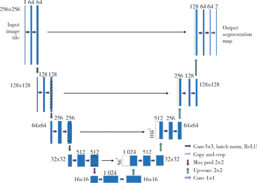
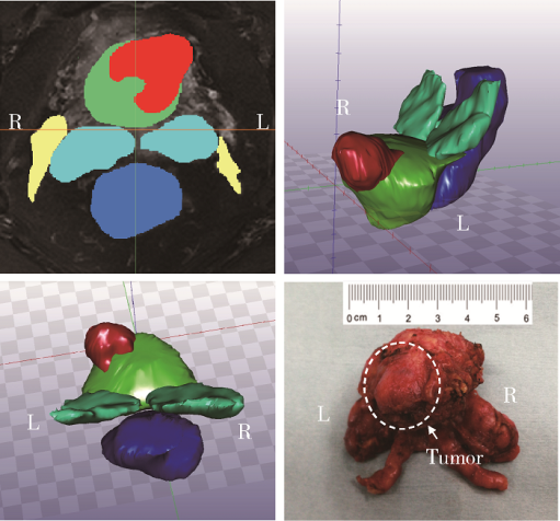
摘要: 目的 探讨基于U型卷积神经网络(U-shaped convolutional neural network, U-net)建立的前列腺磁共振图像自动化分割和重建3D模型对腹腔镜前列腺癌根治术进行术中认知导航的效果。方法 应用含有人工注释的共5 000张前列腺癌磁共振影像训练集,训练U-net,构建了一套以临床需求为导向,稳定高效的全卷积神经网络算法模型,对前列腺磁共振图像进行区域化、多结构和精细自动化分割,并将分割数据使用医学影像处理交互平台(Medical Image Interaction Tool Kit,MITK)自动重建,以STL格式输出建模信息,应用平板电脑在术中展示前列腺模型,进行认知导航。结果 基于201例前列腺癌患者的磁共振图像训练样本,在经典U-net基础上通过适应性改良,建立了一套结构简单、性能优秀的U-net,可以实现对前列腺、肿瘤、精囊腺、直肠等重要结构的单独分割,并进行三维可视化,直观地显示手术关键部位的结构关系和肿瘤侵犯程度。术中通过平板电脑同步展示3D模型,成功进行认知导航。结论 通过改良的U-net可以自动化完成前列腺磁共振图像的结构化分割,通过重建局部解剖部位的3D模型用于术中认知融合导航,可以达到肿瘤可视化、降低手术切缘阳性率、提高手术效果的作用。
中图分类号:
- R737.25


| [1] |
Siegel RL, Miller KD, Jemal A.Cancer statistics, 2018[J]. CA Cancer J Clin, 2018, 68(1): 7-30.
doi: 10.3322/caac.21442 |
| [2] |
Simmons MN, Stephenson AJ, Klein EA.Natural history of biochemical recurrence after radical prostatectomy: risk assessment for secondary therapy[J]. Eur Urol, 2007, 51(5): 1175-1184.
doi: 10.1016/j.eururo.2007.01.015 |
| [3] | Van den Broeck T, van den Bergh R, Arfi N, et al. Prognostic value of biochemical recurrence following treatment with curative intent for prostate cancer: A systematic review [J/OL]. Eur Urol,(2018-10-17) [2019-02-15]. https://doi.org/10.1016/j.eururo.2018.10.011. |
| [4] |
Ukimura O, Aron M, Nakamoto M, et al.Three-dimensional surgical navigation model with TilePro display during robot-assisted radical prostatectomy[J]. J Endourol, 2014, 28(6): 625-630.
doi: 10.1089/end.2013.0749 |
| [5] |
Hughes-Hallett A, Mayer EK, Marcus HJ, et al.Augmented rea-lity partial nephrectomy: examining the current status and future perspectives[J]. Urology, 2014, 83(2): 266-273.
doi: 10.1016/j.urology.2013.08.049 |
| [6] | 王燕, 高旭, 阳青松, 等. 3D打印技术辅助认知融合在前列腺穿刺活检术中的应用[J]. 临床泌尿外科杂志, 2016, 31(2): 104-107. |
| [7] | 邵叶秦, 杨新. 基于随机森林的CT前列腺分割[J]. CT理论与应用研究, 2015, 24(5): 647-655. |
| [8] | Ronneberger O, Fischer P, Brox T.U-net: Convolutional networks for biomedical image segmentation[C]. International Conference on Medical image computing and computer-assisted intervention. Cham: Springer, 2015: 234-241. |
| [9] | 詹曙, 梁植程, 谢栋栋. 前列腺磁共振图像分割的反卷积神经网络方法[J]. 中国图象图形学报, 2017, 22(4): 516-522. |
| [10] | Neher PF, Stieltjes B, Reisert M, et al.MITK global tractography[C]. Proceedings of SPIE: The International Society for Optical Engineering, 2012: 83144D. doi: 10.1117/12.911215. |
| [11] |
Lecun Y, Bottou L, Bengio Y, et al.Gradient-based learning applied to document recognition[J]. Proceedings of the IEEE, 1998, 86(11): 2278-2324.
doi: 10.1109/5.726791 |
| [12] |
Mahapatra D, Buhmann JM.Prostate MRI segmentation using learned semantic knowledge and graph cuts[J]. IEEE Transactions on Biomedical Engineering, 2014, 61(3): 756-764.
doi: 10.1109/TBME.2013.2289306 |
| [13] | Korez R, Likar B, Pernuš F, et al.Model-based segmentation of vertebral bodies from MR images with 3D CNNs[C]. International Conference on Medical Image Computing and Computer-Assisted Intervention. Cham: Springer, 2016: 433-441. |
| [14] |
Brosch T, Tang LY, Yoo Y, et al.Deep 3D convolutional encoder networks with shortcuts for multiscale feature integration applied to multiple sclerosis lesion segmentation[J]. IEEE Transactions on Medical Imaging, 2016, 35(5): 1229-1239.
doi: 10.1109/TMI.2016.2528821 |
| [15] |
Martínez F, Romero E, Dréan G, et al.Segmentation of pelvic structures for planning CT using a geometrical shape model tuned by a multi-scale edge detector[J]. Phys Med Biol, 2014, 59(6): 1471-1484.
doi: 10.1088/0031-9155/59/6/1471 |
| [16] | 凌彤, 杨琬琪, 杨明. 利用多模态U形网络的CT图像前列腺分割[J]. 智能系统学报, 2018, 13(6): 981-988. |
| [17] | Ebbing J, Jäderling F, Collins JW, et al.Comparison of 3D printed prostate models with standard radiological information to aid understanding of the precise location of prostate cancer: A construct validation study[J]. PLoS One, 2018, 13(6): e199477. |
| [18] |
Volonté F, Pugin F, Bucher P, et al.Augmented reality and image overlay navigation with OsiriX in laparoscopic and robotic surgery: not only a matter of fashion[J]. J Hepatobiliary Pancreat Sci, 2011, 18(4): 506-509.
doi: 10.1007/s00534-011-0385-6 |
| [19] |
Teber D, Guven S, Simpfendorfer T, et al.Augmented reality: a new tool to improve surgical accuracy during laparoscopic partial nephrectomy? Preliminary in vitro and in vivo results[J]. Eur Urol, 2009, 56(2): 332-338.
doi: 10.1016/j.eururo.2009.05.017 |
| [20] |
Porpiglia F, Fiori C, Checcucci E, et al.Augmented reality robot-assisted radical prostatectomy: Preliminary experience[J]. Urology, 2018, 115(5): 184.
doi: 10.1016/j.urology.2018.01.028 |
| [21] | Porpiglia F, Checcucci E, Amparore D, et al.Augmented-reality robot-assisted radical prostatectomy using hyper-accuracy three-dimensional reconstruction (HA 3DTM) technology: a radiological and pathological study[J]. BJU international, 2018, 123(5): 834-845. |
| [1] | 白晓强, 袁芷若, 周永胜, 吕珑薇. 动态牵张促进人骨髓间充质干细胞三维培养的成骨分化[J]. 北京大学学报(医学版), 2026, 58(3): 641-649. |
| [2] | FarinEbrahimi, 冯志强, FarazEbrahimi, 韩玮华, 于子杨, 贾宽宽, 安金刚. 上颌药物相关性颌骨坏死的不同分期手术治疗效果[J]. 北京大学学报(医学版), 2026, 58(1): 107-114. |
| [3] | 温奥楠, 张晓会, 杨咏涛, 高梓翔, 李文博, 单珅瑶, 商相宜, 田淯文, 郭殊玮, 王艺蓁, 王勇, 赵一姣. 基于非刚性配准构建三维颜面微笑仿真序列数据的方法[J]. 北京大学学报(医学版), 2026, 58(1): 139-144. |
| [4] | 于录, 吴灵, 刘筱菁, 李自力. 基于数据库相似性检索的正颌外科手术规划技术流程可行性研究: 随机对照试验[J]. 北京大学学报(医学版), 2026, 58(1): 145-152. |
| [5] | 邵梁, 马雯洁, 陈莹, 丁茜, 张磊. 上颌切牙前伸和正中咬合接触解剖特征的数字化测量与分析[J]. 北京大学学报(医学版), 2026, 58(1): 99-106. |
| [6] | 王翠萍, 陈哲, 程永静. 超微血流成像评估与膝骨关节炎临床症状的关联研究[J]. 北京大学学报(医学版), 2025, 57(6): 1096-1100. |
| [7] | 刘艳华, 陆敏, 赵旭阳, 张宽根, 武睿, 梅放, 戴志豪, 由江峰, 裴斐. 肿瘤转移抑制基因LASS2去磷酸化对液泡型ATP酶活性及前列腺癌侵袭性的影响[J]. 北京大学学报(医学版), 2025, 57(6): 1113-1123. |
| [8] | 杨小勇, 张帆, 马潞林, 刘承. 前列腺导管腺癌临床特征及腺外侵犯的影响因素[J]. 北京大学学报(医学版), 2025, 57(5): 956-960. |
| [9] | 宋凤岐, 徐心雨, 刘筱菁, 李自力. 上颌骨前部和整体顺时针旋转改善骨性Ⅲ类牙颌面畸形患者鼻旁凹陷的对比[J]. 北京大学学报(医学版), 2025, 57(5): 980-988. |
| [10] | 肖宇嘉, 毛渤淳, 周彦恒. 姿势性微笑的三维形态学研究[J]. 北京大学学报(医学版), 2025, 57(5): 989-995. |
| [11] | 宁家昕, 王浩然, 罗书航, 敬吉波, 王建业, 侯惠民, 刘明. 氧化应激相关基因与前列腺癌关系的多组学分析[J]. 北京大学学报(医学版), 2025, 57(4): 633-643. |
| [12] | 王泽远, 于栓宝, 郑浩轲, 陶金, 范雅峰, 张雪培. 基于临床特征和多参数MRI的前列腺癌盆腔淋巴结转移的术前预测模型[J]. 北京大学学报(医学版), 2025, 57(4): 684-691. |
| [13] | 宁圆, 张晓盈, 李雪, 李原, 何菁, 金月波. 干燥综合征并发乳腺淋巴瘤1例[J]. 北京大学学报(医学版), 2025, 57(4): 808-811. |
| [14] | 孙建军, 马千权, 尹晓亮, 杨辰龙, 张嘉, 陈素华, 吴超, 谢京城, 韩芸峰, 林国中, 司雨, 杨军, 邬海博, 赵强. 任意维度重建磁共振对骶管囊肿进行精准分型对于指导微创手术和康复的意义[J]. 北京大学学报(医学版), 2025, 57(2): 303-308. |
| [15] | 仇师禹, 练洋, 康一帆, 张雷, 蔡义望, 单小峰, 蔡志刚. 基于下颌骨数据库和全连接神经网络的三维检索模型辅助下的下颌骨个性化重建[J]. 北京大学学报(医学版), 2025, 57(2): 360-368. |
|
||
