Journal of Peking University(Health Sciences) ›› 2020, Vol. 52 ›› Issue (1): 124-128. doi: 10.19723/j.issn.1671-167X.2020.01.020
Previous Articles Next Articles
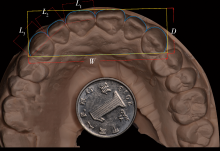
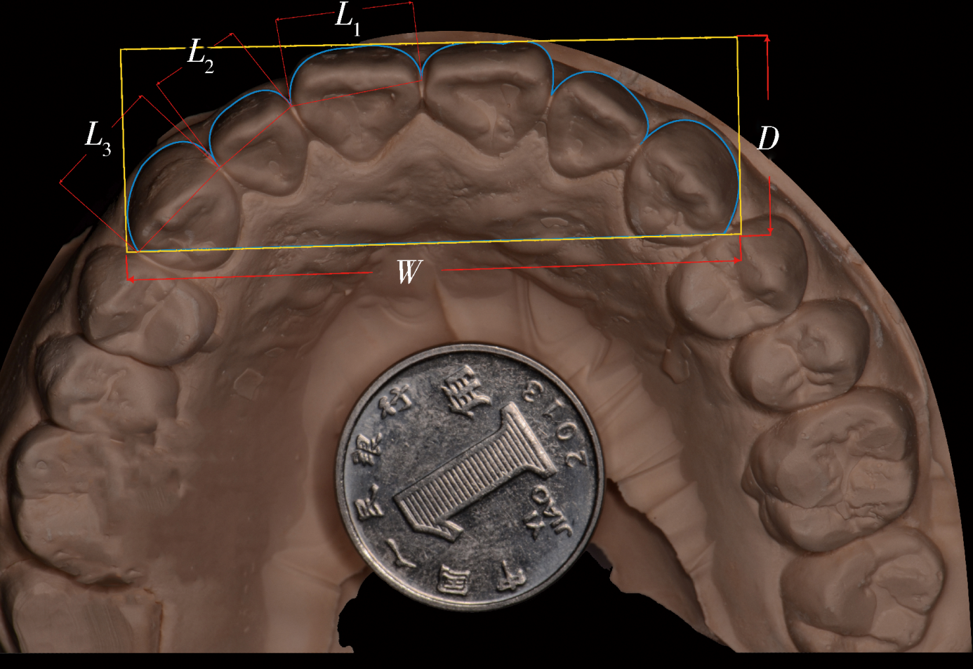
Analysis of the relationship among maxillary anterior teeth width, anterior arch perimeter and anterior segment depth
Peng WANG1,2,Da-jun LI1,3,Jian-zhang LIU( )
)
- 1. Department of Prosthodontics, Peking University School and Hospital of Stomatology & National Clinical Research Center for Oral Diseases & National Engineering Laboratory for Digital and Material Technology of Stomatology & Beijing Key Laboratory of Digital Stomatology, Beijing 100081, China
2. Department of Prosthodontics, Dalian Stomatological Hospital, Dalian 116021, Liaoning, China
3. Yuhua Meinuo Dental Clinic, Shijiazhuang 050000, China
CLC Number:
- R783


| [1] | Liao P, Fan Y, Nathanson D . Evaluation of maxillary anterior teeth width: A systematic review[J]. J Prosthet Dent, 2019,122(3):275-281. |
| [2] | Braun S, Hnat WP, Fender DE , et al. The form of the human dental arch[J]. Angle Orthod, 1998,68(1):29-36. |
| [3] | Noroozi H, Nik TH, Saeeda R . The dental arch form revisited[J]. Angle Orthod, 2001,71(5):386-389. |
| [4] | 宋宇, 周彦恒, 林久祥 . 上颌尖牙间宽度、前牙弓深度与前牙弓周长之间相互关系的二维分析[J]. 现代口腔医学杂志, 2007,21(5):476-478. |
| [5] | Sah SK, Zhang HD, Chang T , et al. Maxillary anterior teeth dimensions and proportions in a central mainland chinese population[J]. Chin J Dent Res, 2014,17(2):117-124. |
| [6] | 王惠芸 . 我国人牙的测量与统计[J]. 中华口腔科杂志, 1959,3(2):149-155. |
| [7] | Agrawal VS, Kapoor S, Bhesania D , et al. Comparative photographic evaluation of various geometric and mathematical proportions of maxillary anterior teeth: A clinical study[J]. Indian J Dent Res, 2016,27(1):32-36. |
| [8] | Ali FM, Jamani KD, Agrabawi J . Geometric and mathematical proportions and their relations to maxillary anterior teeth[J]. J Contemp Dent Pract, 2006,7(5):62-70. |
| [9] | Christian C, Marcelo C . Digital smile design: A tool for treatment planning and communication in esthetic dentistry[J]. Quintessence Dent Technol, 2012,35(3):103-112. |
| [10] | Christian C, Marcelo C, Newton S . Dynamic documentation of the smile and the 2D/3D digital smile design process[J]. Int J Periodontics Restorative Dent, 2017,37(2):183-193. |
| [11] | Garcia PP, da Costa RG, Calgaro M , et al. Digital smile design and mock-up technique for esthetic treatment planning with porcelain laminate veneers[J]. J Conserv Dent, 2018,21(4):455-458. |
| [12] | Veneziani M . Ceramic laminate veneers: clinical procedures with a multidisciplinary approach[J]. Int J Esthet Dent, 2017,12(4):426-448. |
| [13] | Cattoni F, Mastrangelo F, Gherlone EF , et al. A new total digital smile planning technique (3D-DSP) to fabricate CAD-CAM mockups for esthetic crowns and veneers[J]. Int J Dent, 2016,2016:6282587. doi: 10.1155/2016/6282587. |
| [1] | MA Ke-nan,CHEN Hu,SHEN Yan-ru,ZHOU Yong-sheng,WANG Yong,SUN Yu-chun. Finite element analyses of retention of removable partial denture circumferential clasps manufactured by selective laser melting [J]. Journal of Peking University (Health Sciences), 2022, 54(1): 105-112. |
| [2] | Fei SUN,Si-qi LI,Yi-ping WEI,Jin-sheng ZHONG,Cui WANG,Wen-jie HU. Efficacy of combined application of glycine powder air-polishing in non-surgical treatment of peri-implant diseases [J]. Journal of Peking University (Health Sciences), 2022, 54(1): 119-125. |
| [3] | LI Yi,YU Hua-jie,QIU Li-xin. Clinical classification and treatment decision of implant fracture [J]. Journal of Peking University (Health Sciences), 2022, 54(1): 126-133. |
| [4] | LIU Si-min,ZHAO Yi-jiao,WANG Xiao-yan,WANG Zu-hua. In vitro evaluation of positioning accuracy of trephine bur at different depths by dynamic navigation [J]. Journal of Peking University (Health Sciences), 2022, 54(1): 146-152. |
| [5] | LI Yi,WONG Lai U,LIU Xiao-qiang,ZHOU Ti,LYU Ji-zhe,TAN Jian-guo. Marginal features of CAD/CAM laminate veneers with different materials and thicknesses [J]. Journal of Peking University (Health Sciences), 2022, 54(1): 140-145. |
| [6] | QIU Shu-ting,ZHU Yu-jia,WANG Shi-min,WANG Fei-long,YE Hong-qiang,ZHAO Yi-jiao,LIU Yun-song,WANG Yong,ZHOU Yong-sheng. Preliminary clinical application verification of complete digital workflow of design lips symmetry reference plane based on posed smile [J]. Journal of Peking University (Health Sciences), 2022, 54(1): 193-199. |
| [7] | XU Xin-ran,HUO Peng-cheng,HE Lu,MENG Huan-xin,ZHU Yun-xuan,JIN Dong-si-qi. Comparison of initial periodontal therapy and its correlation with white blood cell level in periodontitis patients with or without diabetes mellitus [J]. Journal of Peking University (Health Sciences), 2022, 54(1): 48-53. |
| [8] | WANG Juan,YU Hua-jie,SUN Jing-de,QIU Li-xin. Application evaluation of prefabricated rigid connecting bar in implants immediate impression preparation of edentulous jaw [J]. Journal of Peking University (Health Sciences), 2022, 54(1): 187-192. |
| [9] | SUN Yu-chun,GUO Yu-qing,CHEN Hu,DENG Ke-hui,LI Wei-wei. Independent innovation research, development and transformation of precise bionic repair technology for oral prosthesis [J]. Journal of Peking University (Health Sciences), 2022, 54(1): 7-12. |
| [10] | Yuan LI,Hong LIN,Tie-jun ZHANG. Comparative study on radio-opacity of dental composite resin materials’determination using film imaging and digital imaging [J]. Journal of Peking University (Health Sciences), 2021, 53(5): 995-1001. |
| [11] | Feng LIANG,Min-jie WU,Li-dong ZOU. Clinical observation of the curative effect after 5-year follow-up of single tooth implant-supported restorations in the posterior region [J]. Journal of Peking University (Health Sciences), 2021, 53(5): 970-976. |
| [12] | JIANG You-sheng,FENG Lin,GAO Xue-jun. Influence of base materials on stress distribution in endodontically treated maxillary premolars restored with endocrowns [J]. Journal of Peking University (Health Sciences), 2021, 53(4): 764-769. |
| [13] | LIU Xiao-qiang,YANG Yang,ZHOU Jian-feng,LIU Ming-yue,TAN Jian-guo. Three-dimensional movement of posterior teeth after losing the interproximal and occlusal contacts in adults [J]. Journal of Peking University (Health Sciences), 2021, 53(3): 594-597. |
| [14] | LIU Xiao-qiang,YANG Yang,ZHOU Jian-feng,LIU Jian-zhang,TAN Jian-guo. Blood pressure and heart rate changes of 640 single dental implant surgeries [J]. Journal of Peking University (Health Sciences), 2021, 53(2): 390-395. |
| [15] | MENG Yuan,ZHANG Li-qi,ZHAO Ya-ning,LIU Deng-gao,ZHANG Zu-yan,GAO Yan. Three-dimentional radiographic features of 67 maxillary radicular cysts [J]. Journal of Peking University (Health Sciences), 2021, 53(2): 396-401. |
|