Journal of Peking University (Health Sciences) ›› 2025, Vol. 57 ›› Issue (5): 980-988. doi: 10.19723/j.issn.1671-167X.2025.05.025

Previous Articles     Next Articles

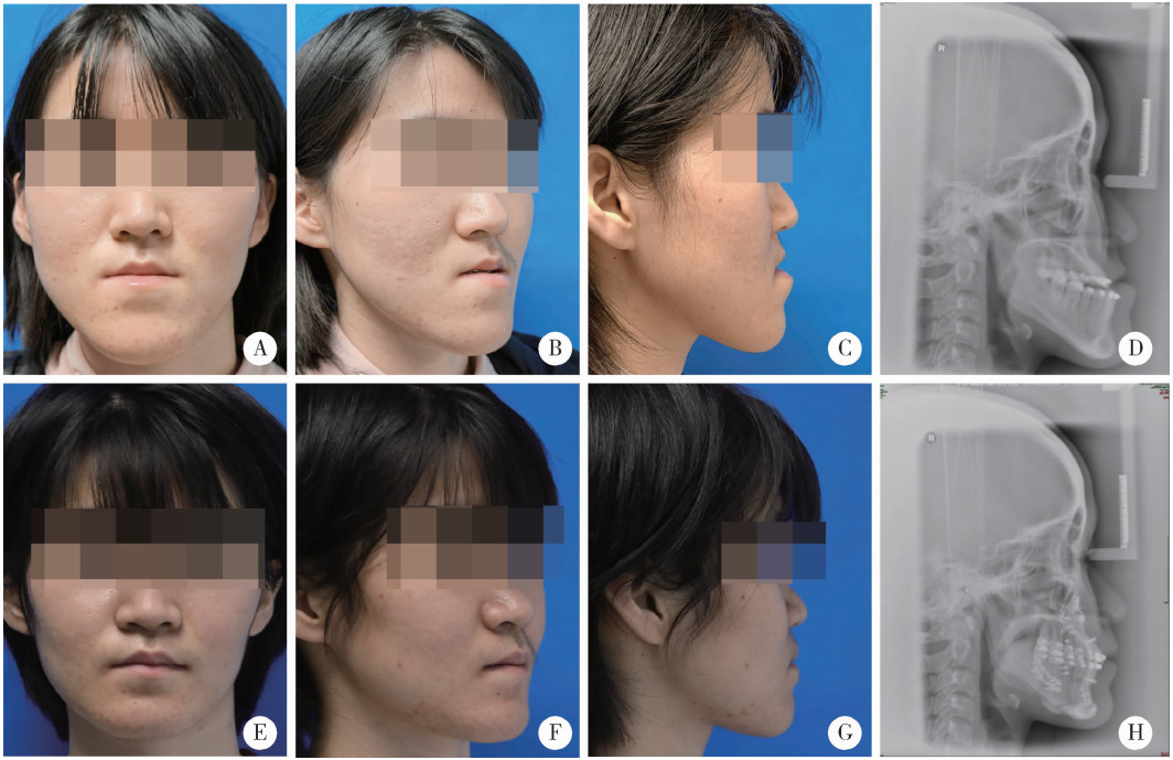
Comparation of anterior maxilla and whole maxilla clockwise rotation to improve paranasal aesthetic defects of skeletal Class Ⅲ maxillofacial deformity

Fengqi SONG, Xinyu XU, Xiaojing LIU, Zili LI*()   

  1. Department of Oral and Maxillofacial Surgery, Peking University School and Hospital of Stomatology & National Center of Stomatology & National Clinical Research Center for Oral Diseases & National Engineering Research Center of Oral Biomaterials and Digital Medical Devices, Beijing 100081, China
  • Received:2022-10-09 Online:2025-10-18 Published:2024-04-03
  • Contact: Zili LI
  • Supported by:
    the National Natural Science Foundation of China(82171012); Capital' s Funds for Health Improvement and Research(CFH2022-2-4104)

RICH HTML


Abstract: Objective: To compare the aesthetic effects of anterior maxilla clockwise rotation combined with segmental Le Fort Ⅰ osteotomy and whole maxilla clockwise rotation on improving paranasal concavity in patients with Class Ⅲ maxillofacial deformity. Methods: A non-randomized controlled trial was designed, and 21 patients diagnosed with skeletal Class Ⅲ maxillofacial deformity were included. In the study, 11 patients in the test group were treated by segmental Le Fort Ⅰ osteotomy combined with anterior maxilla clockwise rotation, and 10 patients in the control group were treated by whole maxilla clockwise rotation. The CBCT and 3D photography of preoperative (T0), 2 weeks postoperative (T1), and 6 months postoperative (T2) were collected respectively, and the three-dimensional cephalometry was carried out. The differences of specific parameters between the two groups were compared by independent sample t-test, including saggital displacement of the cheek mass point (CK) and subalare point (SA), nasolabial angle, occlusal plane angle and labial inclination angle of the upper incisor. Results: There were no significant differences of the parameters on T0 between the two groups. The average sagittal displacement of the upper incisors of the test group was (-0.71±1.67) mm and smaller than that of control group [(2.26±1.68) mm], t=-4.052, P < 0.05. The average angle of the occlusal plane clockwise rotation of the test group was 1.46°±2.38° and smaller than that of the control group (4.31°±1.83°), t=-3.047, P < 0.05. The angle of anterior maxilla clockwise rotation was 11.73°±2.81° during the surgery. The average saggital displacement of the paranasal soft tissue landmarks of the test group from T0 to T2 was larger than that of the control group [CK point, (4.96±1.18) mm vs. (2.01± 1.50) mm, P < 0.05;SA point, (5.19±1.17) mm vs. (2.69±1.45) mm, P < 0.05]. The labial inclination angle of the upper incisor of the test group was 112.15°±5.40° in T2 and significantly smaller than that of the control group (122.38°±8.83°), t=-3.237, P < 0.05. The nasolabial angle of the test group was 106.54°±12.82° in T2 and significantly larger than that of the control group (93.90°±12.46°), t=2.288, P < 0.05. Conclusion: Compared with whole maxilla clockwise rotation, anterior maxilla clockwise rotation combined with segmental Le Fort Ⅰ osteotomy can increase the saggital displacement of the paranasal soft tissue, correct labial inclination of the upper incisors and the acute naso-labial angle and better improve the paranasal aesthetic defects in patients with Class Ⅲ maxillofacial deformity with less changing on the saggital orientation of the upper incisors and the occlusal plane angle.

Key words: Skeletal Class Ⅲ maxillofacial deformity, Paranasal concavity, Segmental Le Fort Ⅰ osteo-tomy, Clockwise rotation, Three dimensional cephalometry

CLC Number: 

  • R782.1

Figure 1

Diagram of main landmarks in three-dimensional cephalometry A, landmarks of hard tissue; B, landmarks of soft tissue. The full names and explanations of all landmarks were listed in Table 1."

Table 1

Definition of the three-dimensional anatomical landmarks and reference planes"

Parameters Abbreviation Definition
Bone and dental landmarks
    Sella S The center of the hypophyseal fossa
    Nasion N The midpoint of the frontonasal suture
    Orbitale OrL, OrR The most inferior point of each infraorbital rim
    Porion PoL, PoR The most superior point of each external acoustic meatus
    Subspinale A The point of maximum concavity in the midline of the dento-alveolar process of the maxilla
    Upper incisor UI The most mesial point of the tip of the crown of the right upper central incisor
    Upper incisor apex UIa The apex of the right upper central incisor
    Upper molar UML, UMR The mesial buccal cusp of the first upper molar
    Supramental B The point of maximum concavity in the midline of the dento-alveolar process of the mandible
    Point h hL, hR Point on the left/right maxilla directly below the soft tissue alar base on axial view
Soft tissue landmarks
    Glabella G The most anterior midpoint on the fronto-orbital soft tissue contour
    Subalare SAL, SAR Labial insertion of each alar base
    Cheek mass CKL, CKR The most anterior point on the mid-pupillary plane (MPP) under infraorbital area and ahead of cornea perpendicular plane (CPP)
    Subnasale Sn The midpoint of the angle at the columella base where the lower border of the nasal septum and the surface of the upper lip meet
    Labiale superius Ls Midpoint of the upper vermillion border
    Columella peak Cp Most superior point of the columella
    Cornea CL, CR The most anterior point of the cornea
Reference planes
    Frankfort horizontal plane FHP The plane passes bilateral Orbitale and the midpoint between bilateral porion
    Midsagittal plane MSP The plane perpendicular to the FHP plane and passing through sella and nasion
    Facial plane FP The plane perpendicular to the FHP and MSP planes and passing through glabella
    Occlusal plane OP The plane passes the midpoint between UI and the midpoint between UML and UMR
    Mid-pupillary plane MPP The plane perpendicular to the FHP and FP planes and passing through cornea
    Cornea perpendicular plane CPP The plane perpendicular to the FHP and MSP planes and passing through cornea

Table 2

General characteristic and preoperative 3D cephalometric values of test group and control group"

Parameters Test group(n=11) Control group(n=10) t P value
Female 9 (81.8) 8 (80.0) -0.101 0.921
Age/years 24.91±6.20 24.00±5.06 0.366 0.719
SNA/(°) 82.47±2.28 81.26±2.89 1.067 0.299
SNB/(°) 85.90±2.46 86.45±2.74 -0.489 0.631
OP-SN/(°) 8.68±5.17 8.33±3.19 0.182 0.857
UI-SN/(°) 122.59±7.56 123.32±7.11 0.229 0.821
SNhR/(°) 72.07±2.91 71.50±4.30 0.364 0.720
SNhL/(°) 71.66±2.68 70.92±4.41 0.468 0.645
CKR-FP/mm -2.22±2.68 -3.04±3.44 0.614 0.546
CKL-FP /mm -3.01±3.26 -3.79±2.93 0.580 0.569
SAR-FP /mm -3.23±3.15 -4.51±3.20 0.921 0.369
SAL-FP /mm -4.16±3.01 -4.87±3.38 0.512 0.615
NL angle/(°) 87.61±14.62 84.63±12.97 0.491 0.629
UI-FP/mm 0.37±4.06 -1.42±3.96 1.020 0.320

Table 3

3D cephalometric values at T2 of test group and control group"

Parameters Test group (n=11) Control group (n=10) t P value
SNA/(°) 84.29±2.66 84.02±2.78 0.225 0.824
SNB/(°) 82.69±1.85 82.84±2.73 -0.142 0.889
OP-SN/(°) 10.26±4.64 11.62±3.92 -0.723 0.479
UI-SN/(°) 112.15±5.40 122.38±8.83 -3.237 0.004**
SNhR/(°) 78.48±2.77 76.16±3.83 1.600 0.126
SNhL/(°) 78.44±2.55 75.97±4.18 1.657 0.114
CKR-FP/mm 2.78±2.92 -1.21±3.31 2.932 0.009**
CKL-FP /mm 2.33±3.20 -1.60±3.25 2.793 0.012*
SAR-FP/mm 2.03±3.17 -1.32±2.95 2.507 0.021
SAL-FP/mm 2.23±3.73 -1.71±2.91 2.682 0.015*
NL angle/(°) 106.54±12.82 93.90±12.46 2.288 0.034*
UI-FP/mm 0.84±4.87 1.79±3.89 -0.494 0.627

Figure 2

Nasolabial angle and UI-SN of T0 and T2 in test group and control group A, variations of nasolabial angle from T0 to T2 of test group and control group; B, variations of UI-SN from T0 to T2 of test group and control group. Student-t test, * P<0.05,* * P<0.01. UI-SN, angle of the axis of the right upper incisor and SN line."

Table 4

Variation of 3D cephalometric values from T0 to T2 of test group and control group"

Parameters Test group (n=11) Control group (n=10) t P value
SNA/(°) 1.82±1.59 2.76±1.87 -1.245 0.228
SNB/(°) -3.21±1.04 -3.62±2.44 0.495 0.629
OP-SN/(°) 1.58±2.91 3.29±1.81 -1.597 0.127
UI-SN/(°) -10.43±4.28 -0.94±4.89 -4.746 <0.001**
SNhR/(°) 6.4±1.88 4.67±1.91 2.098 0.049*
SNhL/(°) 6.79±1.48 5.05±2.23 2.122 0.047*
CKR-FP/mm 4.78±1.22 1.83±1.55 4.886 <0.001**
CKL-FP /mm 5.15±1.74 2.19±1.63 4.005 <0.001**
SAR-FP /mm 5.24±1.71 3.18±1.67 2.782 0.012*
SAL-FP /mm 6.1±1.94 3.17±1.99 3.421 0.003**
NL angle/(°) 12.01±8.17 9.27±13.28 0.576 0.571
UI-FP/mm -0.03±1.95 3.05±2.44 -3.210 0.005**

Figure 3

Saggital displacement of paranasal soft tissue landmarks from T0 to T2 Student-t test, * P<0.05,* * P<0.01, * * * P<0.001. The definition of the parameters were listed under Table 2."

Figure 4

Photographs and X-ray of a patient in test group before (T0) and 6 months after surgery (T2) A-D, frontal photograph, 45° right side photograph, right lateral photograph and cephalometric lateral radiographs of T0; E-H, frontal photograph, 45° right side photograph, right lateral photograph and cephalometric lateral radiographs of T2."

Figure 5

Photographs and X-ray of a patient in control group before (T0) and 6 months after surgery (T2) A-D, frontal photograph, 45° right side photograph, right lateral photograph and cephalometric lateral radiographs of T0; E-H, frontal photograph, 45° right side photograph, right lateral photograph and cephalometric lateral radiographs of T2."

1
Naini FB , Gill DS . Orthognathic surgery: Principles, planning and practice[M]. New York: John Wiley & Sons, 2017: 85- 86.
2
Catherine Z , Scolozzi P . Modified Le Fort Ⅰ step osteotomy for improvement of paranasal flatness in maxillary deficiency: Technical note and series of 24 cases[J]. J Stomatol Oral Maxillofac Surg, 2019, 120 (6): 559- 565.

doi: 10.1016/j.jormas.2019.07.003
3
Lee TY , Chung HY , Dhong ES , et al. Paranasal augmentation using multi-folded expanded polytetrafluorethylene (ePTFE) in the East Asian nose[J]. Aesthet Surg J, 2019, 39 (12): 1319- 1328.

doi: 10.1093/asj/sjz103
4
Park HM , Lee YK , Choi JY , et al. Maxillary incisor inclination of skeletal Class Ⅲ patients treated with extraction of the upper first premolars and two-jaw surgery: Conventional orthognathic surgery vs. surgery-first approach[J]. Angle Orthod, 2014, 84 (4): 720- 729.

doi: 10.2319/072113-529.1
5
Kim DK , Baek SH . Change in maxillary incisor inclination during surgical-orthodontic treatment of skeletal Class Ⅲ malocclusion: Comparison of extraction and nonextraction of the maxillary first premolars[J]. Am J Orthod Dentofacial Orthop, 2013, 143 (3): 324- 335.

doi: 10.1016/j.ajodo.2012.10.014
6
Choi JW , Lee JY , Oh TS , et al. Frontal soft tissue analysis using a 3 dimensional camera following two-jaw rotational orthognathic surgery in skeletal Class Ⅲ patients[J]. J Craniomaxillofac Surg, 2014, 42 (3): 220- 226.

doi: 10.1016/j.jcms.2013.05.004
7
Chemello PD , Wolford LM , Buschang PH . Occlusal plane alteration in orthognathic surgery. Part Ⅱ: Long-term stability of results[J]. Am J Orthod Dentofacial Orthop, 1994, 106 (4): 434- 440.

doi: 10.1016/S0889-5406(94)70066-4
8
Wolford LM , Chemello PD , Hilliard F . Occlusal plane alteration in orthognathic surgery. Part Ⅰ: Effects on function and esthetics[J]. Am J Orthod Dentofacial Orthop, 1994, 106 (3): 304- 316.

doi: 10.1016/S0889-5406(94)70051-6
9
林广贤, 宋震, 范飞. 鼻基底填充术矫正鼻翼基底凹陷的临床应用进展[J]. 中国美容整形外科杂志, 2022, 33 (5): 312- 314.
10
王兴. 正颌外科手术学[M]. 济南: 山东科学技术出版社, 1999: 199- 201.
11
Chen YR , Yeow VK . Multiple-segment osteotomy in maxillofacial surgery[J]. Plast Reconstr Surg, 1999, 104 (2): 381- 388.

doi: 10.1097/00006534-199908000-00009
12
Liu XJ , Li QQ , Pang YJ , et al. Modified method of recording and reproducing natural head position with a multicamera system and a laser level[J]. Am J Orthod Dentofacial Orthop, 2015, 147 (6): 781- 787.

doi: 10.1016/j.ajodo.2015.01.016
13
Wong R , Chau A , Hägg U . 3D CBCT McNamara' s cephalometric analysis in an adult southern Chinese population[J]. Int J Oral Maxillofac Surg, 2011, 40 (9): 920- 925.

doi: 10.1016/j.ijom.2011.03.011
14
Ho CT , Denadai R , Lai HC , et al. Computer-aided planning in orthognathic surgery: A comparative study with the establishment of burstone analysis-derived 3D norms[J]. J Clin Med, 2019, 8 (12): 2106.

doi: 10.3390/jcm8122106
15
Zhang L , Tang MY , Jin R , et al. Classification of nasolabial folds in Asians and the corresponding surgical approaches: By Shanghai 9th People' s Hospital[J]. J Plast Reconstr Aesthet Surg, 2015, 68 (7): 914- 919.

doi: 10.1016/j.bjps.2015.03.023
16
Lai HC , Denadai R , Ho CT , et al. Effect of Le Fort Ⅰ maxillary advancement and clockwise rotation on the anteromedial cheek soft tissue change in patients with skeletal Class Ⅲ pattern and midface deficiency: A 3D imaging-based prediction study[J]. J Clin Med, 2020, 9 (1): 262.

doi: 10.3390/jcm9010262
17
Han MD , Momin MR , Munaretto AM , et al. Three-dimensional cephalometric analysis of the maxilla: Analysis of new landmarks[J]. Am J Orthod Dentofacial Orthop, 2019, 156 (3): 337- 344.

doi: 10.1016/j.ajodo.2018.09.018
18
Lee TS , Park S . Clockwise rotation of the occlusal plane for aesthetic purposes by double jaw surgery without orthodontic treatment[J]. Plast Reconstr Surg, 2019, 144 (6): 1010e- 1013e.

doi: 10.1097/PRS.0000000000006248
19
Jeong S , Sung J , Kim S , et al. Upper airway morphologic changes after mandibular setback surgery in skeletal Class Ⅲ malocclusion patients measured using cone beam computed tomography super-imposition[J]. Int J Oral Maxillofac Surg, 2018, 47 (11): 1405- 1410.

doi: 10.1016/j.ijom.2018.05.002
20
Hernández-Alfaro F , Guijarro-Martínez R . On a definition of the appropriate timing for surgical intervention in orthognathic surgery[J]. Int J Oral Maxillofac Surg, 2014, 43 (7): 846- 855.

doi: 10.1016/j.ijom.2014.02.007
21
Ko EW , Hsu SS , Hsieh HY , et al. Comparison of progressive cephalometric changes and postsurgical stability of skeletal Class Ⅲ correction with and without presurgical orthodontic treatment[J]. J Oral Maxillofac Surg, 2011, 69 (5): 1469- 1477.

doi: 10.1016/j.joms.2010.07.022
22
Sawh-Martinez R , Lin AM , DeSesa CR , et al. Clockwise and counterclockwise Le Fort Ⅰ movements influence nasolabial mor-phology differently[J]. Plast Reconstr Surg, 2018, 142 (6): 1572- 1581.

doi: 10.1097/PRS.0000000000004988
[1] Ziyang YU, Houzuo GUO, Xi JIANG, Weihua HAN, Ye LIN. Imaging study of osteogenesis in maxillary sinus segment of zygomatic implants [J]. Journal of Peking University (Health Sciences), 2025, 57(5): 967-974.
[2] Shiyu QIU, Yang LIAN, Yifan KANG, Lei ZHANG, Yiwang CAI, Xiaofeng SHAN, Zhigang CAI. Personalized mandibular reconstruction assisted by three-dimensional retrieval model based on fully connected neural network and a database of mandibles [J]. Journal of Peking University (Health Sciences), 2025, 57(2): 360-368.
[3] Fei WANG, Xinyue ZHANG, Muqing LIU, Enbo WANG, Denghui DUAN. Clinical application and three-dimensional finite element analysis of along-axis extraction method in mandibular mesial and horizontally impacted third molar surgery [J]. Journal of Peking University (Health Sciences), 2025, 57(1): 106-112.
[4] Yutong SHI, Yiping WEI, Wenjie HU, Tao XU, Haoyun ZHANG. Evaluation of micro crestal flap-alveolar ridge preservation following extraction of mandibular molars with severe periodontitis [J]. Journal of Peking University (Health Sciences), 2025, 57(1): 33-41.
[5] Sui LI,Wenjie MA,Shimin WANG,Qian DING,Yao SUN,Lei ZHANG. Trueness of different digital design methods for incisal guidance of maxillary anterior implant-supported single crowns [J]. Journal of Peking University (Health Sciences), 2024, 56(1): 81-87.
[6] Xiaoqiang LIU,Yin ZHOU. Risk factors of perioperative hypertension in dental implant surgeries with bone augmentation [J]. Journal of Peking University (Health Sciences), 2024, 56(1): 93-98.
[7] Deng-hui DUAN,Hom-Lay WANG,En-bo WANG. Role of collagen membrane in modified guided bone regeneration surgery using buccal punch flap approach: A retrospective and radiographical cohort study [J]. Journal of Peking University (Health Sciences), 2023, 55(6): 1097-1104.
[8] Min ZHEN,Huan-xin MENG,Wen-jie HU,Deng-cheng WU,Yi-ping WEI. Healing of the dento-gingival junction following modified crown lengthening procedure in beagle dogs [J]. Journal of Peking University (Health Sciences), 2022, 54(5): 927-935.
[9] JIANG You-sheng,FENG Lin,GAO Xue-jun. Influence of base materials on stress distribution in endodontically treated maxillary premolars restored with endocrowns [J]. Journal of Peking University (Health Sciences), 2021, 53(4): 764-769.
[10] HUANG Li-dong,GONG Wei-yu,DONG Yan-mei. Effects of bioactive glass on proliferation, differentiation and angiogenesis of human umbilical vein endothelial cells [J]. Journal of Peking University (Health Sciences), 2021, 53(2): 371-377.
[11] WANG Si-wen,YOU Peng-yue,LIU Yu-hua,WANG Xin-zhi,TANG Lin,WANG Mei. Efficacy of two barrier membranes and deproteinized bovine bone mineral on bone regeneration in extraction sockets: A microcomputed tomographic study in dogs [J]. Journal of Peking University (Health Sciences), 2021, 53(2): 364-370.
[12] JIANG Nan,BAO Xu-dong,YUE Lin. Influence of trueness for local finish lines of a full crown preparation on that of complete finish line [J]. Journal of Peking University (Health Sciences), 2021, 53(1): 102-108.
[13] . [J]. Journal of Peking University (Health Sciences), 2020, 52(2): 395-403.
[14] Chang CAO,Fei WANG,En-bo WANG,Yu LIU. Application of β-TCP for bone defect restore after the mandibular third molars extraction: A split-mouth clinical trial [J]. Journal of Peking University(Health Sciences), 2020, 52(1): 97-102.
[15] Qian WANG,Dan LI,Zhi-hui TANG. Sinus floor elevation and simultaneous dental implantation: A long term retrospective study of sinus bone gain [J]. Journal of Peking University(Health Sciences), 2019, 51(5): 925-930.
Viewed
Full text


Abstract

Cited

  Shared   
  Discussed   
No Suggested Reading articles found!