Journal of Peking University (Health Sciences) ›› 2025, Vol. 57 ›› Issue (6): 1165-1173. doi: 10.19723/j.issn.1671-167X.2025.06.022

Previous Articles     Next Articles

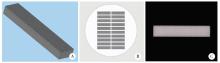
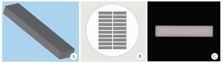
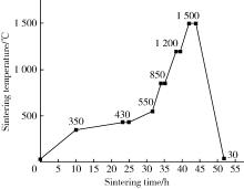
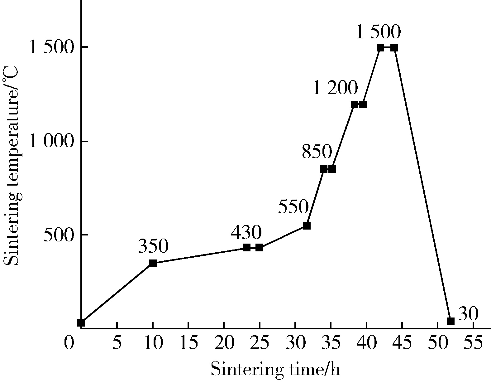
Effect of porous surface structure on fatigue strength of 3D printed zirconia

Jianxiao ZHAO1, Qian DING1, Wenjin LI1, Quanquan MA1, Yixiao LAN2, Lei ZHANG1,*(), Jianmin HAN2,*()   

  1. 1. Department of Prosthodontics, Peking University School and Hospital of Stomatology, Beijing 100081, China
    2. Department of Dental Materials, Peking University School and Hospital of Stomatology & National Center for Stomatology & National Clinical Research Center for Oral Diseases & National Engineering Research Center of Oral Biomaterials and Digital Medical Devices & NMPA Key Laboratory for Dental Materials, Beijing 100081, China
  • Received:2023-04-07 Online:2025-12-18 Published:2024-02-05
  • Contact: Lei ZHANG, Jianmin HAN
  • Supported by:
    the Beijing Natural Science Foundation(7192233)

RICH HTML


Abstract:

Objective: To study the effect of porous surface structure on fatigue strength of zirconia fabricated by stereolithography apparatus (SLA), and to provide reference for the surface design of 3D printed zirconia implants. Methods: Zirconia specimens were fabricated by SLA. According to the surface structure, zirconia specimens were divided into non-porous group, 200 μm group and 400 μm group. The surface morphology was observed by 3D laser morphology microscope and scanning electron microscope, and the surface roughness, pore parameters and grain size were measured. The flexural strength of the specimen was measured by three-point bending test and Weibull analysis was performed. The fatigue strength of the specimens was measured by fatigue test, and the fatigue mechanism was analyzed by fractrography. The crystal phase before and after fatigue test of the specimen was analyzed by X-ray diffraction. Results: The surface roughness of the area between the pores of non-porous group, 200 μm group and 400 μm group was (0.79±0.09) μm, (0.81±0.16) μm and (0.81±0.09) μm, respectively, with no significant difference among them. The surface grain size was (324.11±21.38) nm, (308.06±11.34) nm, (311.62±15.02) nm, respectively, with no significant difference among them. The results of three-point bending test showed that the three-point bending strength of the non-porous group [(1 030.70±111.71) MPa] was significantly higher than that of the porous groups (P < 0.001). The 200 μm group [(272.04±61.16) MPa] was significantly higher than the 400 μm group [(201.21±25.58) MPa] (P < 0.01). The fatigue strength of the non-porous group [(702.29± 21.62) MPa] was significantly higher than that of the porous groups (P < 0.001), and the fatigue strength of the 200 μm group [(159.57±9.30) MPa] was significantly higher than that of the 400 μm group [(125.36±6.11) MPa] (P < 0.001). The fracture analysis results showed that the crack origins were mainly internal defects, air holes, inclusions and the joint of printing layer, etc. There was no significant difference in the content of monoclinic phase before and after fatigue test among all the groups. Conclusion: The surface porous microstructure could significantly reduce the fatigue strength of the zirconia specimens, and the larger pore size showed the lower fatigue strength. In the future, the material and printing process of 3D printing zirconia should be improved, and the surface structure design should be further optimized to improve the mechanical properties of 3D printing zirconia.

Key words: Zirconia, Porous, 3D printing, Fatigue strength

CLC Number: 

  • R783.1

Figure 1

Design and production process of zirconia specimens A, porous surface zirconia specimens, stereolithography file schematic diagram; B, porous surface zirconia specimens, top view of slice file; C, fabrication of zirconia specimen with porous surface."

Figure 2

Sintering curve of the 3D-printed zirconia"

Figure 3

3D printed zirconia specimens A, non-porous group; B, 200 μm group; C, 400 μm group."

Figure 4

Fatigue strength test of zirconia specimen fabricated by stereo-lithography"

Table 1

The roughness of surface between pores of zirconia specimens"

Group Ra/μm Rq/μm Rz/μm
Non-porous 0.79±0.09 0.98±0.21 8.17±1.02
200 μm 0.81±0.16 1.06±0.18 8.79±1.48
400 μm 0.81±0.09 1.05±0.11 8.31±0.89

Table 2

Measurement results of porous parameters of zirconia specimen"

Group Design value of
pore diameter/μm
Measured value of
pore diameter/μm, ${\bar x}$±s
Design value of
pore depth/μm
Measured value of
pore depth/μm, ${\bar x}$±s
Design value of
pore-pitch/μm
Measured value of
pore-pitch/μm,${\bar x}$±s
200 μm 200.00 200.02±6.15 400.00 379.35±11.22** 430.00 429.08±9.31
400 μm 400.00 366.48±6.88** 600.00 572.84±29.35* 860.00 883.99±9.41**

Figure 5

Scanning electron microscope observation on the surface of zirconia specimen"

Table 3

Surface grain size of zirconia specimen"

Group Average grain size/nm, ${\bar x}$±s
Non-porous 324.11±21.38
200 μm 308.06±11.34
400 μm 311.62±15.02

Table 4

Three point flexural strength and fatigue strength of zirconia specimens"

Group Three-point flexural
strength/MPa, ${\bar x}$±s
Fatigue strength/MPa,
${\bar x}$±s
Characteristic
strength/MPa
95%CI Weibull modulus 95%CI
Non-porous 1 030.70±111.71 702.29±21.62 1 080.10 1 027.12-1 135.91 10.61 7.23-15.84
200 μm 272.04±61.16 159.57±9.30 296.40 264.97-331.55 4.80 3.29-6.99
400 μm 201.21±25.58 125.36±6.11 211.68 201.25-222.66 10.50 6.89-15.99

Figure 6

Results of staircase method to determine mean fatigue strength ×, broken; ○, unbroken."

Table 5

Monoclinic phase ratio of zirconia specimen surface"

Group Before fatigue test Unbroken after fatigue test Broken after fatigue test
Xm Vm Xm Vm Xm Vm
Non-porous 9.51% 12.09% 10.78% 13.67% 10.42% 13.23%
200 μm 10.01% 12.72% 11.17% 14.14% 10.43% 13.25%
400 μm 9.64% 12.27% 9.69% 12.34% 10.42% 13.22%

Figure 7

X-ray diffraction spectra of zirconia specimen (before fatigue test) showing monoclinic phase peak (m phase) and tetragonal phase peak (t phase)"

Figure 8

Scanning electron microscope fractography of zirconia specimens White arrows indicate the location of fatigue crack origin."

1
赵祯, 代康, 高勃. 3D打印陶瓷技术在口腔医学领域的研究进展[J]. 中国实用口腔科杂志, 2021, 14 (6): 739- 744.
2
Osman RB , van der Veen AJ , Huiberts D , et al. 3D-printing zirconia implants; a dream or a reality? An in-vitro study evaluating the dimensional accuracy, surface topography and mechanical properties of printed zirconia implant and discs[J]. J Mech Behav Biomed Mater, 2017, 75, 521- 528.

doi: 10.1016/j.jmbbm.2017.08.018
3
Thakur J , Parlani S , Shivakumar S , et al. Accuracy of marginal fit of an implant-supported framework fabricated by 3D printing versus subtractive manufacturing technique: A systematic review and meta-analysis[J]. J Prosthet Dent, 2023, 129 (2): 301- 309.

doi: 10.1016/j.prosdent.2021.05.010
4
Sakthiabirami K , Kang JH , Jang JG , et al. Hybrid porous zirconia scaffolds fabricated using additive manufacturing for bone tissue engineering applications[J]. Mater Sci Eng C Mater Biol Appl, 2021, 123, 111950.

doi: 10.1016/j.msec.2021.111950
5
中国国家标准化管理委员会. 金属平均晶粒度测定方法: GB/T 6394—2017[J]. 北京: 中国标准出版社, 2017, 8- 13.
6
International Organization for Standardization . Dentisry-ceramic materials: ISO 6872: 2015[J]. Switzerland: ISO, 2015, 7- 16.
7
Studart AR , Filser F , Kocher P , et al. Fatigue of zirconia under cyclic loading in water and its implications for the design of dental bridges[J]. Dent Mater, 2007, 23 (1): 106- 114.

doi: 10.1016/j.dental.2005.12.008
8
Zucuni CP , Guilardi LF , Rippe MP , et al. Fatigue strength of yttria-stabilized zirconia polycrystals: Effects of grinding, poli-shing, glazing, and heat treatment[J]. J Mech Behav Biomed Mater, 2017, 75, 512- 520.

doi: 10.1016/j.jmbbm.2017.06.016
9
Ding Q , Zhang L , Bao R , et al. Effects of different surface treatments on the cyclic fatigue strength of one-piece CAD/CAM zirconia implants[J]. J Mech Behav Biomed Mater, 2018, 84, 249- 257.

doi: 10.1016/j.jmbbm.2018.05.002
10
Dehurtevent M , Robberecht L , Hornez JC , et al. Stereolithography: A new method for processing dental ceramics by additive computer-aided manufacturing[J]. Dent Mater, 2017, 33 (5): 477- 485.

doi: 10.1016/j.dental.2017.01.018
11
Zhai Z , Sun J . Research on the low-temperature degradation of dental zirconia ceramics fabricated by stereolithography[J]. J Prosthet Dent, 2023, 130 (4): 629- 638.

doi: 10.1016/j.prosdent.2021.11.012
12
Kohal RJ , Bächle M , Att W , et al. Osteoblast and bone tissue response to surface modified zirconia and titanium implant materials[J]. Dent Mater, 2013, 29 (7): 763- 776.

doi: 10.1016/j.dental.2013.04.003
13
Al Qahtani WMS , Schille C , Spintzyk S , et al. Effect of surface modification of zirconia on cell adhesion, metabolic activity and proliferation of human osteoblasts[J]. Biomed Tech (Berl), 2017, 62 (1): 75- 87.
14
Hafezeqoran A , Koodaryan R . Effect of zirconia dental implant surfaces on bone integration: A systematic review and meta-analysis[J]. Biomed Res Int, 2017, 2017, 9246721.
15
Boyan BD , Hummert TW , Dean DD , et al. Role of material surfaces in regulating bone and cartilage cell response[J]. Biomaterials, 1996, 17 (2): 137- 146.

doi: 10.1016/0142-9612(96)85758-9
16
Hadjicharalambous C , Buyakov A , Buyakova S , et al. Porous alumina, zirconia and alumina/zirconia for bone repair: Fabrication, mechanical and in vitro biological response[J]. Biomed Mater, 2015, 10 (2): 025012.

doi: 10.1088/1748-6041/10/2/025012
17
Cheng A , Humayun A , Cohen DJ , et al. Additively manufactured 3D porous Ti-6Al-4V constructs mimic trabecular bone structure and regulate osteoblast proliferation, differentiation and local factor production in a porosity and surface roughness dependent manner[J]. Biofabrication, 2014, 6 (4): 045007.

doi: 10.1088/1758-5082/6/4/045007
18
Palmero P . Structural ceramic nanocomposites: A review of pro-perties and powders' synthesis methods[J]. Nanomaterials (Basel), 2015, 5 (2): 656- 696.

doi: 10.3390/nano5020656
19
Dos-Santos C , Santos FA , Elias CN . Properties of NAnostructured 3Y-TZP blocks used for CAD/CAM dental restoration[J]. Key Eng Mater, 2008, 396/397/398, 603- 606.
20
Silva CP , Santos C , Silva CRM . Mechanical properties of nanostructured zirconia[J]. Mater Sci Forum, 2010, 660/661, 757- 761.

doi: 10.4028/www.scientific.net/MSF.660-661.757
21
Egilmez F , Ergun G , Cekic-Nagas I , et al. Factors affecting the mechanical behavior of Y-TZP[J]. J Mech Behav Biomed Mater, 2014, 37, 78- 87.

doi: 10.1016/j.jmbbm.2014.05.013
22
Gonzaga CC , Cesar PF , Miranda WG Jr , et al. Slow crack growth and reliability of dental ceramics[J]. Dent Mater, 2011, 27 (4): 394- 406.

doi: 10.1016/j.dental.2010.10.025
23
Lakhdar Y , Tuck C , Binner J , et al. Additive manufacturing of advanced ceramic materials[J]. Prog Mater Sci, 2021, 116, 100736.

doi: 10.1016/j.pmatsci.2020.100736
24
李文利, 周宏志, 刘卫卫, 等. 光固化3D打印陶瓷浆料及流变性研究进展[J]. 材料工程, 2022, 50 (7): 40- 50.
25
Karakoca S , Yilmaz H . Influence of surface treatments on surface roughness, phase transformation, and biaxial flexural strength of Y-TZP ceramics[J]. J Biomed Mater Res B Appl Biomater, 2009, 91 (2): 930- 937.
26
Conti L , Bienenstein D , Borlaf M , et al. Effects of the layer height and exposure energy on the lateral resolution of zirconia parts printed by lithography-based additive manufacturing[J]. Materials (Basel), 2020, 13 (6): 1317.

doi: 10.3390/ma13061317
27
Wang B , Arab A , Xie J , et al. The influence of microstructure on the flexural properties of 3D printed zirconia part via digital light processing technology[J]. Materials (Basel), 2022, 15 (4): 1602.

doi: 10.3390/ma15041602
[1] Kun QIAN, Yihong LIU. Fitness of onlays fabricated with direct and indirect CAD/CAM technology in vitro [J]. Journal of Peking University (Health Sciences), 2025, 57(3): 604-609.
[2] Shuyuan MIN, Yun TIAN. Biocompatibility of 3D printed biodegradable WE43 magnesium alloy scaffolds and treatment of bone defects [J]. Journal of Peking University (Health Sciences), 2025, 57(2): 309-316.
[3] Xinxin ZHAN,Lulu CAO,Dong XIANG,Hao TANG,Dandan XIA,Hong LIN. Effect of printing orientation on physical and mechanical properties of 3D printing prosthodontic base resin materials [J]. Journal of Peking University (Health Sciences), 2024, 56(2): 345-351.
[4] Qian DING,Wen-jin LI,Feng-bo SUN,Jing-hua GU,Yuan-hua LIN,Lei ZHANG. Effects of surface treatment on the phase and fracture strength of yttria- and magnesia-stabilized zirconia implants [J]. Journal of Peking University (Health Sciences), 2023, 55(4): 721-728.
[5] Wei-wei LI,Hu CHEN,Yong WANG,Yu-chun SUN. Research on friction and wear behaviors of silicon-lithium spray coating on zirconia ceramics [J]. Journal of Peking University (Health Sciences), 2023, 55(1): 94-100.
[6] ABUDUREHEMAN Kaidierya,Rong-geng ZHANG,Hao-nan QIAN,Zhen-yang ZOU,YESITAO Danniya,Tian-yuan FAN. Preparation and in vitro evaluation of FDM 3D printed theophylline tablets with personalized dosage [J]. Journal of Peking University (Health Sciences), 2022, 54(6): 1202-1207.
[7] Yi DENG,Yi ZHANG,Bo-wen LI,Mei WANG,Lin TANG,Yu-hua LIU. Effects of different crosslinking treatments on the properties of decellularized small intestinal submucosa porous scaffolds [J]. Journal of Peking University (Health Sciences), 2022, 54(3): 557-564.
[8] SUN Yu-chun,GUO Yu-qing,CHEN Hu,DENG Ke-hui,LI Wei-wei. Independent innovation research, development and transformation of precise bionic repair technology for oral prosthesis [J]. Journal of Peking University (Health Sciences), 2022, 54(1): 7-12.
[9] WANG Zheng,DING Qian,GAO Yuan,MA Quan-quan,ZHANG Lei,GE Xi-yuan,SUN Yu-chun,XIE Qiu-fei. Effect of porous zirconia ceramics on proliferation and differentiation of osteoblasts [J]. Journal of Peking University (Health Sciences), 2022, 54(1): 31-39.
[10] LI Wen-jin,DING Qian,YUAN Fu-song,Sun Feng-bo,ZHENG Jian-qiao,BAO Rui,Zhang Lei. Effects of femtosecond laser treatment on surface characteristics and flexural strength of zirconia [J]. Journal of Peking University (Health Sciences), 2021, 53(4): 770-775.
[11] YANG Xin,LI Rong,YE Hong-qiang,CHEN Hu,WANG Yong,ZHOU Yong-sheng,SUN Yu-chun. Evaluation of fracture strength of two kinds of zirconia all-ceramic crowns with different edge compensation angles [J]. Journal of Peking University (Health Sciences), 2021, 53(2): 402-405.
[12] Miao ZHENG,Ling-lu ZHAN,Zhi-qiang LIU,He-ping LI,Jian-guo TAN. Effect of different plasma treated zirconia on the adhensive behaviour of human gingival fibroblasts [J]. Journal of Peking University(Health Sciences), 2019, 51(2): 315-320.
[13] ZHOU Tuan-feng, WANG Xin-zhi . Clinical observation of the restoration of computer aided designed and manufactured one-piece zirconia posts and cores: a 5-year prospective follow-up study [J]. Journal of Peking University(Health Sciences), 2018, 50(4): 680-684.
[14] ZHU Lin, WANG Yu-dong, DONG Yan-mei,CHEN Xiao-feng. Mesoporous nano-bioactive glass microspheres as a drug delivery system of mino-cycline [J]. Journal of Peking University(Health Sciences), 2018, 50(2): 249-255.
[15] CUI Xin-yue, TONG Dai, WANG Xin-zhi, SHEN Zhi-jian. Comparison of the translucency and color masking effect of the zirconia ceramics made by milling and gel deposition [J]. Journal of Peking University(Health Sciences), 2018, 50(1): 85-90.
Viewed
Full text


Abstract

Cited

  Shared   
  Discussed   
No Suggested Reading articles found!