北京大学学报(医学版) ›› 2026, Vol. 58 ›› Issue (1): 99-106. doi: 10.19723/j.issn.1671-167X.2026.01.013

• 论著 • 上一篇    下一篇

上颌切牙前伸和正中咬合接触解剖特征的数字化测量与分析

邵梁, 马雯洁, 陈莹, 丁茜*(), 张磊*()   

  1. 北京大学口腔医学院·口腔医院修复科, 国家口腔医学中心, 国家口腔疾病临床医学研究中心, 口腔生物材料和数字诊疗装备国家工程研究中心, 口腔数字医学北京市重点实验室, 北京 100081
  • 收稿日期:2025-10-10 出版日期:2026-01-06 发布日期:2026-01-06
  • 通讯作者: 丁茜, 张磊
  • 基金资助:
    北京大学口腔医(学)院临床研究专项基金(PKUSS-2023CRF503); 北京大学口腔医(学)院2025年度临床新技术新疗法项目(PKUSSNCT-25A09); 北京市自然科学基金-海淀原始创新联合基金(L232025)

Digital measurement and analysis of anatomical characteristics of protrusive and intercuspal position occlusal contacts in maxillary incisors

Liang SHAO, Wenjie MA, Ying CHEN, Qian DING*(), Lei ZHANG*()   

  1. Department of Prosthodontics, Peking University School and Hospital of Stomatology & National Center for Stomatology & National Clinical Research Center for Oral Diseases & National Engineering Research Center of Oral Biomaterials and Digital Medical Devices & Beijing Key Laboratory of Digital Stomatology, Beijing 100081, China
  • Received:2025-10-10 Online:2026-01-06 Published:2026-01-06
  • Contact: Qian DING, Lei ZHANG
  • Supported by:
    the Clinical Research Foundation of Peking University School and Hospital of Stomatology(PKUSS-2023CRF503); the Program for New Clinical Techniques and Therapies of Peking University School and Hospital of Stomatology(PKUSSNCT-25A09); Beijing Natural Science Foundation-Haidian Original Innovation Joint Fund Project(L232025)

RICH HTML


摘要:

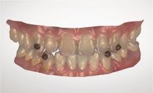
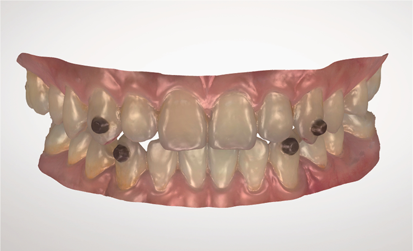
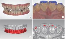
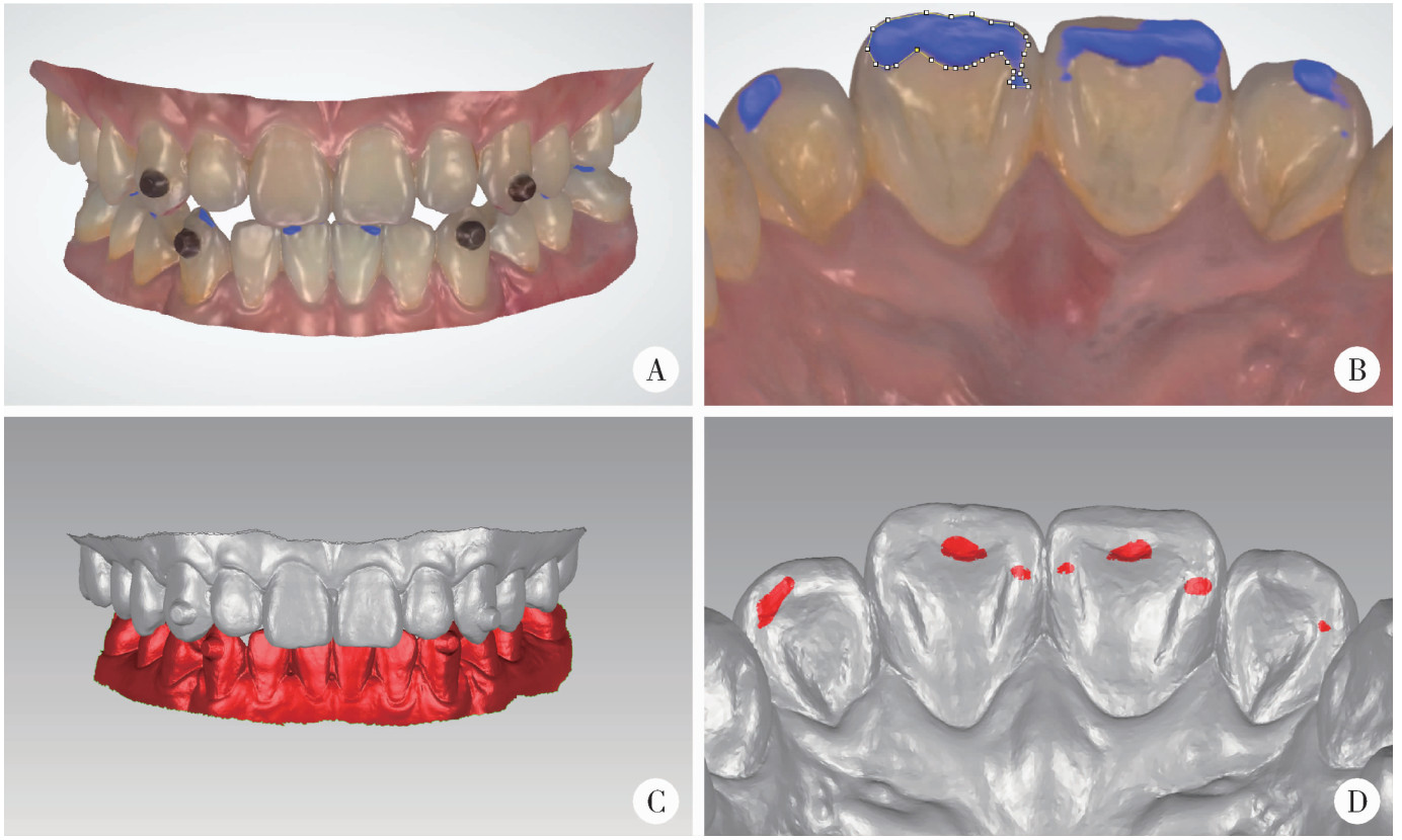
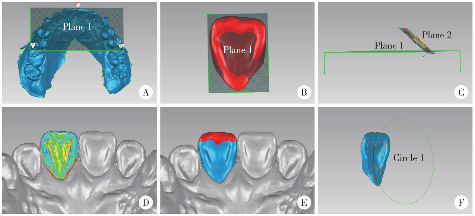
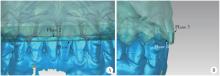
目的: 数字化精准测量分析上颌切牙前伸和正中咬合接触解剖特征, 建立标准化测量流程并获得特征性切导功能相关数据, 为优化修复体的切导设计提供参考。方法: 招募30名切导稳定的正常受试者, 口内扫描获得数字化牙列模型, 并采用改良动态咬合记录法获取前伸运动数据。通过计算机辅助设计软件重现前伸运动中的咬合接触区域并记录其分布, 使用图像分析软件测量各个牙位引导部位面积占比。通过逆向工程软件测量分析牙尖交错位咬合接触与解剖特征, 测量指标包括: 正中咬合接触面积占比与分布、边缘嵴与切嵴面积占比、舌面中央曲率半径、舌面倾斜度与覆牙合、覆盖。以上每项指标重复测量两次, 计算组内相关系数以评价复测信度。结果: 所有指标的复测信度良好, 且在双侧同名牙之间的差异均无统计学意义(P>0.05)。前伸运动时, 中切牙引导部位面积占比显著大于侧切牙(73.4%±12.3% vs. 26.6%±12.3%, P < 0.001), 近远中边缘嵴与切嵴发生咬合接触的频率显著高于舌窝和舌隆突(P < 0.05)。牙尖交错位时, 中切牙与侧切牙咬合接触面积占比差异无统计学意义(48.8%±20.0% vs. 51.2%±20.0%, P=0.758), 近远中边缘嵴发生咬合接触的频率显著高于切嵴、舌窝和舌隆突(P < 0.05)。中切牙的覆牙合、覆盖显著大于侧切牙(P < 0.05), 且近远中边缘嵴面积占比显著小于侧切牙(P < 0.05), 但切嵴面积占比在中切牙和侧切牙之间差异无统计学意义(P>0.05)。不同牙位的舌面倾斜度与舌面中央曲率半径差异均无统计学意义(P>0.05)。结论: 上颌切牙前伸和正中咬合接触解剖特征表现出左右对称性。前伸运动时以上颌中切牙引导为主, 中切牙的引导面积约为侧切牙的3倍, 近远中边缘嵴与切嵴是主要的引导部位; 正中咬合时上颌中切牙与侧切牙有相近的咬合接触面积。

关键词: 牙测量学, 切牙, 牙牙合, 成像, 三维

Abstract:

Objective: To digitally measure and analyze the anatomical characteristics of protrusive and intercuspal position (ICP) occlusal contacts in maxillary incisors, thereby establishing a standardized measurement protocol and obtaining characteristic functional data to optimize the incisal guidance design of prostheses. Methods: Thirty subjects with stable incisal guidance were recruited. Digital dental mo-dels were acquired via intraoral scanning, and protrusive movement data were captured using a modified patient-specific motion (PSM) technique. Computer-aided design software was used to record the distribution of the occlusal contacts during protrusive movement. Image analysis software was employed to measure the area proportion of guiding locations for each tooth. Reverse engineering software was used to measure and analyze the occlusal contacts in ICP and anatomical characteristics. Measured parameters included the area proportion and distribution of occlusal contacts in ICP, the area proportion of marginal ridges and incisal ridges, radius of curvature of lingual surface, lingual surface inclination, overbite, and overjet. Each parameter was measured twice to calculate the intraclass correlation coefficient for the assessment of test-retest reliability. Results: All measured parameters demonstrated good test-retest reliability. No significant differences were found in any parameters between homologous teeth (P>0.05). During protrusive movement, the area proportion of guiding locations was significantly larger for the central incisors than for the lateral incisors (73.4%±12.3% vs. 26.6%±12.3%, P < 0.001). The frequency of occlusal contacts was significantly higher on the mesial and distal marginal ridges and incisal ridges compared with the lingual fossa and cingulum (P < 0.05). In ICP, no significant difference was observed in the occlusal contact area proportion between the central and lateral incisors (48.8%±20.0% vs. 51.2%±20.0%, P=0.758). The frequency of the occlusal contact was significantly higher on the mesial and distal marginal ridges compared with the incisal ridge, lingual fossa, and cingulum (P < 0.05). Central incisors exhibited significantly higher overbite and overjet than lateral incisors (P < 0.05). The area proportion of mesial and distal marginal ridges was significantly smaller for the central incisors than for the lateral incisors (P < 0.05), but no significant difference was observed in the incisal ridge (P>0.05). No significant differences were observed in the lingual surface inclination or radius of curvature among the incisors (P>0.05). Conclusion: The anatomical characteristics of protrusive and ICP occlusal contacts in maxillary incisors demonstrated bilateral symmetry. Protrusive movement was primarily guided by the maxillary central incisors, with the guiding area of the central incisors being approximately three times that of the lateral incisors. The marginal ridges and incisal ridges were the main guiding locations. Central and lateral incisors exhibited comparable occlusal contact area in ICP.

Key words: Odontometry, Incisor, Dental occlusion, Imaging, three-dimensional

中图分类号: 

  • R783

图1

粘接标志物"

图2

前伸和正中咬合接触的测量"

图3

切导相关舌面特征的测量"

图4

覆牙合覆盖的测量"

表1

上颌切牙的前伸和正中咬合接触面积占比(%)"

Items Central incisor Lateral incisor
Left Right t P Left Right t P
Protrusive movement (n=30) 39.3±10.1 34.1±11.2 -1.632 0.113 15.0±7.5 11.7±8.0 -1.953 0.061
Intercuspal position (n=28) 26.2±14.8 22.6±14.5 -0.896 0.378 29.2±15.2 22.0±15.2 -1.332 0.194

表2

舌面解剖结构上的咬合接触分布频率(%,n=30)"

Items Central incisor Lateral incisor
Left Right Left Right
Protrusive movement
  Mesial marginal ridge 86.2 79.3 86.2 79.3
  Distal marginal ridge 75.9 62.1 69.0 44.8
  Incisal ridge 93.1 89.7 86.2 69.0
  Lingual fossa 24.1 24.1 3.5 6.9
  Lingual cingulum 0 3.5 0 0
  No occlusal contact 0 0 3.5 3.5
Intercuspal position
  Mesial marginal ridge 80.0 76.7 73.3 76.7
  Distal marginal ridge 70.0 56.7 53.3 40
  Incisal ridge 13.3 10.0 30.0 26.7
  Lingual fossa 20.0 20.0 13.3 3.3
  Lingual cingulum 3.3 0 0 0
  No occlusal contact 10.0 13.3 13.3 16.7

表3

切导相关上颌切牙的解剖特征(n=30)"

Items Central incisor Lateral incisor F P
Left Right Left Right
Area proportion/%
  Mesial marginal ridge 20.2±3.0 20.2±2.8 25.3±4.3 25.9±4.4 28.263 < 0.001
  Distal marginal ridge 21.5±2.8 20.6±2.7 24.3±4.3 22.9±3.8 9.990 < 0.001
  Incisal ridge 20.6±3.1 21.5±3.2 21.8±3.8 21.8±2.5 1.390 0.252
Lingual surface inclination/(°) 41.87±5.87 41.36±5.87 41.23±6.92 41.76±7.62 0.214 0.886
Radius of curvature of lingual surface/mm 6.97±2.28 7.34±2.70 6.16±2.04 6.40±1.49 2.135 0.102
Overbite/mm 3.05±0.92 3.18±0.84 2.63±0.78 2.60±0.85 10.378 < 0.001
Overjet/mm 3.12±1.06 3.33±1.12 2.66±1.04 2.90±0.91 9.671 < 0.001
1
The glossary of prosthodontic terms 2023: Tenth edition[J]. J Prosthet Dent, 2023, 130(4 Suppl 1): e7-e126.
2
谢秋菲. 临床牙合学——成功修复指导[M]. 北京: 科学出版社, 2012.
3
王美青. 牙合学[M]. 4版 北京: 人民卫生出版社, 2020.
4
Feng Y , Sun X , Li J , et al. Is dynamic occlusal design necessary for anterior guidance recovery in the computer-aided design process? An in vitro study[J]. J Dent, 2024, 142, 104833.

doi: 10.1016/j.jdent.2024.104833
5
Lepidi L , Grande F , Kim BC , et al. Dynamic modelling of the anterior guidance slope using a jaw tracking system in a fully digi-tal CAD-CAM workflow[J]. J Prosthet Dent, 2025, S0022-S3913 (25): 00373- 7.
6
Bauer W , van den Hoven F , Diedrich P . Wear in the upper and lower incisors in relation to incisal and condylar guidance[J]. J Orofac Orthop, 1997, 58 (6): 306- 319.

doi: 10.1007/BF02682671
7
Kulmer S , Ruzicka B , Niederwanger A , et al. Incline and length of guiding elements in untreated naturally grown dentition[J]. J Oral Rehabil, 1999, 26 (8): 650- 660.

doi: 10.1046/j.1365-2842.1999.00430.x
8
Lulla SS , Adaki R , Samant A , et al. Evaluation of influence of incisal guidance on the force exerted at the maximum possible intercuspal position and in protrusion: An in vivo study[J]. J Pharm Bioallied Sci, 2023, 15 (Suppl 2): S931- S933.

doi: 10.4103/jpbs.jpbs_284_23
9
Çakmak G , Cho JH , Choi J , et al. Can deep learning-designed anterior tooth-borne crown fulfill morphologic, aesthetic, and functional criteria in clinical practice?[J]. J Dent, 2024, 150, 105368.

doi: 10.1016/j.jdent.2024.105368
10
Tiwari S , Santosh A , Ramegowda S , et al. An analysis of the palatal surface anatomy of maxillary central incisors: Complexity behind the simplicity[J]. Int J Esthet Dent, 2021, 16 (4): 570- 578.
11
Botzenhart UU , Henningsen A , Quaas S , et al. 3D assisted morphological analysis of lingual upper central and lateral incisor surfaces[J]. Biomed Tech (Berl), 2012, 57 (1): 71- 77.

doi: 10.1515/bmt-2011-0057
12
Donegan SJ , Knap FJ . A study of anterior guidance[J]. J Pros-thodont, 1995, 4 (4): 226- 232.
13
Knösel M , Jung K , Gripp-Rudolph L , et al. Changes in incisor third-order inclination resulting from vertical variation in lingual bracket placement[J]. Angle Orthod, 2009, 79 (4): 747- 754.

doi: 10.2319/072308-385.1
14
Hasegawa Y , Ezura A , Nomintsetseg B . The relationship between the incisor position and lingual surface morphology in normal occlusion[J]. Odontology, 2017, 105 (1): 84- 90.

doi: 10.1007/s10266-016-0240-y
15
Chughtai AM , Jeelani W , Ahmed M , et al. Correlation between faciolingual inclination of maxillary central incisors and molars with maxillary and mandibular morphology[J]. J Ayub Med Coll Abbottabad, 2024, 36 (1): 29- 34.

doi: 10.55519/JAMC-01-12439
16
张瑞, 薛绯, 张勇, 等. 口腔扫描数字化测量法在评估膜龈手术疗效中的可靠性及有效性研究[J]. 实用口腔医学杂志, 2021, 37 (3): 357- 361.
17
金春晓, 娄梦伟, 蔡新杰, 等. 美学区牙齿二维口内照片与三维数字模型的测量对比研究[J]. 中华口腔医学杂志, 2024, 59 (6): 565- 570.
18
Nishine M , Kobayashi H , Ito K , et al. Comparison between interocclusal registration using silicone bite registration material and intraoral scanner on clenching strength[J]. Int J Prosthodont, 2024, 37 (4): 457- 459.

doi: 10.11607/ijp.8445
19
Rovira-Lastra B , Khoury-Ribas L , Flores-Orozco EI , et al. Accuracy of digital and conventional systems in locating occlusal contacts: A clinical study[J]. J Prosthet Dent, 2024, 132 (1): 115- 122.

doi: 10.1016/j.prosdent.2023.06.036
20
Lee YC , Lee C , Shim JS , et al. Comparison between occlusal errors of single posterior crowns adjusted using patient specific motion or conventional methods[J]. Appl Sci (Basel), 2020, 10, 9140.

doi: 10.3390/app10249140
21
Jirajariyavej B , Ounvorawong P , Pornprasertsuk-Damrongsri S , et al. Accuracy of individual mandibular motion records using intra-oral scanner for fixed implant- supported prosthesis designs: A comparative study[J]. BMC Oral Health, 2025, 25 (1): 878.

doi: 10.1186/s12903-025-06282-x
22
Li L , Chen H , Li W , et al. Design of wear facets of mandibular first molar crowns by using patient-specific motion with an intraoral scanner: A clinical study[J]. J Prosthet Dent, 2023, 129 (5): 710- 717.

doi: 10.1016/j.prosdent.2021.06.048
23
Wang H , Shao L , Sun J , et al. A fully digital workflow to design anterior guidance for an implant-supported single crown using a modified patient-specific motion technique[J]. J Prosthet Dent, 2025, 134 (4): 947- 951.

doi: 10.1016/j.prosdent.2024.01.036
24
李穗, 马雯洁, 王时敏, 等. 上前牙种植单冠修复体切导的数字化设计正确度[J]. 北京大学学报(医学版), 2024, 56 (1): 81- 87.

doi: 10.19723/j.issn.1671-167X.2024.01.013
25
Tu Y , Zhao X , Zhao G , et al. Assessment of physiological posterior-tooth displacement under habitual occlusal force by intraoral scanning using implant-supported crowns as the reference[J]. J Prosthet Dent, 2025, 133 (5): 1260- 1267.

doi: 10.1016/j.prosdent.2023.06.019
26
Zhao Z , Wang Q , Li J , et al. Occlusal contact characteristics of molar teeth with food impaction: Insights from a new digital technique[J]. J Dent, 2024, 147, 105133.

doi: 10.1016/j.jdent.2024.105133
27
谢秋菲, 张磊. 牙体解剖与口腔生理学[M]. 3版 北京: 北京大学医学出版社, 2021.
28
Lee W , Lim YJ , Kim MJ , et al. Occlusal consequence of using average condylar guidance settings: An in vitro study[J]. J Prosthet Dent, 2017, 117 (4): 532- 538.

doi: 10.1016/j.prosdent.2016.09.014
29
王富, 牛丽娜, 陈吉华. 数字化咬合分析的方案与效能[J]. 中华口腔医学杂志, 2025, 60 (8): 822- 828.
[1] 吴为良,曾筱,刘晓强,谭建国. 120例中国成年人上前牙美学比例分析[J]. 北京大学学报(医学版), 2020, 52(6): 1130-1134.
[2] 王鹏,李大军,刘建彰. 上颌前牙宽度、前牙弓周长与前牙弓深度的相关性研究[J]. 北京大学学报(医学版), 2020, 52(1): 124-128.
[3] 李琳琳,赵一姣,陈虎,王勇,孙玉春. 转移牙合架固定法三维重建牙尖交错牙合的精度评价[J]. 北京大学学报(医学版), 2020, 52(1): 138-143.
[4] 萧宁,孙玉春,赵一姣,王勇. 三种数字化分析算法测量咬合接触分布及面积的对比研究[J]. 北京大学学报(医学版), 2020, 52(1): 144-151.
[5] 刘敏,章君荡,叶红强,赵一姣,赵旭斌,赵文艳,刘云松,周永胜. Smile Lite MDP便携摄影系统在前牙美学摄影中的应用[J]. 北京大学学报(医学版), 2020, 52(1): 187-192.
[6] 罗强,丁茜,张磊,谢秋菲. 后牙种植冠桥修复后局部咬合变化的定量分析[J]. 北京大学学报(医学版), 2019, 51(6): 1119-1123.
[7] 朱曚曚,李应龙,潘洁. 茶溶液着色离体牛切牙的着色效果[J]. 北京大学学报(医学版), 2018, 50(6): 1083-1087.
[8] 程明轩,姜婷,孙玉春,张皓羽. 比较口内扫描和模型扫描对数字化牙列模型咬合定量分析的影响[J]. 北京大学学报(医学版), 2018, 50(1): 136-140.
[9] 郑旭,胡兴学,马宁,陈晓红. 正畸矫治牙性牙合平面倾斜的新方法——波浪形弓[J]. 北京大学学报(医学版), 2017, 49(1): 176-180.
[10] 杨璇,乐迪,张艳玲,梁凌智,杨刚,胡文杰. 汉族青年上中切牙牙冠外形分类与龈乳头充满的相关性研究[J]. 北京大学学报(医学版), 2016, 48(5): 866-870.
[11] 刘存瑞, 徐啸翔, 曹烨, 谢秋菲. 咬合干扰时间因素对大鼠咀嚼肌机械痛觉敏感的影响[J]. 北京大学学报(医学版), 2016, 48(1): 51-56.
[12] 王智,邹立东△. 锥形束CT对切牙管及其相对位置关系的测量分析[J]. 北京大学学报(医学版), 2015, 47(6): 994-999.
[13] 赵莹,董颖韬,王晓燕,王祖华,李刚,刘木青,傅开元. 4 674颗下颌前牙根管构型的锥形束CT分析[J]. 北京大学学报(医学版), 2014, 46(1): 95-99.
[14] 曹洁, 胡文杰, 张豪, 柳登高, 乐迪. 基于锥形束计算机体层摄影术测量牙龈厚度[J]. 北京大学学报(医学版), 2013, 45(1): 135-139.
[15] 张豪, 乐迪 , 胡文杰 , 曹占强, 张艳玲. 120例中国青年健康上前牙牙龈曲线形状特征分析[J]. 北京大学学报(医学版), 2013, 45(1): 54-58.
Viewed
Full text


Abstract

Cited

  Shared   
  Discussed   
No Suggested Reading articles found!