北京大学学报(医学版) ›› 2026, Vol. 58 ›› Issue (1): 153-159. doi: 10.19723/j.issn.1671-167X.2026.01.020

• 论著 • 上一篇    下一篇

1 812例唾液腺结石患者的人口学特征和临床特点

杨雨婷, 屈留洋, 郑丹妮, 凌晓彤, 许晓韵, 柳登高*()   

  1. 北京大学口腔医学院 · 口腔医院医学影像科,国家口腔医学中心,国家口腔疾病临床医学研究中心,口腔生物材料和数字诊疗装备国家工程研究中心,口腔数字医学北京市重点实验室,国家卫生健康委员会口腔数字医学重点实验室,国家药品监督管理局口腔材料重点实验室,北京 100081
  • 收稿日期:2025-07-22 出版日期:2026-02-18 发布日期:2025-11-28
  • 通讯作者: 柳登高

Demographic characteristic and clinical features in 1 812 patients with salivary gland stones

Yuting YANG, Liuyang QU, Danni ZHENG, Xiaotong LING, Xiaoyun XU, Denggao LIU*()   

  1. Department of Oral and Maxillofacial Radiology, Peking University School and Hospital of Stomatology & National Center for Stomatology & National Clinical Research Center for Oral Diseases & National Engineering Research Center of Oral Biomaterials and Digital Medical Devices & Beijing Key Laboratory of Digital Stomatology & NHC Key Laboratory of Digital Stomatology & NMPA Key Laboratory for Dental Materials, Beijing 100081, China
  • Received:2025-07-22 Online:2026-02-18 Published:2025-11-28
  • Contact: Denggao LIU

RICH HTML


摘要:

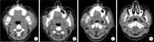
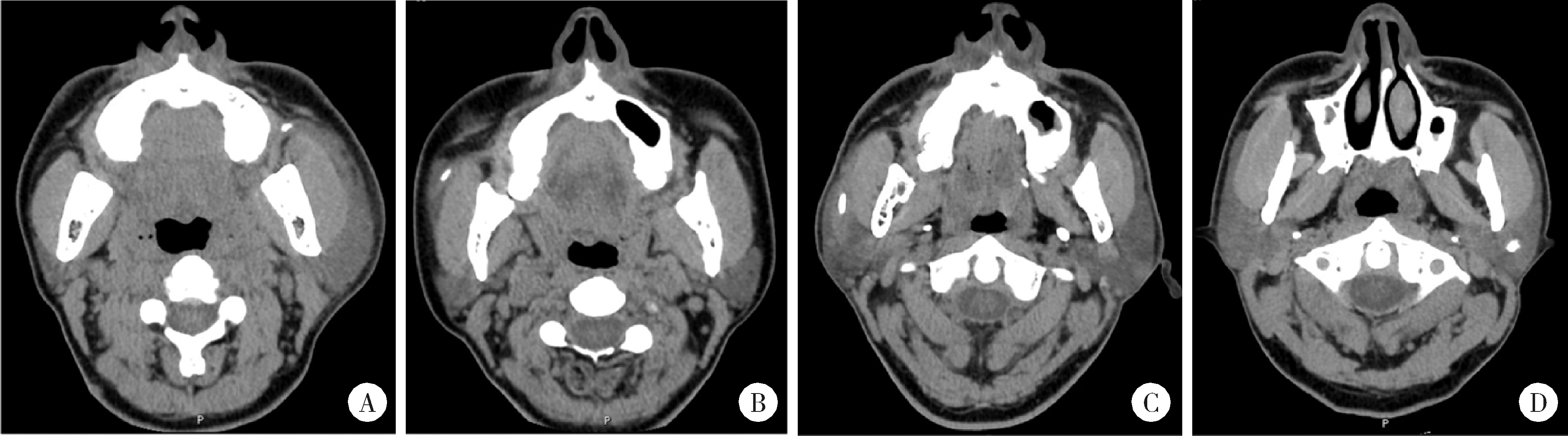
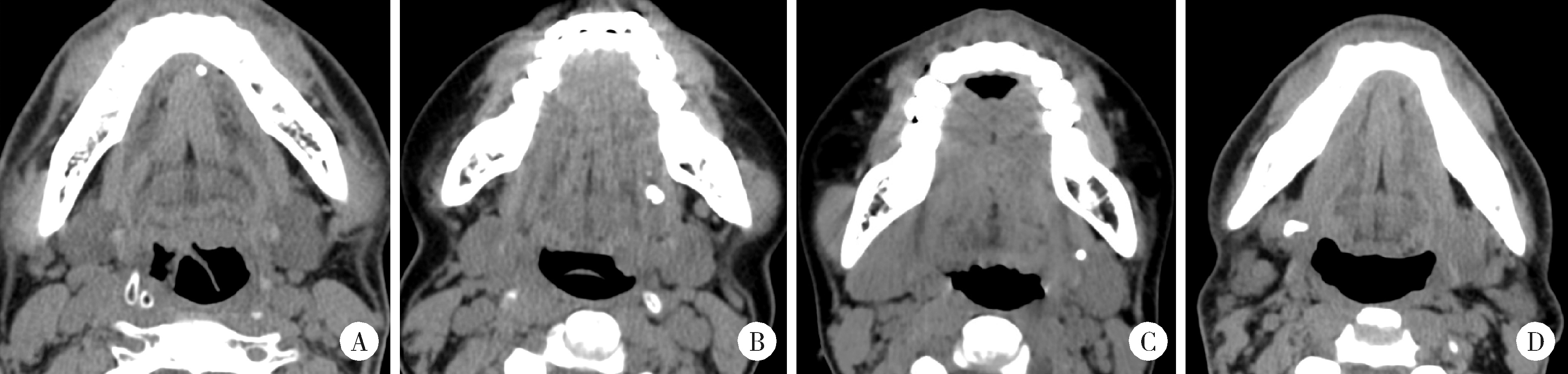
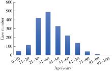
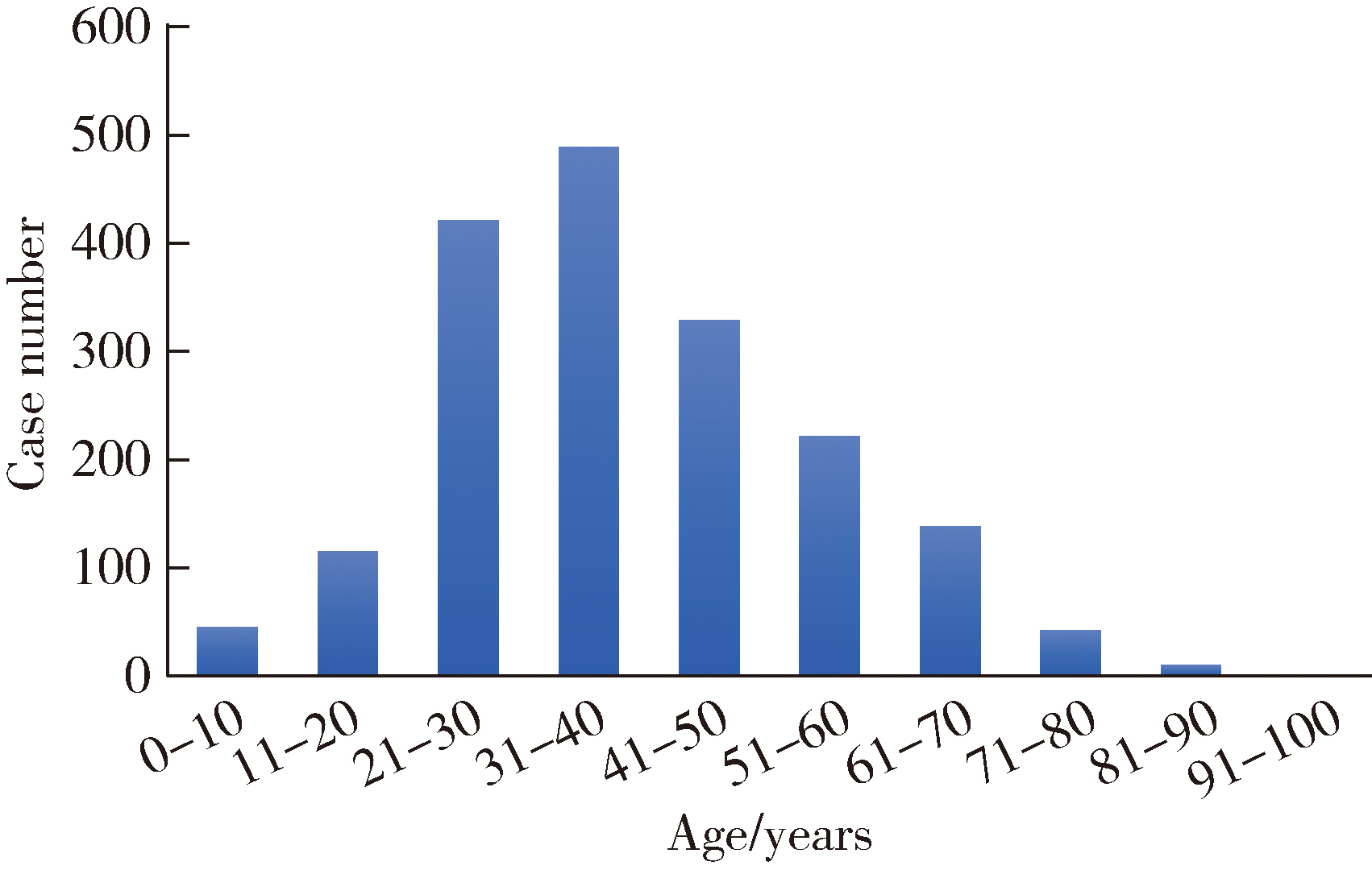
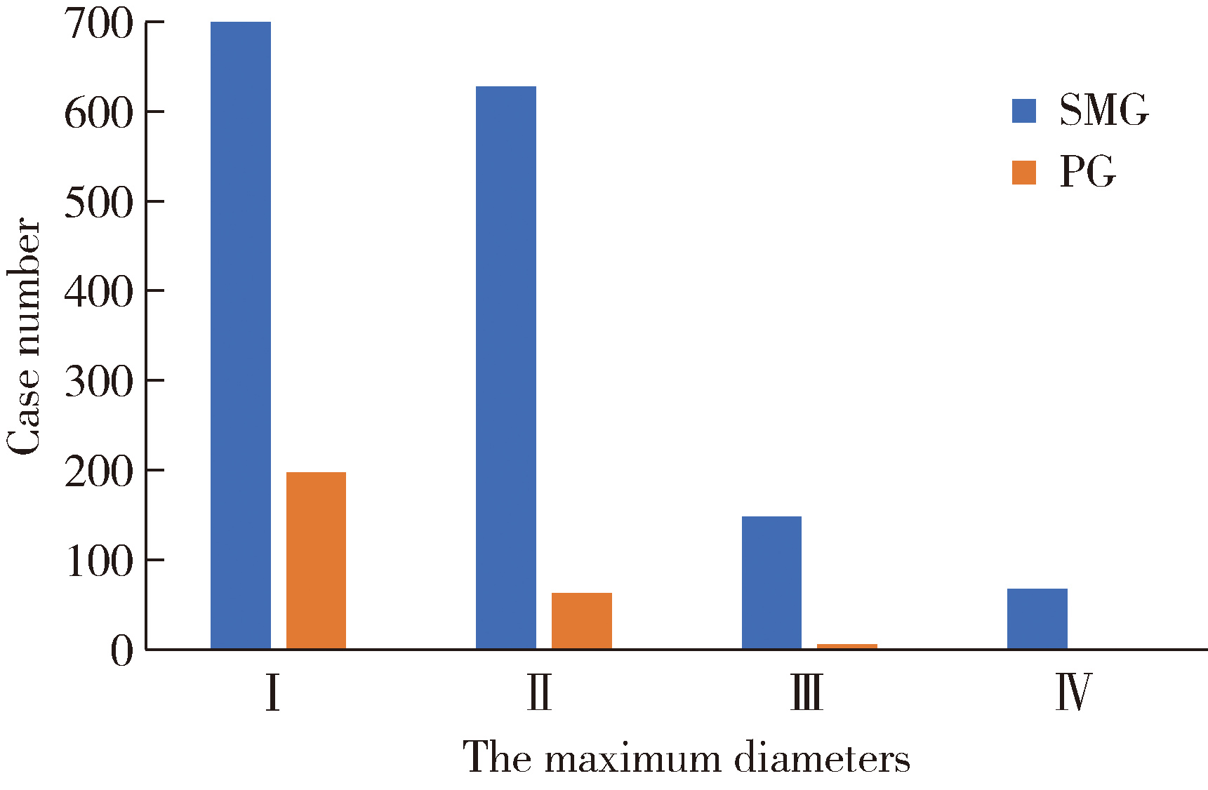
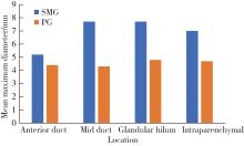
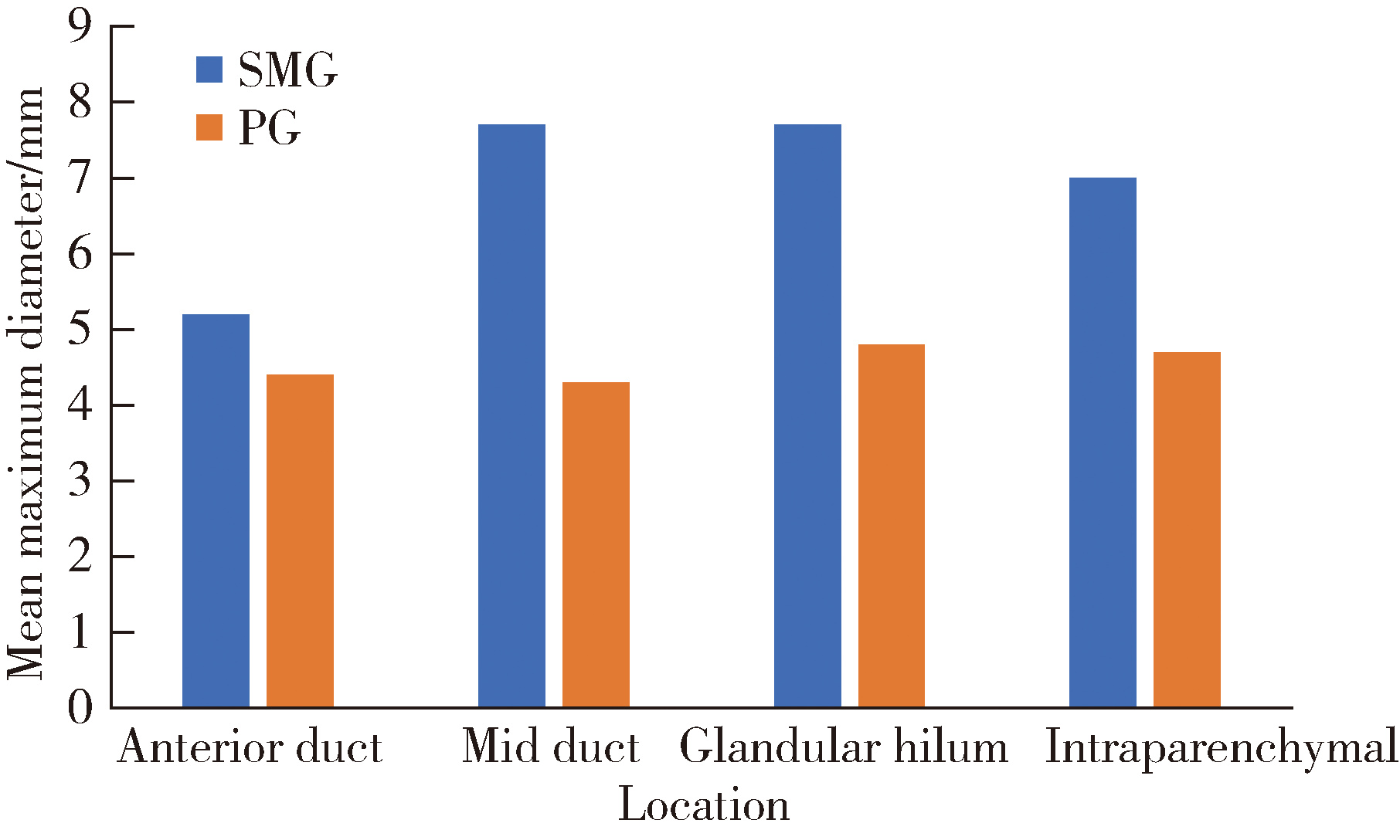
目的: 基于较大样本量及完善的临床与影像学资料,分析唾液腺结石病的人口学特征和临床发病特点。方法: 回顾性收集2020年1月至2024年12月于北京大学口腔医院唾液腺内镜中心就诊的唾液腺结石患者的临床及影像资料,统计患者的性别、年龄、病程及受累腺体,测量结石的数目、大小、位置及长短径之比,分析唾液腺结石病的人群特征、临床与影像学特点。结果: 共收集1 812例唾液腺结石患者,男性855例,女性957例,年龄4~97岁,平均年龄(39.0±15.0)岁,中位病程6.0个月。结石多累及单侧腺体(98.4%),多为单发(64.3%),平均直径(6.5±4.6) mm。本组病例包括下颌下腺结石1 541例(85.0%),腮腺结石267例(14.7%),其余4例患者的下颌下腺及腮腺均受累。下颌下腺结石多位于腺外主导管(50.2%),其次为腺门部(41.5%),仅少数位于腺内(8.2%);腮腺结石大部分位于腺外主导管(75.2%)。下颌下腺结石的平均直径显著大于腮腺结石[(6.9±4.8) mm vs. (4.5±2.5) mm],下颌下腺结石患者的平均年龄显著低于腮腺结石患者[(38.0±15.0)岁vs. (48.0±16.0)岁]。腮腺结石的复发率(3.4%)和下颌下腺结石的复发率(2.1%)都较低,复发间隔2个月至10年不等,平均(33.8±31.4)个月。本组病例中,21例(1.2%)患者的结石来源于异物钙化,包括9例下颌下腺结石和12例腮腺结石患者。结论: 唾液腺结石病好发于中年人群,无明显的性别差异,绝大多数为单侧腺体受累,单发结石较多见;下颌下腺结石与腮腺结石在好发部位、大小、结石形状(长宽比)及患者年龄等方面均存在明显差异;唾液腺结石的复发率较低,少数结石由异物钙化引起;这些发病特点,可为阐明唾液腺结石病的发病机制及优化诊疗策略提供依据。

关键词: 唾液腺结石, 腮腺, 下颌下腺, 影像学表现

Abstract:

Objective: To analyze the demographic and clinical features of sialolithiasis based on a large sample size. Methods: Clinical and imaging data of patients with sialolithiasis treated at the Sialendoscopy Center of Peking University School and Hospital of Stomatology from January 2020 to December 2024 were retrospectively collected. Patient demographics, disease duration, and affected glands were recorded. Moreover, the number, size, location, and long-to-short diameter ratio of the stones were mea-sured. Results: A total of 1 812 patients with salivary gland stones were collected, including 855 males and 957 females. The age range was 4 to 97 years [mean: (39.0±15.0) years] with a median disease duration of 6.0 months (interquartile range: 1.0-24.0). Most stones involved unilateral glands (98.4%), and 64.2% of the affected glands had a single stone. The stone size averaged at a diameter of (6.5±4.6) mm. The cases included 1 541 cases with submandibular gland (SMG) stones (85.0%), 267 cases with parotid gland (PG) stones (14.7%), and 4 cases affecting both SMG and PG. The SMG stones were mostly located in the extra-glandular main duct (50.2%), followed by the hilum (41.5%), and only 8.2% of them were in the intra-glandular area. The majority of PG stones (75.2%) were in the extra-glandular main duct. The average diameter of SMG stones was significantly larger than that of PG stones [(6.9±4.8) mm vs. (4.5±2.5) mm]. The average age of SMG patients was younger than that of PG patients [(38.0±15.0) years vs. (48.0±16.0) years]. There was no significant difference in recurrence rate between PG and SMG stones (3.4% vs. 2.1%). The recurrence intervals ranged from 2 months to 10 years, with an average of (33.8±31.4) months. In 21 patients (1.2%), the stones originated from foreign body calcification, which accounted for 0.6% (9/1 541) of SMG cases and 4.5% (12/267) of PG cases. Conclusion: Among the cases analyzed, sialolithiasis was the most common in middle-aged individuals, with no significant gender difference. Unilateral gland involvement was the most frequent, and 2/3 of the cases had a single stone. Significant differences were observed in the location, size, and patient age between SMG and PG stones. The recurrence rate of sialolithiasis was relatively low. Stones originated from foreign body calcification was occasionally seen. Understanding of these demographic and clinical characteristics provided a basis for clarifying the pathogenesis of sialolithiasis and optimizing the diagnostic and therapeutic strategies.

Key words: Salivary gland calculi, Parotid gland, Submandibular gland, Imaging characteristics

中图分类号: 

  • R781.75

图1

螺旋CT轴位图像显示腮腺导管系统结石"

图2

螺旋CT轴位图像显示下颌下腺导管系统结石"

表1

1 808例唾液腺结石病患者的临床特征"

Items Total (n=1 808)a Type of affected glandsP value
SMG PG
Number 1 808 1 541 267
Gender, n (%) 0.628
  Male 854 (47.2) 732 (47.5) 122 (45.7)
  Female 954 (52.8) 809 (52.5) 145 (54.3)
Age/years, $\bar x \pm s$ 39.0±15.0 38.0±15.0 48.0±16.0 <0.001
Duration of symptoms/month, M (P25, P75) 6.0 (1.0, 24.0) 6.0 (1.0, 24.0) 4.3 (1.0, 24.0) 0.302
Unilateral/Bilateral, n (%) 0.327
  Left 844 (46.7) 724 (47.0) 120 (44.9)
  Right 935 (51.7) 795 (51.6) 140 (52.4)
  Bilateral 29 (1.6) 22 (1.4) 7 (2.6)
Quantity, n (%) 0.062
  Single 1 162 (64.3) 974 (63.2) 188 (70.4)
  Multiple 621 (34.3) 545 (35.4) 76 (28.4)
  Recurrent 25 (1.4) 22 (1.4) 3 (1.1)
Location, n (%) <0.001
  Anterior duct 581 (32.1) 460 (29.8) 121 (45.3)
  Mid duct 394 (21.8) 315 (20.4) 79 (29.6)
  Glandular hilum 669 (37.0) 639 (41.5) 30 (11.2)
  Intraparenchymal 164 (9.1) 127 (8.2) 37 (13.9)
Mean maximum diameter/mm, $\bar x \pm s$ 6.5±4.6 6.9±4.8 4.5±2.5 <0.001
Long-to-short diameter ratio, $\bar x \pm s$ 1.6±1.0 1.6±0.9 1.7±1.1 0.007

图3

1 812例唾液腺结石病患者的年龄分布"

图4

1 808例患者中主要唾液腺结石的最大直径分布情况"

图5

下颌下腺和腮腺结石导管系统内不同位置结石的大小(n=1 808)"

1
Laforgia PD , Favia GF , Chiaravalle N , et al. Clinico-statistical, morphologic and microstructural analysis of 400 cases of sialolithiasis[J]. Minerva Stomatol, 1989, 38 (12): 1329- 1336.
2
Lustmann J , Regev E , Melamed Y . Sialolithiasis: A survey on 245 patients and a review of the literature[J]. Int J Oral Maxillofac Surg, 1990, 19 (3): 135- 138.

doi: 10.1016/S0901-5027(05)80127-4
3
Escudier MP , McGurk M . Symptomatic sialoadenitis and sialoli-thiasis in the English population, an estimate of the cost of hospital treatment[J]. Br Dent J, 1999, 186 (9): 463- 466.

doi: 10.1038/sj.bdj.4800141a
4
Nahlieli O , Eliav E , Hasson O , et al. Pediatric sialolithiasis[J]. Oral Surg Oral Med Oral Pathol Oral Radiol Endod, 2000, 90 (6): 709- 712.

doi: 10.1067/moe.2000.109075a
5
Williams MF . Sialolithiasis[J]. Otolaryngol Clin N Am, 1999, 32 (5): 819- 834.

doi: 10.1016/S0030-6665(05)70175-4
6
Kraaij S , Karagozoglu KH , Forouzanfar T , et al. Salivary stones: Symptoms, aetiology, biochemical composition and treatment[J]. Br Dent J, 2014, 217 (11): E23.

doi: 10.1038/sj.bdj.2014.1054
7
Zheng DN , Zhao YN , Zhang LQ , et al. Comparison of two transcutaneous approaches for the removal of impacted parotid stones[J]. Int J Oral Maxillofac Surg, 2023, 52 (6): 663- 669.

doi: 10.1016/j.ijom.2022.10.007
8
Zhao YN , Zheng DN , Qu LY , et al. Removal of intraglandular calculi in Wharton ' s duct: Clinical outcome and treatment algorithm[J]. Int J Oral Maxillofac Surg, 2024, 53 (5): 382- 388.

doi: 10.1016/j.ijom.2023.12.001
9
Levy DM , Remine WH , Devine KD . Salivary gland calculi. Pain, swelling associated with eating[J]. JAMA, 1962, 181, 1115- 1119.

doi: 10.1001/jama.1962.03050390017005
10
van den Akker HP , Busemann-Sokole E . Submandibular gland function following transoral sialolithectomy[J]. Oral Surg Oral Med Oral Pathol, 1983, 56 (4): 351- 356.

doi: 10.1016/0030-4220(83)90341-9
11
Sigismund PE , Zenk J , Koch M , et al. Nearly 3, 000 salivary stones: Some clinical and epidemiologic aspects[J]. Laryngoscope, 2015, 125 (8): 1879- 1882.

doi: 10.1002/lary.25377
12
Stimec B , Nikolic S , Rakocevic Z , et al. Symmetry of the submandibular glands in humans: A postmortem study assessing the linear morphometric parameters[J]. Oral Surg Oral Med Oral Pathol Oral Radiol Endod, 2006, 102 (3): 391- 394.

doi: 10.1016/j.tripleo.2005.10.063
13
McGurk M , Escudier MP , Brown JE . Modern management of salivary calculi[J]. Br J Surg, 2005, 92 (1): 107- 112.

doi: 10.1002/bjs.4789
14
Zenk J , Constantinidis J , Kydles S , et al. Clinical and diagnostic findings of sialolithiasis[J]. HNO, 1999, 47 (11): 963- 969.

doi: 10.1007/s001060050476
15
Zenk J , Koch M , Klintworth N , et al. Sialendoscopy in the diagnosis and treatment of sialolithiasis: A study on more than 1000 patients[J]. Otolaryngol Head Neck Surg, 2012, 147 (5): 858- 863.

doi: 10.1177/0194599812452837
16
Harrison JD . Causes, natural history, and incidence of salivary stones and obstructions[J]. Otolaryngol Clin North Am, 2009, 42 (6): 927- 947.

doi: 10.1016/j.otc.2009.08.012
17
McGurk M , Escudier MP , Brown E . Modern management of obstructive salivary gland disease[J]. Ann R Australas Coll Dent Surg, 2004, 17, 45- 50.
18
Resende EA , Gomes NR , Abreu LG , et al. The applicability of ultrasound in the diagnosis of inflammatory and obstructive diseases of the major salivary glands: A scoping review[J]. Dentomaxillofac Radiol, 2022, 51 (4): 20210361.

doi: 10.1259/dmfr.20210361
19
伊里夏提·卡米力. 颌下腺导管腺门结石形成的解剖学因素研究[D]. 乌鲁木齐: 新疆医科大学, 2014.
20
Sánchez Barrueco A , López-Acevedo Cornejo MV , Alcalá Rueda I , et al. Sialolithiasis: Mineralogical composition, crystalline structure, calculus site, and epidemiological features[J]. Br J Oral Maxillofac Surg, 2022, 60 (10): 1385- 1390.

doi: 10.1016/j.bjoms.2022.08.005
21
Teymoortash A , Buck P , Jepsen H , et al. Sialolith crystals localized intraglandularly and in the Wharton ' s duct of the human submandibular gland: An X-ray diffraction analysis[J]. Arch Oral Biol, 2003, 48 (3): 233- 236.

doi: 10.1016/S0003-9969(02)00211-X
22
Aldelaimi AA , Enezei HH , Aldelaimi TN , et al. Salivary gland diseases: A retrospective clinicopathological study of 159 cases[J]. Cureus, 2022, 14 (9): e29589.
23
Raksin SZ , Gould SM , Williams AC . Submandibular duct sialolith of unusual size and shape[J]. J Oral Surg, 1975, 33 (2): 142- 145.
24
Gupta A , Rattan D , Gupta R . Giant sialoliths of submandibular gland duct: Report of two cases with unusual shape[J]. Contemp Clin Dent, 2013, 4 (1): 78- 80.

doi: 10.4103/0976-237X.111599
25
Harrill JA , King JS Jr , Boyce WH . Structure and composition of salivary calculi[J]. Trans Am Laryngol Rhinol Otol Soc, 1959, 1959, 496- 507.
26
Capaccio P , Torretta S , Ottaviani F , et al. Modern management of obstructive salivary diseases[J]. Acta Otorhinolaryngol Ital, 2007, 27 (4): 161- 172.
27
Marchal F , Kurt AM , Dulguerov P , et al. Retrograde theory in sialolithiasis formation[J]. Arch Otolaryngol Head Neck Surg, 2001, 127 (1): 66- 68.

doi: 10.1001/archotol.127.1.66
28
Gill AS , Kieliszak CR , Joshi AS . Sialendoscopy as a management tool in patients with foreign body impaction of the salivary gland[J]. Am J Otolaryngol, 2016, 37 (4): 369- 371.

doi: 10.1016/j.amjoto.2016.02.008
29
Xie L , Zheng L , Yu C , et al. Foreign body induced sialolithiasis treated by sialoendoscopic intervention[J]. J Craniofac Surg, 2014, 25 (4): 1372- 1375.

doi: 10.1097/SCS.0000000000000851
30
Fabie JE , Kompelli AR , Naylor TM , et al. Gland-preserving surgery for salivary stones and the utility of sialendoscopes[J]. Head Neck, 2019, 41 (5): 1320- 1327.

doi: 10.1002/hed.25560
[1] 杨源源, 张珊珊, 俞光岩, 杨辉俊, 杨宏宇. 部分下颌下腺切除术治疗下颌下腺良性肿瘤的临床效果[J]. 北京大学学报(医学版), 2025, 57(2): 334-339.
[2] 杨玉淑, 齐晅, 丁萌, 王炜, 郭惠芳, 高丽霞. 抗唾液腺蛋白1抗体联合抗腮腺分泌蛋白抗体对干燥综合征的诊断价值[J]. 北京大学学报(医学版), 2024, 56(5): 845-852.
[3] 柳登高,郑丹妮,赵雅宁,张亚琼,叶欣,张丽琪,谢晓艳,张雷,张祖燕,俞光岩. 疑难唾液腺结石病的治疗研究[J]. 北京大学学报(医学版), 2023, 55(1): 8-12.
[4] 俞光岩,苏家增,柳登高,吴立玲,丛馨. 下颌下腺保存治疗新技术体系的建立与应用[J]. 北京大学学报(医学版), 2022, 54(5): 842-845.
[5] 李炳雨,唐祖南,胡耒豪,章文博,于尧,俞光岩,彭歆. 腮腺微小肿瘤的临床病理研究[J]. 北京大学学报(医学版), 2022, 54(2): 335-339.
[6] 陈超伦,苏家增,俞光岩. 酸刺激对腮腺和下颌下腺唾液流率及成分的影响[J]. 北京大学学报(医学版), 2022, 54(1): 89-94.
[7] 俞光岩,柳登高,李巍,洪霞,张严妍,朱文瑄,张可夫,李潇,栗占国,刘燕鹰,陈艳,高岩,苏家增. 3类新型慢性唾液腺炎的诊断和治疗[J]. 北京大学学报(医学版), 2022, 54(1): 13-17.
[8] 朱忆颖,闵赛南,俞光岩. 局部注射环孢素A对非肥胖糖尿病小鼠下颌下腺分泌功能及炎症的影响[J]. 北京大学学报(医学版), 2021, 53(4): 750-757.
[9] 王怡平,蔡志刚,彭歆,张杰,孙志鹏,李巍,张雷,俞光岩. 下颌下腺质量和体积的实体体外检测[J]. 北京大学学报(医学版), 2021, 53(1): 126-132.
[10] 俞光岩. 多发性唾液腺肿大的鉴别诊断及处理[J]. 北京大学学报(医学版), 2021, 53(1): 1-4.
[11] 李玉冰,孙丽莎,孙志鹏,谢晓艳,张建运,张祖燕,赵燕平,马绪臣. 腮腺CT影像报告与数据系统的初步研究[J]. 北京大学学报(医学版), 2020, 52(1): 83-89.
[12] 丛馨,闵赛南,吴立玲,蔡志刚,俞光岩. 激活毒蕈碱乙酰胆碱受体调控下颌下腺分泌的机制研究[J]. 北京大学学报(医学版), 2019, 51(3): 390-396.
[13] 宿骞,彭歆,周传香,俞光岩. 原发性腮腺淋巴瘤的临床病理特点及预后分析[J]. 北京大学学报(医学版), 2019, 51(1): 35-42.
[14] 俞光岩,洪霞,李巍,张严妍,高岩,陈艳,张祖燕,谢晓艳,栗占国,刘燕鹰,苏家增,朱文瑄,孙志鹏. IgG4相关唾液腺炎的临床病理特点及诊断[J]. 北京大学学报(医学版), 2019, 51(1): 1-3.
[15] 俞光岩,吴立玲,蔡志刚,吕岚,丛馨. 血管化自体下颌下腺移植治疗重症干眼20年研究[J]. 北京大学学报(医学版), 2018, 50(1): 1-4.
Viewed
Full text


Abstract

Cited

  Shared   
  Discussed   
No Suggested Reading articles found!