北京大学学报(医学版) ›› 2020, Vol. 52 ›› Issue (1): 144-151. doi: 10.19723/j.issn.1671-167X.2020.01.023
三种数字化分析算法测量咬合接触分布及面积的对比研究
萧宁,孙玉春,赵一姣( ),王勇()
- 北京大学口腔医学院·口腔医院,口腔医学数字化研究中心,口腔修复教研室 国家口腔疾病临床医学研究中心口腔数字化医疗技术和材料国家工程实验室 口腔数字医学北京市重点实验室,北京 100081
Preliminary study on three digital analysis methods for analyzing the distribution and area of occlusal contacts
Ning XIAO,Yu-chun SUN,Yi-jiao ZHAO(),Yong WANG()
- Center of Digital Dentistry, Department of Prosthodontics, Peking University School and Hospital of Stomatology & National Clinical Research Center for Oral Diseases & National Engineering Laboratory for Digital and Material Technology of Stomatology & Beijing Key Laboratory of Digital Stomatology, Beijing 100081, China
摘要:
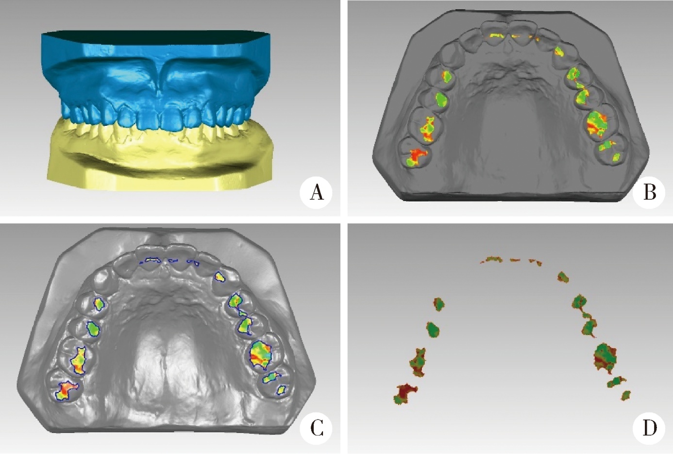
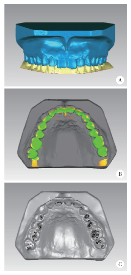
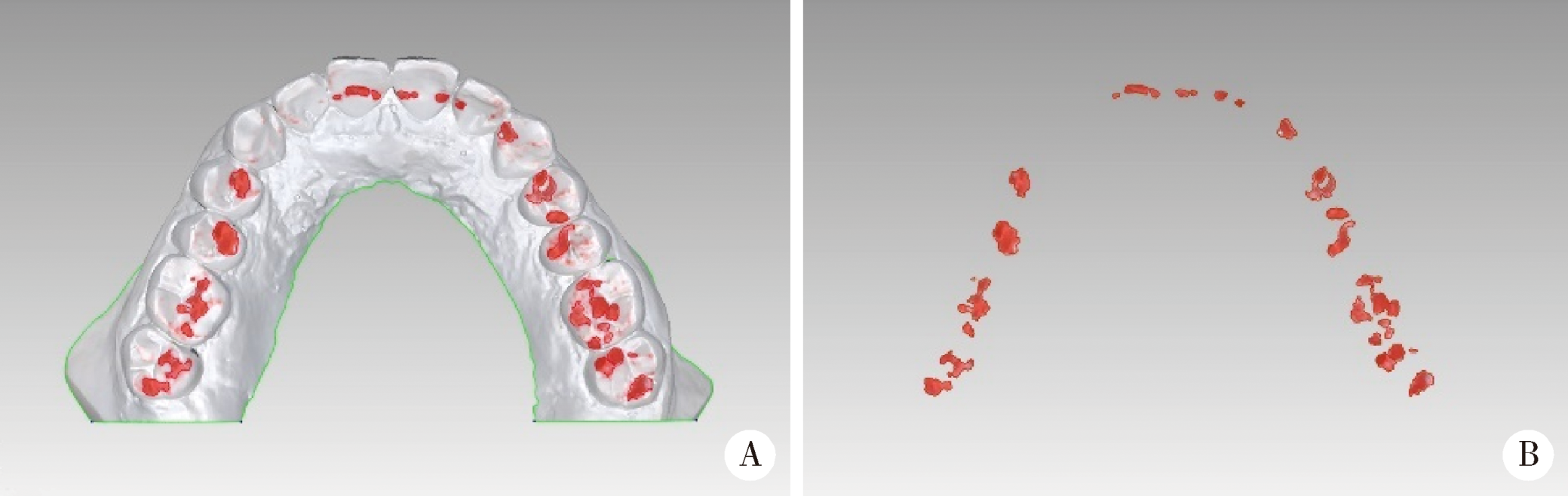
目的:研究三种数字化分析算法测量石膏牙颌模型三维咬合接触分布及面积的检测效果,并与传统咬合分析方法进行比较,探究各数字化分析算法的特点和应用。方法:选取一副正常受试者的上、下颌石膏牙颌模型,使用3shape E4牙颌模型三维扫描仪进行数字化扫描得到数字模型,在三维测量分析软件Geomagic Studio 2013及Geomagic Qualify 2013中采用“三维偏差色阶图法”、“点云统计分析法”和“虚拟咬合纸法”三种数字化分析算法获得相应的三维咬合接触分布及面积,同时使用牙合记录硅橡胶法及咬合纸扫描法两种传统咬合分析方法获得咬合接触分布和面积。各方法的咬合检测阈值为100 μm,量化评价各数字化分析算法与传统咬合分析方法的检测结果。结果:上述五种方法所得的全牙列咬合接触分布的定性评价结果基本一致,三维偏差色阶图法、点云统计分析法、虚拟咬合纸法、牙合记录硅橡胶法和咬合纸扫描法所得到的总咬合接触面积分别为133.10 mm 2、142.08 mm 2、128.95 mm 2、163.31 mm 2、100.55 mm 2。三种数字化分析算法间的检测结果差异性不大,数字化方法与传统方法检测的总咬合接触面积有一定差异。结论:三种数字化分析算法均可提供较为可靠、准确的牙颌模型咬合接触分布及面积量化分析结果,可为口腔临床修复体数字化设计制作及咬合分析提供参考。
中图分类号:
- R783



| [1] | 马斐斐, 胡秀莲, 林野 . 口腔种植修复与咬合[J]. 实用口腔医学杂志, 2013,29(1):121-123. |
| [2] | Foz AM, Artese HP, Horliana AC , et al. Occlusal adjustment associated with periodontal therapy: A systematic review[J]. J Dent, 2012,40(12):1025-1035. |
| [3] | 曾艳, 王嘉德 . 牙体牙髓病临床问题解析Ⅱ. 牙齿的慢性损伤性疾病[J]. 中华口腔医学杂志, 2009,44(7):441-443. |
| [4] | 谢秋菲 . 牙体解剖与口腔生理学 [M]. 北京: 北京大学医学出版社, 2013. |
| [5] | Abduo J, Bennamoun M, Tennant M , et al. Effect of prosthodontic planning on intercuspal occlusal contacts: Comparison of digital and conventional planning[J]. Comput Biol Med, 2015,60:143-150. |
| [6] | Moreno-Hay I, Okeson JP . Does altering the occlusal vertical dimension produce temporomandibular disorders? A literature review[J]. J Oral Rehabil, 2015,42(11):875-882. |
| [7] | 韩科, 张豪 . 牙合学理论与临床实践 [M]. 北京: 人民军医出版社, 2014. |
| [8] | 宋倩, 王辉, 冯春雷 , 等. 咬合纸指导调牙合可靠性的定量研究[J]. 牙体牙髓牙周病学杂志, 2016,26(2):86-90. |
| [9] | Koos B, Godt A, Schille C , et al. Precision of an instrumentation-based method of analyzing occlusion and its resulting distribution of forces in the dental arch[J]. J Orofac Orthop, 2010,71(6):403-410. |
| [10] | Forrester SE, Presswood RG, Toy AC , et al. Occlusal measurement method can affect SEMG activity during occlusion[J]. J Oral Rehabil, 2011,38(9):655-660. |
| [11] | 赵一姣, 王勇, 吕培军 . 一种基于数字化牙颌模型的三维咬合分析方法[J]. 北京大学学报(医学版), 2008,40(1):109-111. |
| [12] | Gintaute A, Keeling AJ, Osnes CA , et al. Precision of maxillo-mandibular registration with intraoral scanners in vitro [J]. J Prosthodont Res, 2019, pii: S1883- 1958(19) 30145-8. doi: 10.1016/j.jpor.2019.05.006.[Epub ahead of print]. |
| [13] | Lee H, Cha J, Chun YS , et al. Comparison of the occlusal contact area of virtual models and actual models: a comparative in vitro study on Class Ⅰ and Class Ⅱ malocclusion models[J]. Bmc Oral Health, 2018,18(1):109. |
| [14] | 陈磊, 张豪, 冯海兰 , 等. 正常受试者单侧咀嚼运动中的牙合接触模式[J]. 北京大学学报(医学版), 2009,41(1):90-94. |
| [15] | Abduo J . Geometrical effects of conventional and digital prosthodontic planning wax-ups on lateral occlusal contact number, contact area, and steepness[J]. J Oral Sci, 2017,59(3):431-438. |
| [16] | Iwase Y, Saitoh I, Okamoto A , et al. Do occlusal contact areas of maximum closing position during gum chewing and intercuspal position coincide?[J]. Arch Oral Biol, 2011,56(12):1616-1623. |
| [17] | Schelb E, Kaiser DA, Brukl CE . Thickness and marking characteristics of occlusal registration strips[J]. J Prosthet Dent, 1985,54(1):122-126. |
| [18] | Cohen-Levy J, Cohen N . Computerized analysis of occlusal contacts after lingual orthodontic treatment in adults[J]. Int Orthod, 2011,9(4):410-431. |
| [19] | Qadeer S, Kerstein R, Kim RJ , et al. Relationship between arti-culation paper mark size and percentage of force measured with compu-terized occlusal analysis[J]. J Adv Prosthodont, 2012,4(1):7-12. |
| [20] | Kerstein RB . Articulating paper mark misconceptions and compu-terized occlusal analysis technology: A clinical brief[J]. Dent Implantol Update, 2008,19(6):41-46. |
| [21] | Toledo MF, Jóias RP, Marques-Iasi YS , et al. Thickness and marking quality of different occlusal contact registration strips[J]. J Appl Oral Sci, 2014,22(6):516-521. |
| [22] | Malta Barbosa J, Urtula AB, Hirata R , et al. Thickness evaluation of articulating papers and foils[J]. J Esthet Restor Dent, 2018,30(1):70-72. |
| [23] | Saraçoġlu A, Ozpinar B . In vivo and in vitro evaluation of occlusal indicator sensitivity[J]. J Prosthet Dent, 2002,88(5):522-526. |
| [24] | Matsui Y, Ohno K, Michi K , et al. A computerized method for evaluating balance of occlusal load[J]. J Oral Rehabil, 1996,23(8):530-535. |
| [25] | Imamura Y, Sato Y, Kitagawa N , et al. Influence of occlusal loading force on occlusal contacts in natural dentition[J]. J Prosthodont Res, 2015,59(2):113-120. |
| [26] | Augusti D, Augusti G, Re D , et al. Effect of different dental articulating papers on SEMG activity during maximum clenching[J]. J Electromyogr Kinesiol, 2015,25(4):612-618. |
| [27] | Sharma A, Rahul GR, Poduval ST , et al. History of materials used for recording static and dynamic occlusal contact marks: a literature review[J]. J Clin Exp Dent, 2013,5(1):e48-e53. |
| [28] | 易新竹 . 牙合学[M]. 北京: 人民卫生出版社, 2012. |
| [29] | Makino E, Nomura M, Motegi E , et al. Effect of orthodontic treatment on occlusal condition and masticatory function[J]. Bull Tokyo Dent Coll, 2014,55(4):185-197. |
| [30] | Horie T, Kanazawa M, Komagamine Y , et al. Association between near occlusal contact areas and mixing ability[J]. J Oral Rehabil, 2014,41(11):829-835. |
| [31] | 刘洋 . 调牙合——临床实用技术图解 [M]. 南京: 江苏凤凰科学技术出版社, 2018. |
| [32] | Brizuela-Velasco A, Álvarez-Arenal Á, Ellakuria-Echevarria J , et al. Influence of articulating paper thickness on occlusal contacts registration: A preliminary report[J]. Int J Prosthodont, 2015,28(4):360-362. |
| [33] | Komiyama O, Obara R, Iida T , et al. Comparison of direct and indirect occlusal contact examinations with different clenching intensities[J]. J Oral Rehabil, 2015,42(3):185-191. |
| [34] | 程明轩, 姜婷, 孙玉春 , 等. 比较口内扫描和模型扫描对数字化牙列模型咬合定量分析的影响[J]. 北京大学学报(医学版), 2018,50(1):136-140. |
| [35] | Ayuso-Montero R, Mariano-Hernandez Y, Khoury-Ribas L , et al. Reliability and validity of T-scan and 3D intraoral scanning for measuring the occlusal contact area[J]. J Prosthodont, 2019. doi: 10.1111/jopr.13096. |
| [36] | Gupta S, Tarannum F, Gupta NK , et al. Effect of head posture on tooth contacts in dentate and complete denture wearers using computerized occlusal analysis system[J]. J Indian Prosthodont Soc, 2017,17(3):250-254. |
| [37] | Nishimori H, Iida T, Kamiyama H , et al. Comparing the occlusal contact area of individual teeth during low-level clenching[J]. J Oral Sci, 2017,59(3):337-342. |
| [1] | 白晓强, 袁芷若, 周永胜, 吕珑薇. 动态牵张促进人骨髓间充质干细胞三维培养的成骨分化[J]. 北京大学学报(医学版), 2026, 58(3): 641-649. |
| [2] | 温奥楠, 张晓会, 杨咏涛, 高梓翔, 李文博, 单珅瑶, 商相宜, 田淯文, 郭殊玮, 王艺蓁, 王勇, 赵一姣. 基于非刚性配准构建三维颜面微笑仿真序列数据的方法[J]. 北京大学学报(医学版), 2026, 58(1): 139-144. |
| [3] | 邵梁, 马雯洁, 陈莹, 丁茜, 张磊. 上颌切牙前伸和正中咬合接触解剖特征的数字化测量与分析[J]. 北京大学学报(医学版), 2026, 58(1): 99-106. |
| [4] | 王翠萍, 陈哲, 程永静. 超微血流成像评估与膝骨关节炎临床症状的关联研究[J]. 北京大学学报(医学版), 2025, 57(6): 1096-1100. |
| [5] | 宋凤岐, 徐心雨, 刘筱菁, 李自力. 上颌骨前部和整体顺时针旋转改善骨性Ⅲ类牙颌面畸形患者鼻旁凹陷的对比[J]. 北京大学学报(医学版), 2025, 57(5): 980-988. |
| [6] | 肖宇嘉, 毛渤淳, 周彦恒. 姿势性微笑的三维形态学研究[J]. 北京大学学报(医学版), 2025, 57(5): 989-995. |
| [7] | 王泽远, 于栓宝, 郑浩轲, 陶金, 范雅峰, 张雪培. 基于临床特征和多参数MRI的前列腺癌盆腔淋巴结转移的术前预测模型[J]. 北京大学学报(医学版), 2025, 57(4): 684-691. |
| [8] | 宁圆, 张晓盈, 李雪, 李原, 何菁, 金月波. 干燥综合征并发乳腺淋巴瘤1例[J]. 北京大学学报(医学版), 2025, 57(4): 808-811. |
| [9] | 孙建军, 马千权, 尹晓亮, 杨辰龙, 张嘉, 陈素华, 吴超, 谢京城, 韩芸峰, 林国中, 司雨, 杨军, 邬海博, 赵强. 任意维度重建磁共振对骶管囊肿进行精准分型对于指导微创手术和康复的意义[J]. 北京大学学报(医学版), 2025, 57(2): 303-308. |
| [10] | 仇师禹, 练洋, 康一帆, 张雷, 蔡义望, 单小峰, 蔡志刚. 基于下颌骨数据库和全连接神经网络的三维检索模型辅助下的下颌骨个性化重建[J]. 北京大学学报(医学版), 2025, 57(2): 360-368. |
| [11] | 马丽娟, 腾雍辉, 王勇, 赵一姣, 张馨月, 秦庆钊, 尹东. 乳牙缺失数字化丝圈间隙保持器的三维有限元分析[J]. 北京大学学报(医学版), 2025, 57(2): 376-383. |
| [12] | 朱玉佳, 沈华, 温奥楠, 高梓翔, 秦庆钊, 单珅瑶, 李文博, 傅湘玲, 赵一姣, 王勇. 三维颌面对称参考平面智能构建的深度学习算法[J]. 北京大学学报(医学版), 2025, 57(1): 113-120. |
| [13] | 徐昕恺, 赵建江, 田素坤, 刘中宁, 赵晓一, 赵晓波, 江腾飞, 陈晓军, 马超, 孙玉春. 集成压缩气流系统扫描头辅助获取液体干扰表面三维数据精度评价[J]. 北京大学学报(医学版), 2025, 57(1): 121-127. |
| [14] | 吴灵, 方嘉琨, 刘筱菁, 李自力, 李阳, 王晓霞. 基于牙颌面畸形患者三维颅面特征相似性度量模型的建立及评估[J]. 北京大学学报(医学版), 2025, 57(1): 128-135. |
| [15] | 吴晓冉, 金逸凡, 肖睿思, 廖培雯, 王媛媛. 20例混合牙列个别正常  |
|
||
