北京大学学报(医学版) ›› 2021, Vol. 53 ›› Issue (2): 364-370. doi: 10.19723/j.issn.1671-167X.2021.02.022
两种可吸收生物膜联合去蛋白牛骨基质植入犬拔牙窝成骨的影像学评价
王思雯,尤鹏越,刘玉华( ),王新知,唐琳,王梅
- 北京大学口腔医学院·口腔医院,修复科 国家口腔疾病临床医学研究中心 口腔数字化医疗技术和材料国家工程实验室 口腔数字医学北京市重点实验室,北京 100081
Efficacy of two barrier membranes and deproteinized bovine bone mineral on bone regeneration in extraction sockets: A microcomputed tomographic study in dogs
WANG Si-wen,YOU Peng-yue,LIU Yu-hua(),WANG Xin-zhi,TANG Lin,WANG Mei
- Department of Prosthodontics, Peking University School and Hospital of Stomatology & National Clinical Research Center for Oral Diseases & National Engineering Laboratory for Digital and Material Technology of Stomatology & Beijing Key Laboratory of Digital Stomatology, Beijing 100081, China
摘要:
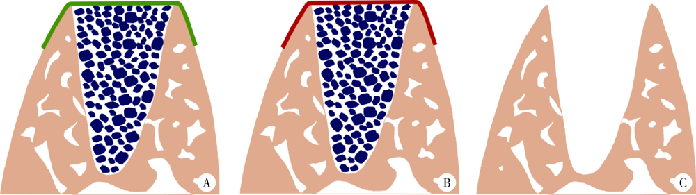
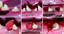
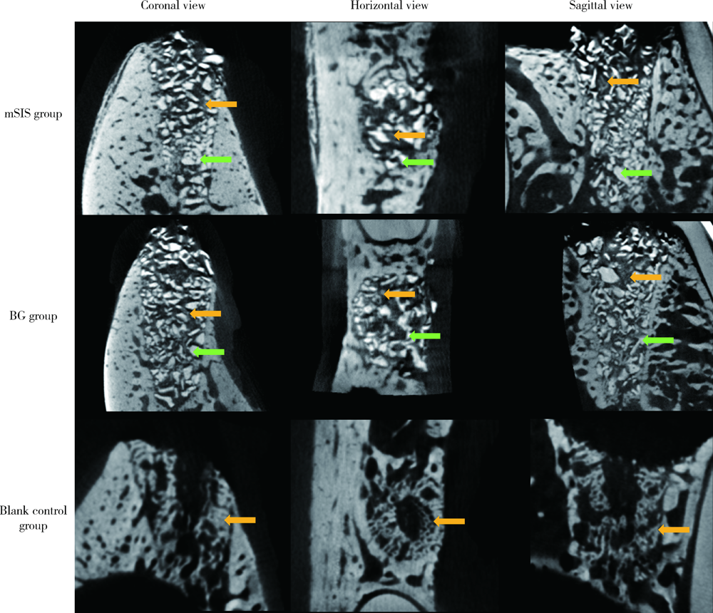
目的: 建立犬拔牙窝模型,采用影像学分析方法评价拔牙窝内植入去蛋白牛骨基质骨粉颗粒Bio-Oss®(简称Bio-Oss骨粉)并覆盖复层猪小肠黏膜下层膜(multilaminated small intestinal submucosa membrane, mSIS)或可吸收胶原膜Bio-Gide® (简称Bio-Gide膜), 愈合4周和12周后的牙槽窝内成骨效果。方法: 拔除3只比格犬双侧上下颌共计18颗前磨牙的远中根,得到18个拔牙窝,随机平均分为3大组,并分别对各拔牙窝组进行以下操作:(1)植入Bio-Oss骨粉并覆盖mSIS膜(mSIS组),(2)植入Bio-Oss骨粉并覆盖Bio-Gide膜(BG组),(3)自然愈合(空白对照组)。每大组各随机平均分为2个小组,分别于手术后4周和12周取样进行微计算机体层扫描(micro-computed tomograph, Micro-CT), 检测评价各组牙槽窝内新骨的生长情况,比较mSIS膜和Bio-Gide膜对拔牙窝内骨再生的影响。结果: Micro-CT分析显示,mSIS组和BG组在术后4周和12周的新生骨容积比均显著高于空白对照组(P<0.05),其中mSIS组略高于BG组,但两组间差异无统计学意义(P>0.05)。术后4周mSIS组和BG组的牙槽窝冠1/3区域新生骨容积比例显著高于中1/3及根1/3区域(P<0.05)。术后4周各组的新生骨密度值相近(P>0.05),术后12周时mSIS组和BG组的新生骨密度值均显著高于对照组(P<0.05)。术后4周和12周mSIS组和BG组的新生骨小梁的数量以及排列紧凑程度明显优于空白对照组(P<0.05),而mSIS略优于BG组,但两组间差异无统计学意义(P>0.05)。各组间骨小梁厚度的差异无统计学意义(P>0.05)。结论: 两种屏障膜联合去蛋白牛骨基质植入拔牙窝内有利于新骨再生,mSIS膜与Bio-Gide膜的应用效果相似。
中图分类号:
- R782.1


| [1] |
Ersanli S, Olgac V, Leblebicioglu B. Histologic analysis of alveolar bone following guided bone regeneration[J]. J Periodontol, 2004,75(5):750-756.
doi: 10.1902/jop.2004.75.5.750 pmid: 15212358 |
| [2] | Chiapasco M, Zaniboni M. Clinical outcomes of GBR procedures to correct peri-implant dehiscences and fenestrations: a systematic review[J]. Clin Oral Implants Res, 2009,20(Suppl 4):113-123. |
| [3] |
Oikarinen KS, Sandor GK, Kainulainen VT, et al. Augmentation of the narrow traumatized anterior alveolar ridge to facilitate dental implant placement[J]. Dent Traumatol, 2003,19(1):19-29.
pmid: 12656851 |
| [4] |
Amler MH. The time sequence of tissue regeneration in human extraction wounds[J]. Oral Surg Oral Med Oral Pathol, 1969,27(3):309-318.
pmid: 5251474 |
| [5] |
Nyman S, Lang NP, Buser D, et al. Bone regeneration adjacent to titanium dental implants using guided tissue regeneration: a report of two cases[J]. Int J Oral Maxillofac Implants, 1990,5(1):9-14.
pmid: 2391139 |
| [6] |
MacBeth N, Trullenque-Eriksson A, Donos N, et al. Hard and soft tissue changes following alveolar ridge preservation: a syste-matic review[J]. Clin Oral Implants Res, 2017,28(8):982-1004.
doi: 10.1111/clr.12911 pmid: 27458031 |
| [7] | 詹雅琳, 胡文杰, 甄敏, 等. 去蛋白牛骨基质与可吸收胶原膜的磨牙拔牙位点保存效果影像学评价[J]. 北京大学学报(医学版), 2015,47(1):19-26. |
| [8] |
Kim JJ, Schwarz F, Song HY, et al. Ridge preservation of extraction sockets with chronic pathology using Bio-Gide® Collagen with or without collagen membrane: an experimental study in dogs[J]. Clin Oral Implants Res, 2017,28(6):727-733.
doi: 10.1111/clr.12870 pmid: 27194177 |
| [9] |
Wu W, Li B, Liu Y, et al. Effect of multilaminate small intestinal submucosa as a barrier membrane on bone formation in a rabbit mandible defect model[J]. Biomed Res Int, 2018,2018:3270293.
pmid: 30018978 |
| [10] | 吴唯伊, 李博文, 刘玉华, 等. 复层猪小肠黏膜下层可吸收膜的降解性能[J]. 北京大学学报(医学版), 2020,52(3):564-569. |
| [11] |
Eitel F, Klapp F, Jacobson W, et al. Bone regeneration in animals and in man. A contribution to understanding the relative value of animal experiments to human pathophysiology[J]. Arch Orthop Trauma Surg, 1981,99(1):59-64.
pmid: 7316703 |
| [12] |
Lindhe J, Araujo MG, Bufler M, et al. Biphasic alloplastic graft used to preserve the dimension of the edentulous ridge: an experimental study in the dog[J]. Clin Oral Implants Res, 2013,24(10):1158-1163.
pmid: 22804845 |
| [13] |
Naenni N, Sapata V, Bienz SP, et al. Effect of flapless ridge preservation with two different alloplastic materials in sockets with buccal dehiscence defects-volumetric and linear changes[J]. Clin Oral Investig, 2018,22(6):2187-2197.
doi: 10.1007/s00784-017-2309-6 pmid: 29280075 |
| [14] | 詹雅琳, 胡文杰, 徐涛, 等. 罹患重度牙周炎磨牙拔除后应用去蛋白牛骨基质与可吸收胶原膜进行位点保存的组织学研究[J]. 北京大学学报(医学版), 2017,49(1):169-175. |
| [15] |
Benic GI, Thoma DS, Sanz-Martin I, et al. Guided bone regene-ration at zirconia and titanium dental implants: a pilot histological investigation[J]. Clin Oral Implants Res, 2017,28(12):1592-1599.
doi: 10.1111/clr.13030 pmid: 28653343 |
| [16] |
Wang F, Li Q, Wang Z. A comparative study of the effect of Bio-Gide® in combination with concentrated growth factors or bone marrow-derived mesenchymal stem cells in canine sinus grafting[J]. J Oral Pathol Med, 2017,46(7):528-536.
pmid: 27682609 |
| [17] |
Turri A, Elgali I, Vazirisani F, et al. Guided bone regeneration is promoted by the molecular events in the membrane compartment[J]. Biomaterials, 2016,84:167-183.
pmid: 26828682 |
| [18] |
Bouxsein ML, Boyd SK, Christiansen BA, et al. Guidelines for assessment of bone microstructure in rodents using micro-computed tomography[J]. J Bone Miner Res, 2010,25(7):1468-1486.
pmid: 20533309 |
| [19] | Leventis M, Fairbairn P, Mangham C, et al. Bone healing in rabbit calvaria defects using a synthetic bone substitute: A histological and micro-CT comparative study[J]. Materials (Basel), 2018,11(10):1-13. |
| [20] |
Sun Y, Wang CY, Wang ZY, et al. Test in canine extraction site preservations by using mineralized collagen plug with or without membrane[J]. J Biomater Appl, 2016,30(9):1285-1299.
pmid: 26721867 |
| [21] |
Omar O, Dahlin A, Gasser A, et al. Tissue dynamics and rege-nerative outcome in two resorbable non-cross-linked collagen memb-ranes for guided bone regeneration: A preclinical molecular and histological study in vivo[J]. Clin Oral Implants Res, 2018,29(1):7-19.
pmid: 28703398 |
| [1] | 高若凡, 马天宇, 王润楷, 殷雨辰, 李芮迪, 王丹丹, 夏斌. 细胞膜囊泡递送靶向肿瘤坏死因子-α的小干扰RNA对牙髓干细胞的抗炎作用[J]. 北京大学学报(医学版), 2026, 58(1): 22-29. |
| [2] | 孙菲, 王翠, 李思琪, 危伊萍, 余日月, 胡文杰. 赤藓糖醇喷砂与超声治疗对种植体周黏膜炎疗效的随机对照临床研究[J]. 北京大学学报(医学版), 2026, 58(1): 37-42. |
| [3] | 池彦廷, 蒋鸿杰, 陈艳, 徐志秀, 李斌斌. 直接免疫荧光在口腔黏膜寻常型天疱疮诊断中的价值: 基于多指标联合分析的回顾性研究[J]. 北京大学学报(医学版), 2026, 58(1): 68-73. |
| [4] | 彭嘉婧, 崔莉. 以眼部病变首发的结节病的临床特征及预后[J]. 北京大学学报(医学版), 2025, 57(6): 1061-1066. |
| [5] | 高雅静, 李正芳, 马梦思, 武丽君. SII和SIRI对白塞病葡萄膜炎的风险预测及疾病活动度和预后的评估[J]. 北京大学学报(医学版), 2025, 57(6): 1067-1073. |
| [6] | 吕雪冰, 俞烜华, 张伟桢, 刘昌泉, 林互涵, 曾珊婷, 黄惠娟, 吴月萍. 类风湿关节炎合并坏死性筋膜炎1例[J]. 北京大学学报(医学版), 2025, 57(6): 1198-1202. |
| [7] | 杨菊, 徐婧, 戴菊华, 石连杰. Lumican蛋白在类风湿关节炎患者血清中的表达及其与疾病和免疫活动的相关性[J]. 北京大学学报(医学版), 2025, 57(5): 911-918. |
| [8] | 冷汶远, 高端, 李晓宇, 左炜, 胡伟民, 朱振鹏, 徐纯如, 林健, 李学松. 口腔黏膜补片与脱细胞真皮基质补片治疗长段尿道狭窄的疗效和安全性对比[J]. 北京大学学报(医学版), 2025, 57(5): 975-979. |
| [9] | 刘杰, 马茗微, 王庆安, 石明, 尹金鹏, 王占平, 申静涛, 高献书. 基于锥形束CT的前列腺癌放射治疗两种体位固定方式摆位误差比较[J]. 北京大学学报(医学版), 2025, 57(4): 692-697. |
| [10] | 卞雯, 周文君, 吴天晨, 朱培静, 陈一诺, 原鹏波, 王学举, 王颖, 魏瑗, 赵扬玉. 单绒毛膜双羊膜囊双胎妊娠双胎之一胎死宫内对妊娠结局的影响[J]. 北京大学学报(医学版), 2025, 57(3): 592-598. |
| [11] | 朱慧, 闵赛南, 苏家增, 陈艳, 彭歆, 于尧, 俞光岩. 口腔黏膜嗜酸性溃疡的临床病理分析[J]. 北京大学学报(医学版), 2025, 57(3): 620-625. |
| [12] | 付玮, 宁静, 付伟伟, 张静, 丁士刚. CMTM6对幽门螺杆菌感染的胃上皮细胞中PD-L1的作用[J]. 北京大学学报(医学版), 2025, 57(2): 245-252. |
| [13] | 王菲, 张馨月, 刘木清, 王恩博, 段登辉. 顺牙长轴拔牙法在下颌近中与水平智齿拔除术中的应用及三维有限元分析[J]. 北京大学学报(医学版), 2025, 57(1): 106-112. |
| [14] | 刘爱春, 赵慧萍, 武蓓, 郑姝颖, 左力, 王梅. 腹膜透析相关性腹膜炎拔管患者导管内细菌生物膜的形成[J]. 北京大学学报(医学版), 2025, 57(1): 161-165. |
| [15] | 聂骏男, 董佳芸, 路瑞芳. 骨瓣重建颌骨区域角化黏膜增量术后软组织愈合效果分析[J]. 北京大学学报(医学版), 2025, 57(1): 57-64. |
|
||
