北京大学学报(医学版) ›› 2025, Vol. 57 ›› Issue (3): 604-609. doi: 10.19723/j.issn.1671-167X.2025.03.027

• 论著 • 上一篇    下一篇

基于直接法和间接法数字印模制作的高嵌体适合性评价的体外研究

钱锟, 刘亦洪*()   

  1. 北京大学口腔医学院·口腔医院综合科,国家口腔医学中心,国家口腔疾病临床医学研究中心,口腔生物材料和数字诊疗装备国家工程研究中心,口腔数字医学北京市重点实验室,国家卫生健康委员会口腔医学计算机应用工程技术研究中心,国家药品监督管理局口腔材料重点实验室,北京 100081
  • 收稿日期:2022-10-06 出版日期:2025-06-18 发布日期:2025-06-13
  • 通讯作者: 刘亦洪
  • 基金资助:
    国家自然科学基金(52111530189)

Fitness of onlays fabricated with direct and indirect CAD/CAM technology in vitro

Kun QIAN, Yihong LIU*()   

  1. Department of General Dentistry, Peking University School and Hospital of Stomatology & National Center for Stomatology & National Clinical Research Center for Oral Diseases & National Engineering Research Center of Oral Biomaterials and Digital Medical Devices & Beijing Key Laboratory of Digital Stomatology & NHC Research Center of Engineering and Technology for Computerized Dentistry & NMPA Key Laboratory for Dental Materials, Beijing 100081, China
  • Received:2022-10-06 Online:2025-06-18 Published:2025-06-13
  • Contact: Yihong LIU
  • Supported by:
    National Natural Science Foundation of China(52111530189)

RICH HTML


摘要:

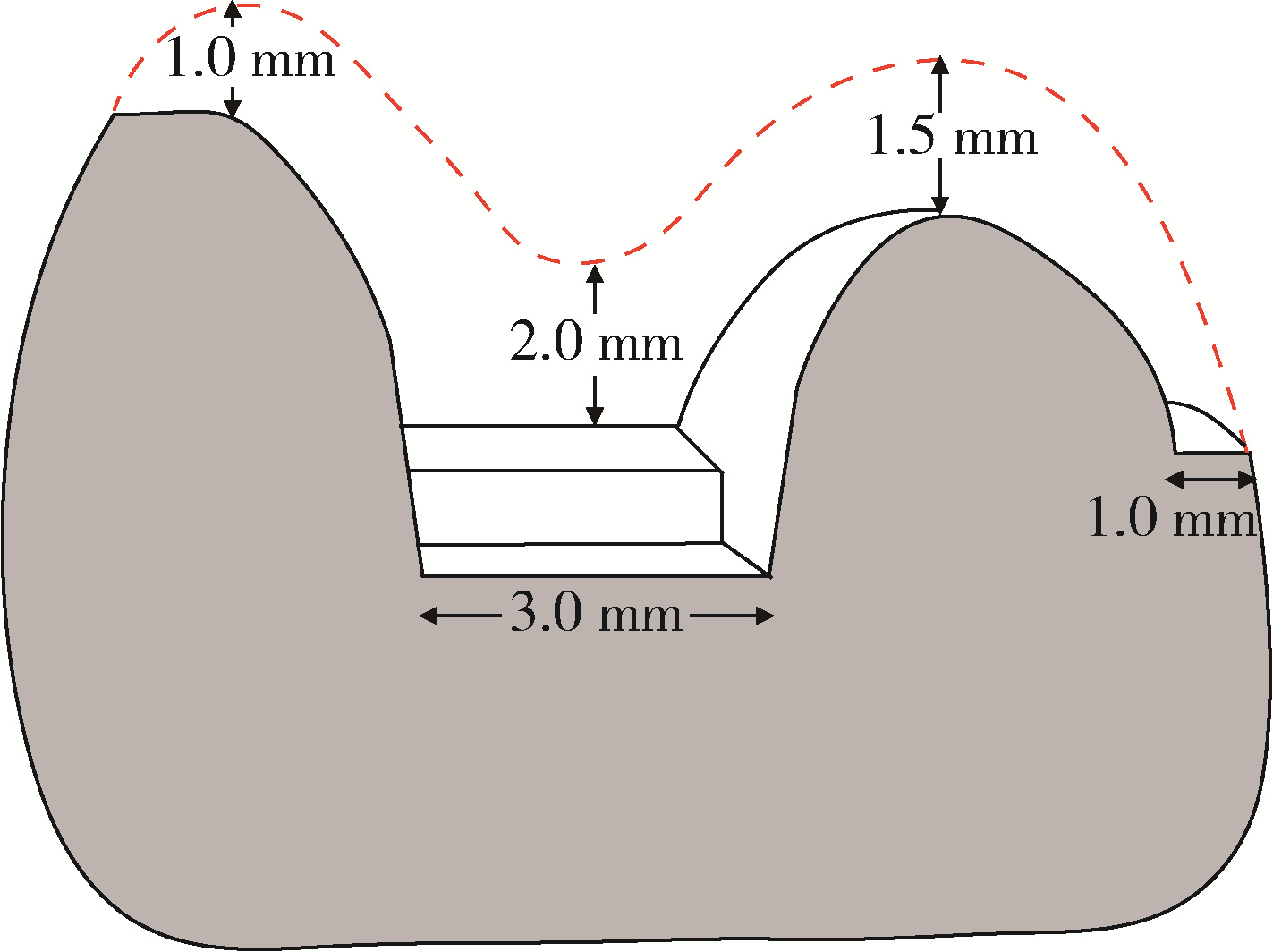
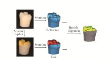
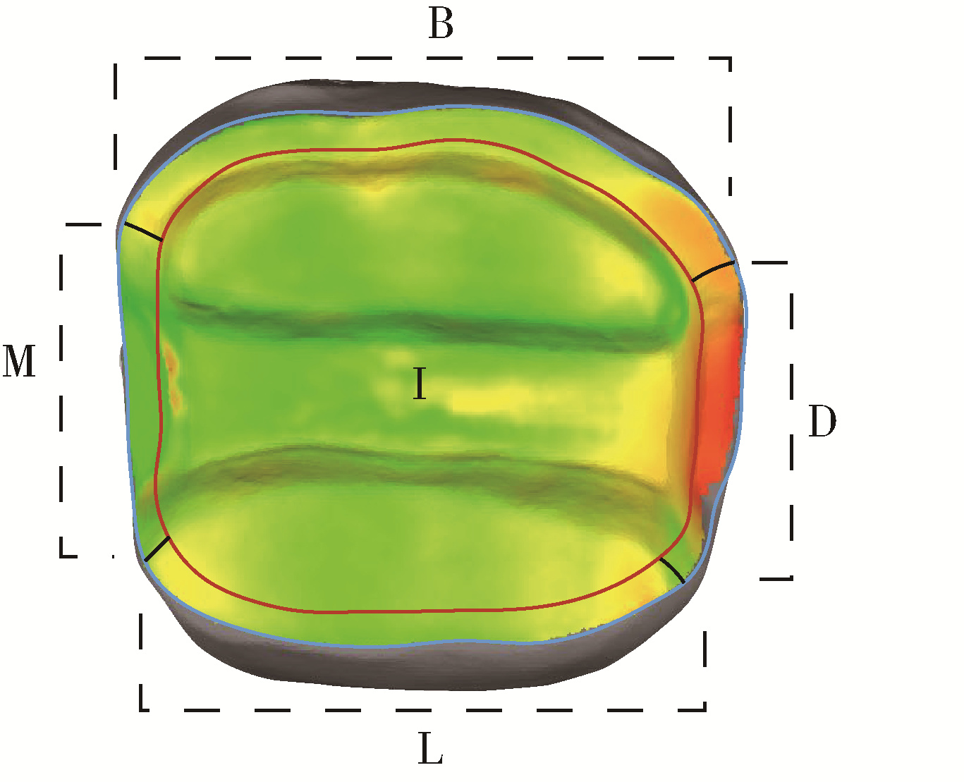
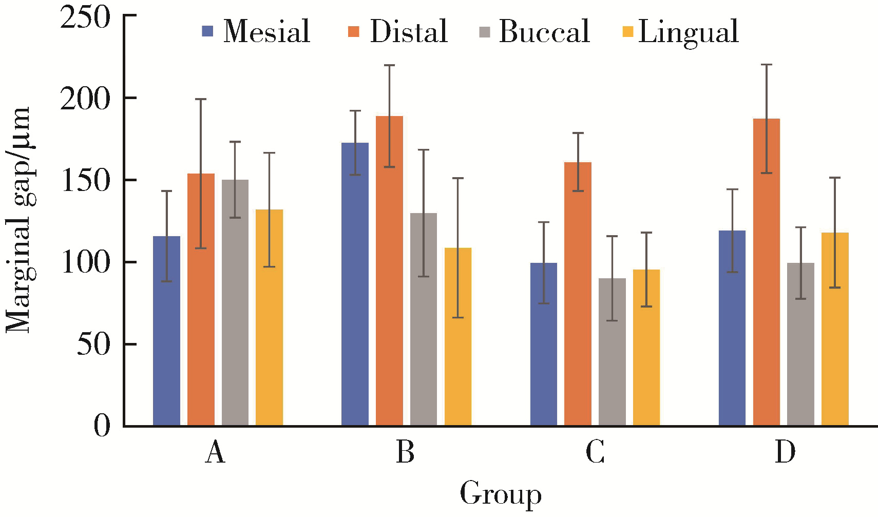
目的: 探索体外环境下,基于直接法及间接法数字印模技术制作的氧化锆及二硅酸锂增强玻璃陶瓷高嵌体的适合性。方法: 36牙位树脂人工牙48颗,随机分成A、B、C、D 4组,每组12颗,体外完成人工牙标准近中牙合远中高嵌体牙体预备,显微镜下检查预备质量。仿头模固定于牙科治疗椅,将标准人工牙列模型固定于仿头模内。将A、B组树脂人工牙依次置于人工牙列内相应位置,口内扫描仪获取36牙位数字印模。成品托盘,聚醚硅橡胶制取C、D组树脂牙列印模,灌注石膏模型,使用模型扫描仪扫描C、D组石膏模型。4组均使用数字化设计软件完成高嵌体设计,送加工部门完成高嵌体制作(A、C组使用氧化锆材料,B、D组使用二硅酸锂增强玻璃陶瓷材料),使用三维配准方法评价高嵌体边缘间隙及内部间隙。结果: 基于间接法数字印模技术制作的高嵌体的边缘间隙及内部间隙均小于直接法数字印模组(P < 0.05),各处理组远中边缘间隙值最大(P < 0.05),不同制作材料对高嵌体边缘及内部适合性均无显著影响(P>0.05)。结论: 基于间接法数字印模技术制作的高嵌体的边缘适合性及内部适合性均优于直接法,氧化锆及二硅酸锂增强玻璃陶瓷两种修复材料对高嵌体适合性没有影响。

关键词: 牙科印模技术, 牙瓷料, 氧化锆, 二硅酸锂, 高嵌体

Abstract:

Objective: To analyze the fitness of zirconia and lithium disilicate glass-ceramic onlays fabricated with direct and indirect computer-aided design and computer-aided manufacturing (CAD/CAM) technology in vitro. Methods: In the study, 48 standardised typodont left mandibular first molars received standardised onlay preparation. Then, all the specimens were randomly divided into 4 groups. There were 12 specimens in each group. The preparation quality was checked under the stereomicroscope. All the specimens were fixed in typodonts. Subsequently, the typodonts were fixed in the dental simulators to simulate the oral conditions. In groups A and B, the digital impressions were obtained by using the intraoral scanner. In groups C and D, conventional impressions of polyether impression material were obtained according to the instructions of the manufacturer using individual trays. The stone casts were made with type Ⅳ gypsum later. Then, all casts were digitized with the model scanner. Based on the data obtained from the scan, onlay restorations of all the groups were designed using the corresponding software, the simulated cement thickness was set to 50 μm. Then, the final onlays restorations of all the groups were machined with the milling machines in lab. The fabrication materials were different in groups. The specimens of groups A and C were fabricated with zirconia. While, the specimens of groups B and D were fabricated with lithium disilicate glass-ceramic. The marginal gap and internal gap of all restorations were analyzed by 3D replica technique, for each measurement, the specimen was digitised using the model scanner. Results: The marginal gap of the onlays fabricated with indirect digital impressions were smaller than that with direct digital impressions (P < 0.05). At the same time, the internal gap of the onlays fabricated with indirect digital impressions were smaller than that with direct digi-tal impressions (P < 0.05). The marginal gap was larger in distal gingival than that in the other regions in all the groups (P < 0.05). Different fabrication materials, zirconia or lithium disilicate reinforced glass-ceramic, had no effect on onlay marginal and internal fit (P>0.05). Conclusion: The marginal and internal adaptation of the onlays fabricated with indirect digital impressions was better than with direct digital impressions. Zirconia and lithium disilicate reinforced glass-ceramic had no effect on the onlay adaptation.

Key words: Dental impression technique, Dental porcelain, Zirconia, Lithium disilicate, Onlay

中图分类号: 

  • R783.3

图1

高嵌体设计尺寸示意图"

图2

三维测量方法流程图"

图3

测量区域示意图"

表1

各组高嵌体内部及边缘间隙值"

Group Method Material Internal gap values/μm Marginal gap values/μm
A (n=12) Direct Zirconia 135.54±14.69 144.30±20.44
B (n=12) Direct Ceramic 124.16±22.04 135.53±23.44
C (n=12) Indirect Zirconia 103.77±13.61 104.55±19.02
D (n=12) Indirect Ceramic 108.49±17.83 122.78±23.36

表2

内部间隙双因素方差分析统计结果"

Source Type Ⅲ sum of squares df Mean square F P
Corrected model 5 251.241a 2 2 625.621 8.717 0.001
Intercept 515 841.350 1 515 841.350 1 712.556 < 0.001
Method 5 211.009 1 5 211.009 17.300 < 0.001
Material 40.232 1 40.232 0.134 0.717

表3

边缘间隙多因素方差分析统计结果"

Source Type Ⅲ sum of squares df Mean square F P
Corrected model 150 406.021a 15 10 027.068 9.374 < 0.001
Intercept 2 532 324.909 1 2 532 324.909 2 367.339 < 0.001
Method 13 937.982 1 13 937.982 13.030 < 0.001
Position 84 163.215 3 28 054.405 26.227 < 0.001
Material 3 819.499 1 3 819.499 3.571 0.061
Method×Position 12 588.477 3 4 196.159 3.923 0.010
Method×Material 3 140.300 1 3 140.300 2.936 0.089
Position×Material 14 902.929 3 4 967.643 4.644 0.004
Method×Position×Material 9 945.476 3 3 315.159 3.099 0.029

图4

各组4个位置边缘间隙值"

1
Rekow ED . Digital dentistry: The new state of the art. Is it disruptive or destructive?[J]. Dent Mater, 2020, 36 (1): 9- 24.

doi: 10.1016/j.dental.2019.08.103
2
Papaspyridakos P , Gallucci GO , Chen CJ , et al. Digital versus conventional implant impressions for edentulous patients: Accuracy outcomes[J]. Clin Oral Implants Res, 2016, 27 (4): 465- 472.

doi: 10.1111/clr.12567
3
Ender A , Zimmermann M , Attin T . In vivo precision of conventional and digital methods for obtaining quadrant dental impressions[J]. Clin Oral Invest, 2016, 20 (7): 1495- 1504.

doi: 10.1007/s00784-015-1641-y
4
Güth JF , Edelhoff D , Schweiger J . A new method for the evaluation of the accuracy of full-arch digital impressions in vitro[J]. Clin Oral Invest, 2016, 20 (7): 1487- 1494.

doi: 10.1007/s00784-015-1626-x
5
Rhee YK , Huh YH , Cho LR . Comparison of intraoral scanning and conventional impression techniques using 3-dimensional superimposition[J]. J Adv Prosthodont, 2015, 7 (6): 460- 467.

doi: 10.4047/jap.2015.7.6.460
6
Wismeijer D , Mans R , van Genuchten M . Patients' preferences when comparing analogue implant impressions using a polyether impression material versus digital impressions (intraoral scan) of dental implants[J]. Clin Oral Implants Res, 2014, 25 (10): 1113- 1118.

doi: 10.1111/clr.12234
7
Yuzbasioglu E , Kurt H , Turunc R . Comparison of digital and conventional impression techniques: Evaluation of patients' perception, treatment comfort, effectiveness and clinical outcomes[J]. BMC Oral Health, 2014, 14, 10.

doi: 10.1186/1472-6831-14-10
8
Holmes JR , Bayne SC , Holland GA , et al. Considerations in measurement of marginal fit[J]. J Prosthet Dent, 1989, 62 (4): 405- 408.

doi: 10.1016/0022-3913(89)90170-4
9
Ahrberg D , Lauer HC , Ahrberg M , et al. Evaluation of fit and efficiency of CAD/CAM fabricated all-ceramic restorations based on direct and indirect digitalization: A double-blinded, randomized clinical trial[J]. Clin Oral Invest, 2016, 20 (2): 291- 300.

doi: 10.1007/s00784-015-1504-6
10
Kale E , Seker E , Yilmaz B , et al. Effect of cement space on the marginal fit of CAD-CAM-fabricated monolithic zirconia crowns[J]. J Prosthet Dent, 2016, 116 (6): 890- 895.

doi: 10.1016/j.prosdent.2016.05.006
11
Abdel-Azim T , Rogers K , Elathamna E . Comparison of the marginal fit of lithium disilicate crowns fabricated with CAD/CAM technology by using conventional impressions and two intraoral di-gital scanners[J]. J Prosthet Dent, 2015, 114 (4): 554- 559.

doi: 10.1016/j.prosdent.2015.04.001
12
Zeltner M , Sailer I , Mühlemann S , et al. Randomized controlled within-subject evaluation of digital and conventional workflows for the fabrication of lithium disilicate single crowns. Part Ⅲ: Margi-nal and internal fit[J]. J Prosthet Dent, 2017, 117 (3): 354- 362.

doi: 10.1016/j.prosdent.2016.04.028
13
Agm A , Ham B , Pc C , et al. Marginal and internal fit of CAD-CAM inlay/onlay restorations: A systematic review of in vitro stu-dies[J]. J Prosthet Dent, 2019, 121 (4): 590- 597.

doi: 10.1016/j.prosdent.2018.06.006
14
Liu Y , Ye H , Yong W , et al. Three-dimensional analysis of internal adaptations of crowns cast from resin patterns fabricated using computer-aided design/computer-assisted manufacturing technologies[J]. Int J Prosthodont, 2018, 31 (4): 386- 393.
15
DeLong R , Pintado MR , Ko CC , et al. Factors influencing optical 3D scanning of vinyl polysiloxane impression materials[J]. J Prosthodont, 2001, 10 (2): 78- 85.

doi: 10.1111/j.1532-849X.2001.00078.x
16
Abduo J , Elseyoufi M . Accuracy of intraoral scanners: A syste-matic review of influencing factors[J]. Eur J Prosthodont Restor Dent, 2018, 26 (3): 101- 121.
17
Rudolph H , Luthardt RG , Walter MH . Computer-aided analysis of the influence of digitizing and surfacing on the accuracy in den-tal CAD/CAM technology[J]. Comput Biol Med, 2007, 37 (5): 579- 587.

doi: 10.1016/j.compbiomed.2006.05.006
18
姜楠, 包旭东, 岳琳. 全冠预备体终止线局部扫描正确度对整体的影响[J]. 北京大学学报(医学版), 2021, 53 (1): 102- 108.
19
Kim DY , Kim JH , Kim HY , et al. Comparison and evaluation of marginal and internal gaps in cobalt-chromium alloy copings fabricated using subtractive and additive manufacturing[J]. J Pros-thodont Res, 2018, 62 (1): 56- 64.

doi: 10.1016/j.jpor.2017.05.008
20
McLean JW , von Fraunhofer JA . The estimation of cement film thickness by an in vivo technique[J]. Brit Dent J, 1971, 131 (3): 107- 111.

doi: 10.1038/sj.bdj.4802708
21
Guess PC , Vagkopoulou T , Zhang Y . Marginal and internal fit of heat pressed versus CAD/CAM fabricated all-ceramic onlays after exposure to thermo-mechanical fatigue[J]. J Dent, 2014, 42 (2): 199- 209.

doi: 10.1016/j.jdent.2013.10.002
22
Bindl A , Mörmann WH . Clinical and SEM evaluation of all-ceramic chair-side CAD/CAM generated partial crowns[J]. Eur J Oral Sci, 2003, 111 (2): 163- 169.

doi: 10.1034/j.1600-0722.2003.00022.x
23
Merrill TC , Mackey T , Luc R , et al. Effect of chairside CAD/CAM restoration type on marginal fit accuracy: A comparison of crown, inlay and onlay restorations[J]. Eur J Prosthodont Restor Dent, 2021, 29 (2): 119- 127.
24
Colpani JT , Borba M , Della Bona A . Evaluation of marginal and internal fit of ceramic crown copings[J]. Dent Mater, 2013, 29 (2): 174- 180.

doi: 10.1016/j.dental.2012.10.012
25
Mously HA , Finkelman M , Zandparsa R , et al. Marginal and internal adaptation of ceramic crown restorations fabricated with CAD/CAM technology and the heat-press technique[J]. J Prosthet Dent, 2014, 112 (2): 249- 256.

doi: 10.1016/j.prosdent.2014.03.017
26
Lins L , Bemfica V , Queiroz C , et al. In vitro evaluation of the internal and marginal misfit of CAD/CAM zirconia copings[J]. J Prosthet Dent, 2015, 113 (3): 205- 211.

doi: 10.1016/j.prosdent.2014.09.010
27
Alfaro DP , Ruse ND , Carvalho RM , et al. Assessment of the internal fit of lithium disilicate crowns using micro-CT[J]. J Prosthodont, 2015, 24 (5): 381- 386.

doi: 10.1111/jopr.12274
28
Bosch G , Ender A , Mehl A . A 3-dimensional accuracy analysis of chairside CAD/CAM milling processes[J]. J Prosthet Dent, 2014, 112 (6): 1425- 1431.

doi: 10.1016/j.prosdent.2014.05.012
29
Akalin ZF , Ozkan YK , Ekerim A . Effects of implant angulation, impression material, and variation in arch curvature width on implant transfer model accuracy[J]. Int J Oral Maxillofac Implants, 2013, 28 (1): 149- 157.

doi: 10.11607/jomi.2070
30
Persson A , A Odén , Andersson M , et al. Digitization of simulated clinical dental impressions: Virtual three-dimensional analysis of exactness[J]. Dent Mater, 2009, 25 (7): 929- 936.

doi: 10.1016/j.dental.2009.01.100
31
Duqum IS , Brenes C , Mendonca G , et al. Marginal fit evaluation of CAD/CAM all ceramic crowns obtained by two digital workflows: An in vitro study using micro-CT technology[J]. J Pros-thodont, 2019, 28 (9): 1037- 1043.
32
Francesco R , Massimo A , Renato L , et al. In vitro evaluation of the marginal fit and internal adaptation of zirconia and lithium disilicate single crowns: Micro-CT comparison between different manufacturing procedures[J]. Open Dent J, 2018, 12 (1): 160- 172.

doi: 10.2174/1874210601812010160
33
Fuzzi M , Tricarico MG , Cagidiaco EF , et al. Nanoleakage and internal adaptation of zirconia and lithium disilicate single crowns with feather edge preparation[J]. Journal of Osseointegration, 2017, 9 (2): 250- 262.
34
Jemt T , Hjalmarsson L . In vitro measurements of precision of fit of implant-supported frameworks. A comparison between "virtual" and "physical" assessments of fit using two different techniques of measurements[J]. Clin Implant Dent R, 2012, 14 (Suppl 1): e175- e175.
35
Schaefer O , Watts DC , Sigusch BW , et al. Marginal and internal fit of pressed lithium disilicate partial crowns in vitro: A three-dimensional analysis of accuracy and reproducibility[J]. Dent Mater, 2012, 28 (3): 320- 326.

doi: 10.1016/j.dental.2011.12.008
36
Kuhn K , Ostertag S , Ostertag M . Comparison of an analog and digital quantitative and qualitative analysis for the fit of dental copings[J]. Comput Biol Med, 2015, 57, 32- 41.

doi: 10.1016/j.compbiomed.2014.11.017
37
Qian K , Li B , Pu T , et al. Fitness of self-glazed zirconia onlays using conventional and digital impressions[J]. Adv Appl Ceram, 2022, 121 (3): 93- 100.

doi: 10.1080/17436753.2022.2069988
[1] 吴美辰,许桐楷,安伟,刘中宁,姜婷. 后牙高嵌体和贴面修复的4年临床随访[J]. 北京大学学报(医学版), 2024, 56(1): 88-92.
[2] 丁茜,李文锦,孙丰博,谷景华,林元华,张磊. 表面处理对氧化钇和氧化镁稳定的氧化锆种植体晶相及断裂强度的影响[J]. 北京大学学报(医学版), 2023, 55(4): 721-728.
[3] 吴思妤,李娅宁,张晓,吕珑薇,刘云松,叶红强,周永胜. 上颌中切牙全瓷冠牙体预备学习曲线的预测、分析与应用[J]. 北京大学学报(医学版), 2023, 55(1): 108-113.
[4] 李伟伟,陈虎,王勇,孙玉春. 氧化锆陶瓷表面硅锂喷涂层的摩擦磨损性能[J]. 北京大学学报(医学版), 2023, 55(1): 94-100.
[5] 王鹃,尉华杰,孙井德,邱立新. 预成刚性连接杆用于无牙颌种植即刻印模制取的应用评价[J]. 北京大学学报(医学版), 2022, 54(1): 187-192.
[6] 王铮,丁茜,高远,马全诠,张磊,葛兮源,孙玉春,谢秋菲. 氧化锆多孔表面显微形貌对成骨细胞增殖及分化的影响[J]. 北京大学学报(医学版), 2022, 54(1): 31-39.
[7] 李文锦,丁茜,原福松,孙丰博,郑剑桥,鲍蕊,张磊. 飞秒激光表面处理对氧化锆表面特征及弯曲强度的影响[J]. 北京大学学报(医学版), 2021, 53(4): 770-775.
[8] 杨欣,李榕,叶红强,陈虎,王勇,周永胜,孙玉春. 不同刃状边缘补偿角度的两种氧化锆全瓷冠断裂强度的评价[J]. 北京大学学报(医学版), 2021, 53(2): 402-405.
[9] 徐啸翔,曹烨,赵一姣,贾璐,谢秋菲. 数字化个齿托盘制取下颌全牙列全冠预备体印模的体外评价[J]. 北京大学学报(医学版), 2021, 53(1): 54-61.
[10] 郑苗,詹凌璐,刘志强,李和平,谭建国. 不同等离子体处理氧化锆对人牙龈成纤维细胞黏附能力的影响[J]. 北京大学学报(医学版), 2019, 51(2): 315-320.
[11] 周团锋,王新知. 计算机辅助设计与制作的一体化氧化锆全瓷桩核5年临床观察[J]. 北京大学学报(医学版), 2018, 50(4): 680-684.
[12] 焦洋, 王继德, 邓久鹏. 不同表面处理对氧化锆晶相结构及性能的影响[J]. 北京大学学报(医学版), 2018, 50(1): 49-52.
[13] 廖宇,刘晓强,陈立,周建锋,谭建国. 不同表面处理方法对氧化锆与树脂水门汀粘接强度的影响[J]. 北京大学学报(医学版), 2018, 50(1): 53-57.
[14] 张皓羽,姜婷,程明轩,张玉玮. 类瓷树脂及玻璃陶瓷牙合贴面疲劳实验前后的磨耗及表面粗糙度的变化[J]. 北京大学学报(医学版), 2018, 50(1): 73-77.
[15] 崔新悦,佟岱,王新知,沈志坚. 对比切削与胶态成型工艺制作的氧化锆陶瓷的透光性与遮色效果[J]. 北京大学学报(医学版), 2018, 50(1): 85-90.
Viewed
Full text


Abstract

Cited

  Shared   
  Discussed   
No Suggested Reading articles found!