北京大学学报(医学版) ›› 2025, Vol. 57 ›› Issue (4): 684-691. doi: 10.19723/j.issn.1671-167X.2025.04.009

• 论著 • 上一篇    下一篇

基于临床特征和多参数MRI的前列腺癌盆腔淋巴结转移的术前预测模型

王泽远, 于栓宝, 郑浩轲, 陶金, 范雅峰, 张雪培*()   

  1. 郑州大学第一附属医院泌尿外科, 郑州 450000
  • 收稿日期:2025-02-27 出版日期:2025-08-18 发布日期:2025-08-02
  • 通讯作者: 张雪培
  • 基金资助:
    河南省青年科学基金(232300420254); 河南省科技攻关项目(242102311074)

A preoperative prediction model for pelvic lymph node metastasis in prostate cancer: Integrating clinical characteristics and multiparametric MRI

Zeyuan WANG, Shuanbao YU, Haoke ZHENG, Jin TAO, Yafeng FAN, Xuepei ZHANG*()   

  1. Department of Urology, the First Affiliated Hospital of Zhengzhou University, Zhengzhou 450000, China
  • Received:2025-02-27 Online:2025-08-18 Published:2025-08-02
  • Contact: Xuepei ZHANG
  • Supported by:
    the Youth Science Fund of Henan Province(232300420254); the Key Scientific and Technological Projects of Henan Province(242102311074)

RICH HTML


摘要:

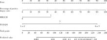
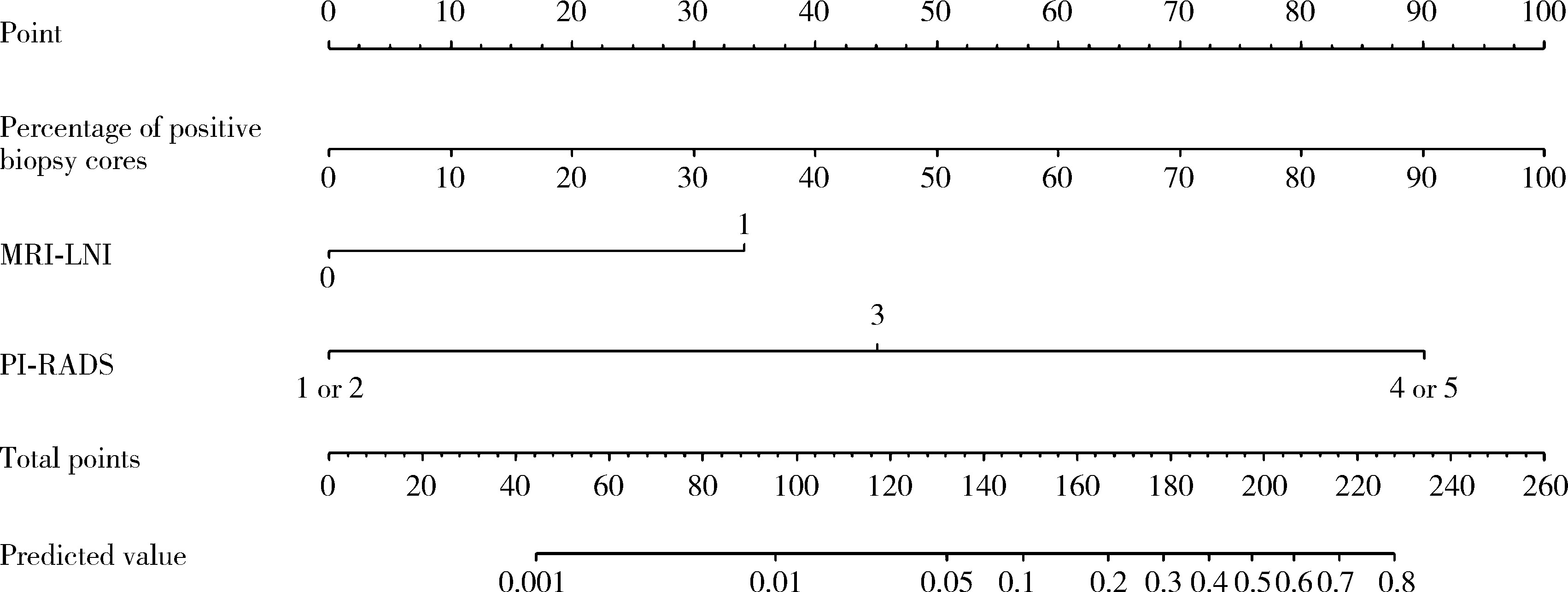
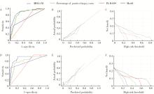
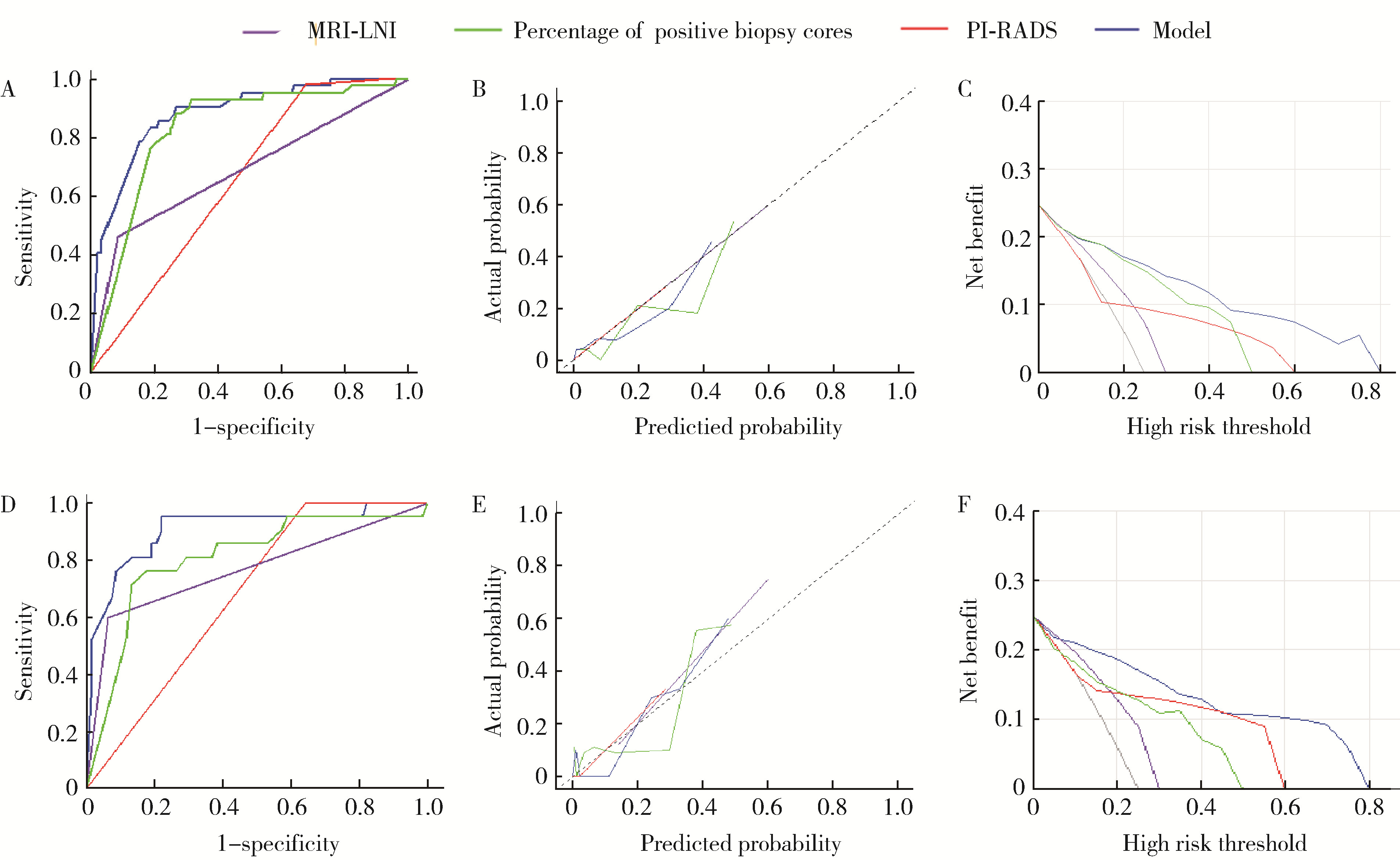
目的: 分析与前列腺癌患者盆腔淋巴结转移(pelvic lymph node metastasis, PLNM)相关的临床特征, 构建PLNM的术前预测模型, 以减少不必要的扩大盆腔淋巴结清扫(extended pelvic lymph node dissection, ePLND)。方法: 根据纳入与排除标准, 回顾性收集2014—2024年间在郑州大学第一附属医院接受前列腺癌根治术和ePLND的344例患者, 其中77例(22.4%)患者淋巴结阳性。收集患者的临床特征、MRI报告和组织病理结果, 将数据随机分为训练集(241例, 70%)和验证集(103例, 30%), 采用单因素和多因素Logistic回归分析构建PLNM的术前预测模型。结果: 单因素Logistic回归分析表明, 总前列腺特异性抗原(total prostate specific antigen, tPSA) (P=0.021)、游离前列腺特异性抗原(free prostate specific antigen, fPSA) (P=0.002)、fPSA/tPSA (P=0.011)、穿刺阳性针数百分比(P < 0.001)、前列腺影像报告和数据系统(prostate imaging reporting and data system, PI-RADS)评分(P=0.004)、穿刺病理Gleason评分≥8 (P=0.005)、临床T分期(P < 0.001)和MRI显示的淋巴结受累(MRI-indicated lymph node involvement, MRI-LNI) (P < 0.001)是预测PLNM的显著因素。多因素Logistic回归分析表明, 穿刺阳性针数百分比(OR=91.24, 95%CI: 13.34~968.68)、PI-RADS评分(OR=7.64, 95%CI: 1.78~138.06)和MRI-LNI (OR=4.67, 95%CI: 1.74~13.24)是预测PLNM的独立危险因素。基于此构建列线图, 多因素模型的预测效果[曲线下面积(area under curve, AUC)=0.883]显著优于单一指标[阳性针数百分比(AUC=0.806)、PI-RADS评分(AUC=0.679)和MRI-LNI(AUC=0.768)]。校准曲线和决策曲线表明, 多因素模型具有较高的预测准确度和显著的净收益, 在6%的截断值下只漏检了约5.2%的PLNM(4/77), 而减少了约53%的ePLND(139/267), 显示出较好的预测效果。结论: 穿刺阳性针数百分比、PI-RADS评分和MRI-LNI是PLNM的独立危险因素, 构建多因素模型可显著提高预测效果, 为指导临床ePLND策略提供了有价值的参考。

关键词: 前列腺肿瘤, 淋巴转移, 多参数磁共振成像, 活组织检查

Abstract:

Objective: To analyze the clinical features associated with pelvic lymph node metastasis (PLNM) in prostate cancer and to construct a preoperative prediction model for PLNM, thereby reducing unnecessary extended pelvic lymph node dissection (ePLND). Methods: Based on predefined inclusion and exclusion criteria, 344 patients who underwent radical prostatectomy and ePLND at the First Affiliated Hospital of Zhengzhou University between 2014 and 2024 were retrospectively enrolled, among whom, 77 patients (22.4%) were pathologically confirmed to have lymph node-positive disease. The clinical characteristics, MRI reports, and pathological results were collected. The data were then randomly divi-ded into a training cohort (241 cases, 70%) and a validation cohort (103 cases, 30%). Univariate and multivariate Logistic regression analysis were employed to construct a preoperative prediction model for PLNM. Results: Univariate Logistic regression analysis revealed that total prostate specific antigen (tPSA) (P=0.021), free prostate specific antigen (fPSA) (P=0.002), fPSA to tPSA ratio (fPSA/tPSA) (P=0.011), percentage of positive biopsy cores (P < 0.001), prostate imaging reporting and data system (PI-RADS) score (P=0.004), biopsy Gleason score ≥8 (P=0.005), clinical T stage (P < 0.001), and MRI-indicated lymph node involvement (MRI-LNI) (P < 0.001) were significant predictors of PLNM. Multivariate Logistic regression analysis demonstrated that the percentage of positive biopsy cores (OR=91.24, 95%CI: 13.34-968.68), PI-RADS score (OR=7.64, 95%CI: 1.78-138.06), and MRI-LNI (OR=4.67, 95%CI: 1.74-13.24) were independent risk factors for PLNM. And a novel nomogram for predicting PLNM was developed by integrating all these three variables. Compared with the individual predictors: percentage of positive biopsy cores [area under curve (AUC)=0.806], PI-RADS score (AUC=0.679), and MRI-LNI (AUC=0.768), the multivariate model incorporating all three variables demonstrated significantly superior predictive performance (AUC=0.883). Consistently, calibration curves and decision curve analyses confirmed that the multivariable model had high predictive accuracy and provided significant net clinical benefit relative to single-variable models. And using a cutoff of 6%, the multiparameter model missed only approximately 5.2% of PLNM cases (4/77), while reducing approximately 53% of ePLND procedures (139/267), demonstrating favorable predictive efficacy. Conclusion: Percentage of positive biopsy cores, PI-RADS score and MRI-LNI are independent risk factors for PLNM. The constructed multivariate model significantly improves predictive efficacy, offering a valuable tool to guide clinical decisions on ePLND.

Key words: Prostatic neoplasms, Lymphatic metastasis, Multiparametric magnetic resonance imaging, Biopsy

中图分类号: 

  • R737.25

表1

盆腔淋巴结转移组和非转移组的临床资料比较"

Parameter Total (n=344) Pelvic lymph node metastasis Statistic P
Positive (n=77) Negative (n=267)
Age/years, M (P25, P75) 68 (64, 73) 68 (64, 73) 68 (64, 75) Z=-0.39 0.699
BMI/(kg/m2), M (P25, P75) 24.9 (23.0, 26.7) 25.4 (23.7, 26.6) 24.8 (22.9, 26.8) Z=-0.71 0.475
tPSA/(ng/mL), M (P25, P75) 19.5 (11.6, 50.4) 48.5 (24.2, 100.0) 16.8 (10.1, 34.3) Z=-6.44 < 0.001
fPSA/(ng/mL), M (P25, P75) 2.28 (1.13, 5.57) 5.28 (2.71, 11.90) 1.68 (0.99, 4.02) Z=-6.03 < 0.001
fPSA/tPSA, M (P25, P75) 0.103 (0.074, 0.150) 0.121 (0.082, 0.175) 0.098 (0.071, 0.148) Z=-2.59 0.01
PSAD/(ng/mL2), M (P25, P75) 0.62 (0.33, 1.24) 1.08 (0.61, 2.18) 0.54 (0.28, 0.95) Z=-4.90 < 0.001
Volume/mL, M (P25, P75) 40 (29, 59) 46 (34, 71) 38 (28, 55) Z=-2.30 0.021
Positive biopsy cores/%, M (P25, P75) 0.63 (0.38, 1.00) 1.00 (0.92, 1.00) 0.53 (0.29, 0.85) Z=-7.84 < 0.001
PI-RADS score, n (%) χ2=31.77 < 0.001
  ≤2 25 (7.27) 0 (0) 25 (9.36)
  3 65 (18.90) 1 (1.30) 64 (23.97)
  ≥4 254 (73.84) 76 (98.70) 178 (66.67)
Biopsy Gleason score, n (%) χ2=42.01 < 0.001
  3+3 48 (13.95) 3 (3.90) 45 (16.85)
  3+4 65 (18.90) 7 (9.09) 58 (21.72)
  4+3 74 (21.51) 7 (9.09) 67 (25.09)
  ≥8 157 (45.64) 60 (77.92) 97 (36.33)
Clinical T stage, n (%) χ2=47.16 < 0.001
  ≤T2 249 (72.38) 32 (41.56) 217 (81.27)
  >T2 95 (27.62) 45 (58.44) 50 (18.73)
MRI-LNI, n (%) χ2=75.97 < 0.001
  N0 284 (82.56) 38 (49.35) 246 (92.13)
  N1 60 (17.44) 39 (50.65) 21 (7.87)

表2

训练集与验证集的基线特征比较"

Characteristic Training cohort (n=241) Validation cohort (n=103) Statistic P
Age/years, M (P25, P75) 69 (64, 73) 68 (64, 74) Z=-0.05 0.959
BMI/(kg/m2), M (P25, P75) 25.0 (22.8, 26.8) 4.5 (23.5, 25.9) Z=-0.35 0.726
tPSA/(ng/mL), M (P25, P75) 19.1 (11.1, 47.7) 20.2 (13.1, 51.7) Z=-0.88 0.376
fPSA/(ng/mL), M (P25, P75) 2.41 (1.07, 5.39) 2.20 (1.28, 6.12) Z=-0.52 0.606
Positive biopsy cores/%, M (P25, P75) 0.62 (0.39, 1.00) 0.63 (0.38, 0.92) Z=-0.78 0.433
Volume/mL, M (P25, P75) 39.0 (29.0, 55.0) 43.0 (27.5, 61.5) Z=-0.66 0.511
PSAD/(ng/mL2), M (P25, P75) 0.63 (0.32, 1.23) 0.62 (0.33, 1.25) Z=-0.09 0.929
fPSA/tPSA, M (P25, P75) 0.10 (0.08, 0.14) 0.11 (0.07, 0.16) Z=-0.001 0.999
PI-RADS score, n (%) χ2=0.09 0.955
  ≤2 17 (7.05) 8 (7.77)
  3 45 (18.67) 20 (19.42)
  ≥4 179 (74.27) 75 (72.82)
Biopsy Gleason score, n (%) χ2=3.17 0.365
  3+3 29 (12.03) 19 (18.45)
  3+4 49 (20.33) 16 (15.53)
  4+3 51 (21.16) 23 (22.33)
  ≥8 112 (46.47) 45 (43.69)
Clinical T stage, n (%) χ2=2.06 0.152
  ≤T2 169 (70.12) 80 (77.67)
  >T2 72 (29.88) 23 (22.33)
MRI-LNI, n (%) χ2=0.30 0.583
  N0 189 (78.42) 78 (75.73)
  N1 52 (21.58) 25 (24.27)

表3

单因素与多因素Logistic回归分析训练集中预测PLNM的临床参数"

Characteristic Univariate analysis Multivariable analysis
OR (95%CI) Z P Coefficient Z OR (95%CI) P
Age 1.006 (0.957, 1.057) 0.231 0.817
BMI 1.093 (0.924, 1.294) 1.039 0.299
tPSA 1.007 (1.001, 1.014) 2.315 0.021
fPSA 1.099 (1.037, 1.166) 3.171 0.002
fPSA/tPSA 591.8 (4.3, 81 857.0) 2.538 0.011
PSAD 1.111 (0.926, 1.333) 1.136 0.256
PV 1.008 (0.998, 1.019) 1.556 0.120
Positive biopsy cores 232.4 (30.3, 1 780.0) 5.245 < 0.001 4.513 4.188 91.24 (13.34, 968.68) < 0.001
PI-RADS score 18.30 (2.56, 130.60) 2.898 0.004 2.034 2.078 7.64 (1.78, 138.06) 0.038
Biopsy Gleason score
  3+3 1.000 (Reference)
  3+4 0.880 (0.138, 5.606) -0.135 0.893
  4+3 1.149 (0.197, 6.692) 0.154 0.877
≥8 8.413 (1.904, 37.180) 2.809 0.005
Clinical T stage 0.150 (0.077, 0.291) -5.594 < 0.001
MRI-LNI 9.268 (4.386, 19.580) 5.834 < 0.001 1.540 2.995 4.67 (1.74, 13.24) 0.003

图1

预测前列腺癌PLNM的列线图"

图2

MRI-LNI、PI-RADS、穿刺阳性针数百分比和多因素模型对于可疑PLNM的ROC、校准图和决策曲线分析"

表4

多因素模型在不同截断值时对PLNM的诊断表现"

Cutoff value Below the cutoff (PLND not recommended), n (%) Above the cutoff (PLND recommended), n (%)
Total Without PLNM With PLNM Total Without PLNM With PLNM
2% 92 (26.7) 90 (97.8) 2 (2.2) 252 (73.3) 177 (70.2) 75 (29.8)
3% 111 (32.3) 108 (97.3) 3 (2.7) 233 (67.7) 159 (68.2) 74 (31.8)
4% 120 (34.9) 116 (96.7) 4 (3.3) 224 (65.1) 151 (67.4) 73 (32.6)
5% 129 (37.5) 125 (96.9) 4 (3.1) 215 (62.5) 142 (66.0) 73 (34.0)
6% 143 (41.6) 139 (97.2) 4 (2.8) 201 (58.4) 128 (63.7) 73 (36.3)
7% 146 (42.4) 141 (96.6) 5 (3.4) 198 (57.6) 126 (63.6) 72 (36.4)
8% 159 (46.2) 152 (95.6) 7 (4.4) 185 (53.7) 115 (62.2) 70 (37.8)
9% 167 (48.5) 159 (95.2) 8 (4.8) 177 (51.5) 108 (61.0) 69 (39.0)
10% 182 (52.9) 174 (95.6) 8 (4.4) 162 (47.1) 93 (57.4) 69 (42.6)
1
Briganti A, Larcher A, Abdollah F, et al. Updated nomogram predicting lymph node invasion in patients with prostate cancer undergoing extended pelvic lymph node dissection: the essential importance of percentage of positive cores[J]. Eur Urol, 2012, 61(3): 480- 487.
2
Sandhu S, Moore CM, Chiong E, et al. Prostate cancer[J]. Lancet, 2021, 398(10305): 1075- 1090.
3
Briganti A, Blute ML, Eastham JH, et al. Pelvic lymph node dissection in prostate cancer[J]. Eur Urol, 2009, 55(6): 1251- 1265.
4
Haiquel L, Cathelineau X, Sanchez-Salas R, et al. Pelvic lymph node dissection in high-risk prostate cancer[J]. Int Braz J Urol, 2022, 48(1): 54- 66.
5
Ploussard G, Briganti A, de la Taille A, et al. Pelvic lymph node dissection during robot-assisted radical prostatectomy: Efficacy, limitations, and complications: A systematic review of the literature[J]. Eur Urol, 2014, 65(1): 7- 16.
6
Dong B, Zhan H, Luan T, et al. The role and controversy of pelvic lymph node dissection in prostate cancer treatment: A focused review[J]. World J Surg Oncol, 2024, 22(1): 68.
7
Fossati N, Willemse PM, Van den Broeck T, et al. The benefits and harms of different extents of lymph node dissection during radical prostatectomy for prostate cancer: A systematic review[J]. Eur Urol, 2017, 72(1): 84- 109.
8
Touijer K, Fuenzalida RP, Rabbani F, et al. Extending the indications and anatomical limits of pelvic lymph node dissection for prostate cancer: Improved staging or increased morbidity?[J]. BJU Int, 2011, 108(3): 372- 377.
9
Woo S, Suh CH, Kim SY, et al. The diagnostic performance of MRI for detection of lymph node metastasis in bladder and prostate cancer: An updated systematic review and diagnostic meta-analysis[J]. AJR Am J Roentgenol, 2018, 210(3): W95- W109.
10
Hövels AM, Heesakkers RA, Adang EM, et al. The diagnostic accuracy of CT and MRI in the staging of pelvic lymph nodes in patients with prostate cancer: A meta-analysis[J]. Clin Radiol, 2008, 63(4): 387- 395.
11
Yang B, Dong H, Zhang S, et al. PSMA PET vs. mpMRI for lymph node metastasis of prostate cancer: A systematic review and head-to-head comparative meta-analysis[J]. Acad Radiol, 2024, 32(5): 2797- 2814.
12
Hou Y, Bao ML, Wu CJ, et al. A machine learning-assisted decision-support model to better identify patients with prostate cancer requiring an extended pelvic lymph node dissection[J]. BJU Int, 2019, 124(6): 972- 983.
13
van Leenders GJLH, van der Kwast TH, Grignon DJ, et al. The 2019 International Society of Urological Pathology (ISUP) Consensus Conference on grading of prostatic carcinoma[J]. Am J Surg Pathol, 2020, 44(8): e87- e99.
14
Eastham JA, Auffenberg GB, Barocas DA, et al. Clinically loca-lized prostate cancer: AUA/ASTRO guideline, Part Ⅰ: Introduction, risk assessment, staging, and risk-based management[J]. J Urol, 2022, 208(1): 10- 18.
15
Mottet N, van den Bergh RCN, Briers E, et al. EAU-EANM-ESTRO-ESUR-SIOG guidelines on prostate cancer-2020 update. Part 1: Screening, diagnosis, and local treatment with curative intent[J]. Eur Urol, 2021, 79(2): 243- 262.
16
Mohler JL, Antonarakis ES, Armstrong AJ, et al. Prostate cancer, version 2.2019, NCCN clinical practice guidelines in oncology[J]. J Natl Compr Canc Netw, 2019, 17(5): 479- 505.
17
Cagiannos I, Karakiewicz P, Eastham JA, et al. A preoperative nomogram identifying decreased risk of positive pelvic lymph nodes in patients with prostate cancer[J]. J Urol, 2003, 170(5): 1798- 1803.
18
Diamand R, Oderda M, Al Hajj Obeid W, et al. A multicentric study on accurate grading of prostate cancer with systematic and MRI/US fusion targeted biopsies: Comparison with final histopathology after radical prostatectomy[J]. World J Urol, 2019, 37(10): 2109- 2117.
19
Gandaglia G, Fossati N, Zaffuto E, et al. Development and internal validation of a novel model to identify the candidates for extended pelvic lymph node dissection in prostate cancer[J]. Eur Urol, 2017, 72(4): 632- 640.
20
Giorgio G, Guillaume P, Massimo V, et al. A novel nomogram to identify candidates for extended pelvic lymph node dissection among patients with clinically localized prostate cancer diagnosed with magnetic resonance imaging-targeted and systematic biopsies[J]. Eur Urol, 2019, 75(3): 506- 514.
21
Yusuke G, Takanobu U, Masafumi M, et al. Development and validation of novel nomogram to identify the candidates for exten-ded pelvic lymph node dissection for prostate cancer patients in the robotic era[J]. Int J Urol, 2023, 30(8): 659- 665.
22
Makoto K, Shin E, Tomoyuki T, et al. A nomogram for predicting prostate cancer with lymph node involvement in robot-assisted radical prostatectomy era: A retrospective multicenter cohort study in Japan (The MSUG94 Group)[J]. Diagnostics (Basel), 2022, 12(10): 2545.
23
Li Z, Huang Y, Zhao D, et al. Development and internal validation of a novel nomogram for predicting lymph node invasion for prostate cancer patients undergoing extended pelvic lymph node dissection[J]. Front Oncol, 2023, 13, 1186319.
24
Qin C, Goldberg O, Kakar G, et al. MRI fat fraction imaging of nodal and bone metastases in prostate cancer[J]. Eur Radiol, 2023, 33(8): 5851- 5855.
25
Heidenreich A, Pfister D, Thüer D, et al. Percentage of positive biopsies predicts lymph node involvement in men with low-risk prostate cancer undergoing radical prostatectomy and extended pelvic lymphadenectomy[J]. BJU Int, 2011, 107(2): 220- 225.
26
Benidir T, Lone Z, Nguyen JK, et al. The combination of prostate MRI PI-RADS scoring system and a genomic classifier is associated with pelvic lymph node metastasis at the time of radical prostatectomy[J]. Br J Radiol, 2023, 96(1144): 20220663.
27
Eastham JA, Auffenberg GB, Barocas DA, et al. Clinically loca-lized prostate cancer: AUA/ASTRO guideline, Part Ⅱ: Principles of active surveillance, principles of surgery, and follow-up[J]. J Urol, 2022, 208(1): 19- 25.
28
Cornford P, van den Bergh RCN, Briers E, et al. EAU-EANM-ESTRO-ESUR-ISUP-SIOG guidelines on prostate cancer-2024 update. Part Ⅰ: Screening, diagnosis, and local treatment with curative intent[J]. Eur Urol, 2024, 89(2): 148- 163.
[1] 宁家昕, 王浩然, 罗书航, 敬吉波, 王建业, 侯惠民, 刘明. 氧化应激相关基因与前列腺癌关系的多组学分析[J]. 北京大学学报(医学版), 2025, 57(4): 633-643.
[2] 李志存, 吴天俣, 梁磊, 范宇, 孟一森, 张骞. 穿刺活检单针阳性前列腺癌术后病理升级的危险因素分析及列线图模型构建[J]. 北京大学学报(医学版), 2024, 56(5): 896-901.
[3] 邢念增,王明帅,杨飞亚,尹路,韩苏军. 前列腺免活检创新理念的临床实践及其应用前景[J]. 北京大学学报(医学版), 2024, 56(4): 565-566.
[4] 田宇轩,阮明健,刘毅,李德润,吴静云,沈棋,范宇,金杰. 双参数MRI改良PI-RADS评分4分和5分病灶的最大径对临床有意义前列腺癌的预测效果[J]. 北京大学学报(医学版), 2024, 56(4): 567-574.
[5] 姚凯烽,阮明健,李德润,田宇轩,陈宇珂,范宇,刘毅. 靶向穿刺联合区域系统穿刺对PI-RADS 4~5分患者的前列腺癌诊断效能[J]. 北京大学学报(医学版), 2024, 56(4): 575-581.
[6] 欧俊永,倪坤明,马潞林,王国良,颜野,杨斌,李庚午,宋昊东,陆敏,叶剑飞,张树栋. 肌层浸润性膀胱癌合并中高危前列腺癌患者的预后因素[J]. 北京大学学报(医学版), 2024, 56(4): 582-588.
[7] 薛蔚,董樑,钱宏阳,费笑晨. 前列腺癌新辅助治疗与辅助治疗的现状及进展[J]. 北京大学学报(医学版), 2023, 55(5): 775-780.
[8] 刘毅,袁昌巍,吴静云,沈棋,肖江喜,赵峥,王霄英,李学松,何志嵩,周利群. 靶向穿刺+6针系统穿刺对PI-RADS 5分患者的前列腺癌诊断效能[J]. 北京大学学报(医学版), 2023, 55(5): 812-817.
[9] 毛海,张帆,张展奕,颜野,郝一昌,黄毅,马潞林,褚红玲,张树栋. 基于MRI前列腺腺体相关参数构建腹腔镜前列腺癌术后尿失禁的预测模型[J]. 北京大学学报(医学版), 2023, 55(5): 818-824.
[10] 袁昌巍,李德润,李志华,刘毅,山刚志,李学松,周利群. 多参数磁共振成像中动态对比增强状态在诊断PI-RADS 4分前列腺癌中的应用[J]. 北京大学学报(医学版), 2023, 55(5): 838-842.
[11] 田聪,刘军,杨波,乔佳佳,黄晓波,许清泉. 经皮肾镜取石术中异常肾盂黏膜活检结果分析[J]. 北京大学学报(医学版), 2023, 55(5): 948-952.
[12] 郑丹枫,李君禹,李佳曦,张英爽,钟延丰,于淼. 青少年特发性脊柱侧凸椎旁肌的病理特征[J]. 北京大学学报(医学版), 2023, 55(2): 283-291.
[13] 武颖超,蔡云龙,戎龙,张继新,刘金,汪欣. 早期胃癌淋巴结转移规律及内镜黏膜下剥离术治疗早期胃癌的疗效评价[J]. 北京大学学报(医学版), 2020, 52(6): 1093-1097.
[14] 刘毅,刘志坚,沈棋,吴静云,范宇,李德润,虞巍,何志嵩. 14例恶性潜能未定的前列腺间质肿瘤病例分析[J]. 北京大学学报(医学版), 2020, 52(4): 621-624.
[15] 郝一昌,颜野,张帆,邱敏,周朗,刘可,卢剑,肖春雷,黄毅,刘承,马潞林. 穿刺活检单针阳性的前列腺癌手术策略选择及经验总结[J]. 北京大学学报(医学版), 2020, 52(4): 625-631.
Viewed
Full text


Abstract

Cited

  Shared   
  Discussed   
No Suggested Reading articles found!