北京大学学报(医学版) ›› 2025, Vol. 57 ›› Issue (4): 692-697. doi: 10.19723/j.issn.1671-167X.2025.04.010

• 论著 • 上一篇    下一篇

基于锥形束CT的前列腺癌放射治疗两种体位固定方式摆位误差比较

刘杰, 马茗微*(), 王庆安, 石明, 尹金鹏, 王占平, 申静涛, 高献书   

  1. 北京大学第一医院放射治疗科, 北京 100034
  • 收稿日期:2025-01-03 出版日期:2025-08-18 发布日期:2025-08-02
  • 通讯作者: 马茗微
  • 基金资助:
    国家重点研发计划项目(2022YFC2404606); 北京市自然科学基金(L232126)

Comparison of setup errors between two immobilization methods in prostate cancer radiotherapy based on cone-beam computed tomography

Jie LIU, Mingwei MA*(), Qing'an WANG, Ming SHI, Jinpeng YIN, Zhanping WANG, Jingtao SHEN, Xianshu GAO   

  1. Department of Radiation Oncology, Peking University First Hospital, Beijing 100034, China
  • Received:2025-01-03 Online:2025-08-18 Published:2025-08-02
  • Contact: Mingwei MA
  • Supported by:
    the National Key Research and Development Program of China(2022YFC2404606); the Beijing Natural Science Foundation(L232126)

RICH HTML


摘要:

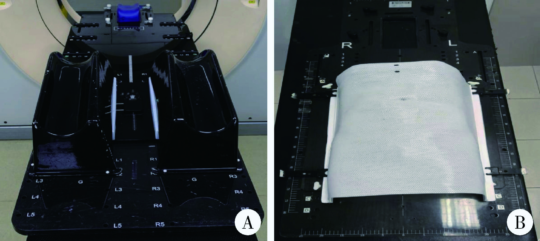
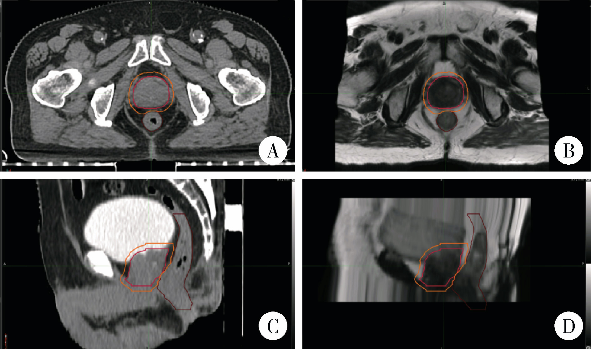
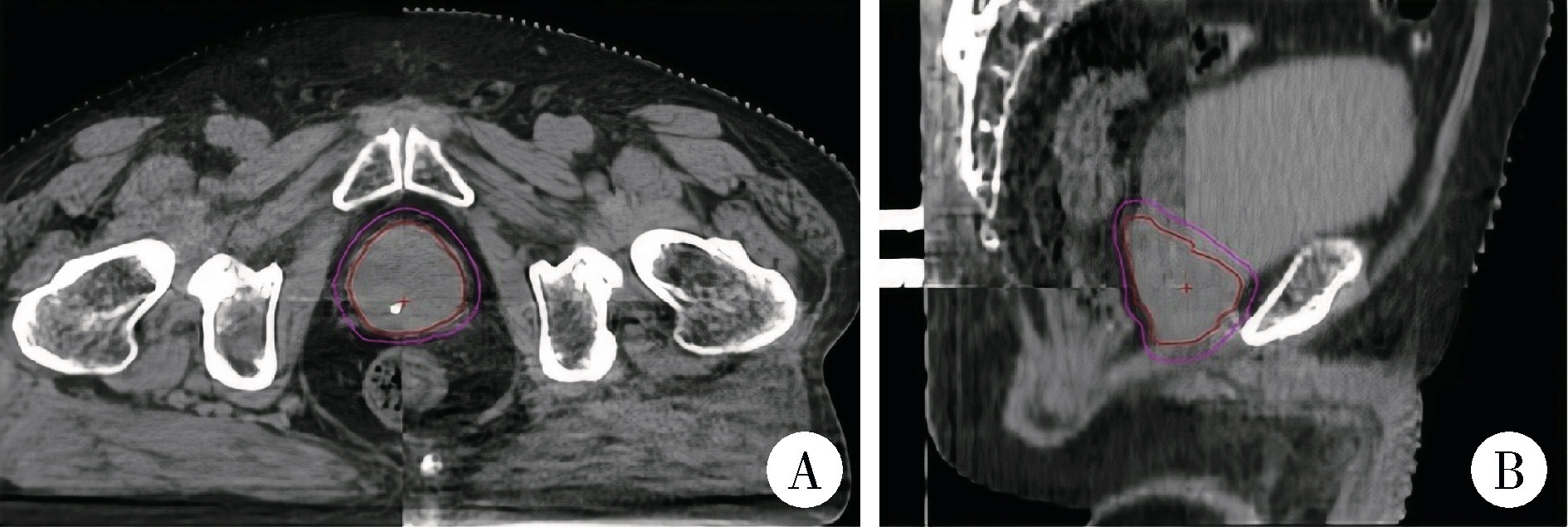
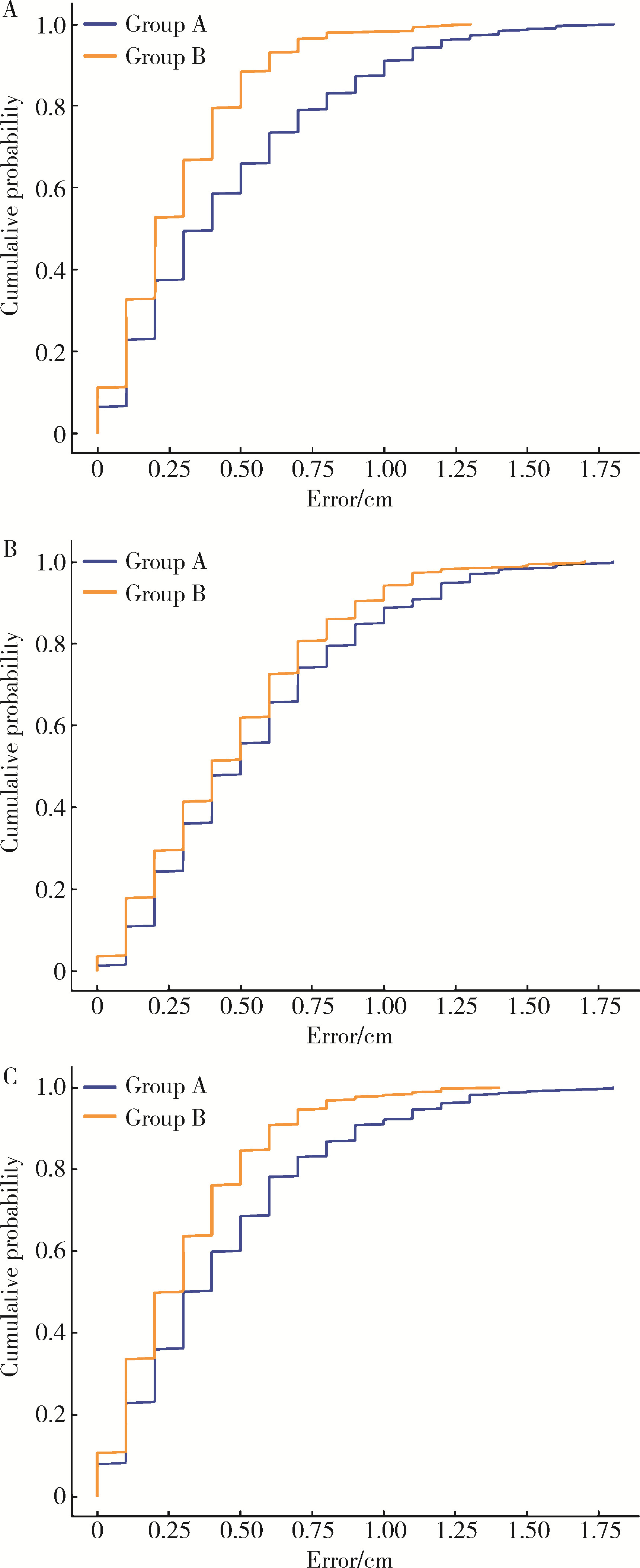
目的: 分析并比较前列腺癌根治性放射治疗中两种体位固定方式(截石位碳纤维全身固定架与常规碳纤维人体固定架结合热塑膜)的分次间摆位误差, 并计算两种方式下的临床靶区(clinical target volume, CTV)-计划靶区(planning target volume, PTV)外扩距离(margin for planning target volume, MPTV), 以优化体位固定方式和放射治疗流程。方法: 连续性纳入2021年8月至2023年3月在北京大学第一医院接受前列腺癌根治性放射治疗的37例患者的病例资料进行回顾性分析, 按体位固定方式分为两组[A组采用截石位碳纤维全身固定架18例, 共450组锥形束CT(cone-beam computed tomography, CBCT)图像, B组采用常规碳纤维人体固定架结合热塑膜19例, 共461组CBCT图像]。所有患者均行每日锥形束CT图像引导, 采用骨配准结合手动配准方法获取左右(X轴)、头脚(Y轴)及前后(Z轴)方向上的摆位误差数据。采用独立样本t检验和Mann-Whitney U检验和卡方检验比较两组摆位误差, 计算平均摆位误差、系统摆位误差(Σ)和随机摆位误差(δ), 并利用公式MPTV=2.5Σ+0.7δ计算CTV-PTV外扩距离。结果: 两组患者在三维方向上的摆位误差差异均有统计学意义(P均 < 0.01), A组XYZ轴方向误差绝对值的M (P25, P75)分别为0.40(0.20, 0.70) cm、0.50(0.30, 0.80) cm和0.35(0.20, 0.60) cm, 而B组相应数值显著降低至0.20(0.10, 0.40) cm、0.40(0.20, 0.70) cm和0.20(0.10, 0.40) cm。Mann-Whitney U检验结果显示各方向差异具有统计学意义(X轴: z=-6.86;Y轴: z=-2.76;Z轴: z=-5.71)。在摆位误差位移量≤0.5 cm的累积分布中, A组和B组的比例分别为X轴297(66.0%)和408(88.5%)(P0.01)、Y轴250(55.6%)和285(61.8%)(P=0.055)、Z轴308(68.4%)和391(84.8%)(P < 0.01)。两组的CTV-PTV外扩距离分别为X轴0.66 cm和0.35 cm, Y轴0.67 cm和0.45 cm, Z轴0.54 cm和0.42 cm。结论: 常规碳纤维人体固定架结合热塑膜能显著减少摆位误差。在XYZ轴三个方向上, 截石位碳纤维全身固定架的摆位误差较常规碳纤维人体固定架结合热塑膜更大, 因此需要进一步优化其摆位流程。由于截石位碳纤维全身固定架固定方法是经会阴超声引导技术中不可或缺的体位固定方式, 未来需针对其进行深入研究和改进, 以实现更高的摆位精度。

关键词: 前列腺癌, 锥形束CT, 摆位误差, 体位固定, 热塑膜

Abstract:

Objective: To analyze and compare the interfractional setup errors between two body positioning fixation methods (lithotomy position with carbon fiber full-body fixation frame vs. conventional carbon fiber body fixation frame combined with thermoplastic membrane) in radical radiotherapy for prostate cancer, and to calculate the clinical target volume (CTV) to planning target volume (PTV) margin (MPTV) for both methods to optimize immobilization techniques and radiotherapy workflows. Methods: A retrospective analysis was conducted on 37 consecutive patients who underwent radical prostate radiotherapy at Peking University First Hospital between August 2021 and March 2023. The patients were divided into two groups based on the immobilization method: Group A (18 patients, 450 CBCT image sets) used a carbon fiber whole-body fixator in the lithotomy position, while Group B (19 patients, 461 CBCT image sets) used a conventional carbon fiber fixator combined with a thermoplastic mask. All the patients underwent daily cone-beam computed tomography (CBCT) image guidance. Bone registration combined with manual registration was used to obtain the setup error data in the left-right (X), cranio-caudal (Y) and anterior-posterior (Z) directions. The positioning errors of the two groups were compared by using the independent sample t-test, the Mann-Whitney U test and the chi-square test. The average positioning error, systematic positioning error (Σ) and random positioning error (δ) were calculated, and the CTV-PTV extension distance was calculated by using the (MPTV=2.5Σ+0.7δ). Results: The analysis of the setup errors in the three-dimensional direction showed significant differences between the two groups (all P < 0.01). Specifically, the median (quartile) absolute values of the errors in the X, Y, and Z directions of group A were [0.40 (0.20, 0.70) cm, 0.50 (0.30, 0.80) cm, and 0.35 (0.20, 0.60) cm], respectively. In group B, the corresponding values were significantly reduced to [0.20 (0.10, 0.40) cm, 0.40 (0.20, 0.70) cm and 0.20 (0.10, 0.40) cm]. The results of Mann-Whitney U test showed that the differences in each direction were highly statistically significant (X: z=-6.86; Y: z=-2.76; Z: z=-5.71). The cumulative distribution ratio of the setup error displacement within 0.5 cm in the X, Y, and Z directions in group A and group B were 297 (66.0%) and 408 (88.5%) (P < 0.01), 250 (55.6%) and 285 (61.8%) (P=0.055), 308 (68.4%) and 391 (84.8%) (P < 0.01), respectively. The CTV-PTV margins in three directions were X 0.66 cm in group A and 0.35 cm in group B; Y 0.67 cm and 0.45 cm; Z 0.54 cm and 0.42 cm. Conclusion: Conventional carbon fiber human body fixator combined with thermoplastic film can significantly reduce the setup error. However, the carbon fiber whole-body fixator in the lithotomy position demonstrated larger setup errors in the X, Y, and Z directions compared with the conventional fixator combined with a thermoplastic mask, indicating the need for further workflow optimization. Given that the lithotomy position is essential for transperineal ultrasound-guided techniques, further research and improvements are required to achieve higher positioning accuracy.

Key words: Prostate cancer, Cone-beam computed tomography, Setup error, Immobilization, Thermoplastic mask

中图分类号: 

  • R737.25

图1

前列腺癌放射治疗体位固定装置"

图2

前列腺癌放射治疗患者的定位CT与MRI图像及靶区"

图3

前列腺癌放射治疗患者的CBCT匹配图像"

表1

A组与B组摆位误差比较"

Parameters Group A Group B z P
X (left-right)/cm 0.40 (0.20, 0.70) 0.20 (0.10, 0.40) -6.86 < 0.001
Y (cranio-caudal)/cm 0.50 (0.30, 0.80) 0.40 (0.20, 0.70) -2.76 0.006
Z (anterior-posterior)/cm 0.35 (0.20, 0.60) 0.20 (0.10, 0.40) -5.71 < 0.001

表2

A组与B组不同方向摆位误差位移量在0.5 cm内的累积分布比例"

Parameters Group A (n=450) Group B (n=461) P
X (left-right) 297 (66.0) 408 (88.5) < 0.001
Y (cranio-caudal) 250 (55.6) 285 (61.8) 0.055
Z (anterior-posterior) 308 (68.4) 391 (84.8) < 0.001

图4

A、B组摆位误差的累积分布"

表3

A组与B组CTV-PTV外放边界及分次间摆位误差的Σ、δ"

Parameters MPTV/cm Average setup error/cm Σ/cm δ/cm
X (left-right)
  Group A 0.66 0.46 0.175 0.321
  Group B 0.35 0.285 0.084 0.206
Y (cranio-caudal)
  Group A 0.67 0.552 0.178 0.314
  Group B 0.45 0.48 0.094 0.311
Z (anterior-posterior)
  Group A 0.54 0.438 0.126 0.321
  Group B 0.42 0.302 0.107 0.218
1
Wolff RF, Ryder S, Bossi A, et al. A systematic review of randomised controlled trials of radiotherapy for localised prostate cancer[J]. Eur J Cancer, 2015, 51(16): 2345- 2367.
2
Butler WM, Merrick GS, Reed JL, et al. Intrafraction displacement of prone versus supine prostate positioning monitored by real-time electromagnetic tracking[J]. J Appl Clin Med Phys, 2013, 14(2): 4141.
3
Daşu A. Is the alpha/beta value for prostate tumours low enough to be safely used in clinical trials[J]. Clin Oncol (R Coll Radiol), 2007, 19(5): 289- 301.
4
van Herk M. Errors and margins in radiotherapy[J]. Semin Radiat Oncol, 2004, 14(1): 52- 64.
5
Pommer T, Oh JH, Rosenschöld PMA, et al. Simulating intrafraction prostate motion with a random walk model[J]. Adv Radiat Oncol, 2017, 2(3): 429- 436.
6
Aluwini S, Pos F, Schimmel E, et al. Hypofractionated versus conventionally fractionated radiotherapy for patients with prostate cancer (HYPRO): Late toxicity results from a randomised, non-inferiority, phase 3 trial[J]. Lancet Oncol, 2016, 17(4): 464- 474.
7
Aluwini S, Pos F, Schimmel E, et al. Hypofractionated versus conventionally fractionated radiotherapy for patients with prostate cancer (HYPRO): Acute toxicity results from a randomised non-inferiority phase 3 trial[J]. Lancet Oncol, 2015, 16(3): 274- 283.
8
Dearnaley D, Syndikus I, Mossop H, et al. Conventional versus hypofractionated high-dose intensity-modulated radiotherapy for prostate cancer: 5-year outcomes of the randomised, non-inferiority, phase 3 CHHiP trial[J]. Lancet Oncol, 2016, 17(8): 1047- 1060.
9
King CR, Freeman D, Kaplan I, et al. Stereotactic body radiotherapy for localized prostate cancer: Pooled analysis from a multi-institutional consortium of prospective phase Ⅱ trials[J]. Radio-ther Oncol, 2013, 109(2): 217- 221.
10
马广栋, 洪莉. 宫颈癌的图像引导放疗研究综述[J]. 中国医疗设备, 2018, 33(5): 117-120, 129.
11
于松茂, 孟繁里, 康加阜, 等. 真空垫制作方式对肺癌立体定向放疗摆位精度影响研究[J]. 医疗卫生装备, 2019, 40(12): 33-35, 40.
12
马茗微, 王淑莲, 覃仕瑞, 等. 面罩及乳腺托架固定下乳腺癌保乳术后放疗锁骨上下区摆位误差分析[J]. 中华放射肿瘤学杂志, 2019, 28(3): 217- 221.
13
Murray J, Griffin C, Gulliford S, et al. A randomised assessment of image guided radiotherapy within a phase 3 trial of conventional or hypofractionated high dose intensity modulated radiotherapy for prostate cancer[J]. Radiother Oncol, 2020, 142, 62- 71.
14
高研, 高献书, 马茗微, 等. 前列腺癌放疗时CBCT使用频率和匹配策略的剂量学分析[J]. 中华放射肿瘤学杂志, 2024, 33(8): 733- 739.
15
Ghilezan M, Yan D, Martinez A. Adaptive radiation therapy for prostate cancer[J]. Semin Radiat Oncol, 2010, 20(2): 130- 137.
[1] 田素青, 孙海涛, 赵田地, 王巍. 6D治疗床辅助影像引导下放射治疗头颈部肿瘤摆位误差分析[J]. 北京大学学报(医学版), 2024, 56(6): 1097-1100.
[2] 黄教悌,胡菁,韩博. 治疗相关神经内分泌前列腺癌机制研究与靶向治疗新进展[J]. 北京大学学报(医学版), 2024, 56(4): 557-561.
[3] 邢念增,王明帅,杨飞亚,尹路,韩苏军. 前列腺免活检创新理念的临床实践及其应用前景[J]. 北京大学学报(医学版), 2024, 56(4): 565-566.
[4] 颜野,李小龙,夏海缀,朱学华,张羽婷,张帆,刘可,刘承,马潞林. 前列腺癌根治术后远期膀胱过度活动症的危险因素[J]. 北京大学学报(医学版), 2024, 56(4): 589-593.
[5] 于书慧,韩佳凝,钟丽君,陈聪语,肖云翔,黄燕波,杨洋,车新艳. 术前盆底肌电生理参数对前列腺癌根治性切除术后早期尿失禁的预测价值[J]. 北京大学学报(医学版), 2024, 56(4): 594-599.
[6] 薄士仕,高承志. 基于卷积神经网络实现锥形束CT牙齿分割及牙位标定[J]. 北京大学学报(医学版), 2024, 56(4): 735-740.
[7] 章锦花,潘洁,孙志鹏,王霄. 不同根管内容物对口腔颌面锥形束CT诊断牙根纵裂准确性的影响[J]. 北京大学学报(医学版), 2023, 55(2): 333-338.
[8] 叶佳学,梁宇红. 牙髓专科医师应用锥形束CT的现况调查[J]. 北京大学学报(医学版), 2023, 55(1): 114-119.
[9] 潘孟乔,刘建,徐莉,徐筱,侯建霞,李小彤,王晓霞. 牙周-正畸-正颌联合治疗骨性安氏Ⅲ类错畸形患者下前牙牙周表型的长期观察[J]. 北京大学学报(医学版), 2023, 55(1): 52-61.
[10] 高娟,吕航苗,马慧敏,赵一姣,李小彤. 锥形束CT三维体积测量评估骨性Ⅲ类错正畸正颌治疗后的牙根吸收[J]. 北京大学学报(医学版), 2022, 54(4): 719-726.
[11] 刘圣杰,侯惠民,吕政通,丁鑫,王璐,张磊,刘明. 双极雄激素序贯免疫检查点抑制剂治疗转移性去势抵抗性前列腺癌4例[J]. 北京大学学报(医学版), 2022, 54(4): 766-769.
[12] 刘伟涛,王怡然,王雪东,周彦恒. 锥形束CT研究上颌反复扩缩前方牵引后上颌骨缝的三维变化[J]. 北京大学学报(医学版), 2022, 54(2): 346-355.
[13] 白杲琛,宋毅,金杰,虞巍,何志嵩. 多西他赛联合卡铂治疗转移性去势抵抗性前列腺癌的临床疗效[J]. 北京大学学报(医学版), 2021, 53(4): 686-691.
[14] 孟圆,张丽琪,赵雅宁,柳登高,张祖燕,高岩. 67例上颌根尖周囊肿的三维影像特点分析[J]. 北京大学学报(医学版), 2021, 53(2): 396-401.
[15] 徐涛,韩敬丽,姚伟娟. 雄激素剥夺治疗相关心血管疾病的机制与临床对策[J]. 北京大学学报(医学版), 2020, 52(4): 607-609.
Viewed
Full text


Abstract

Cited

  Shared   
  Discussed   
No Suggested Reading articles found!