Journal of Peking University (Health Sciences) ›› 2026, Vol. 58 ›› Issue (1): 184-189. doi: 10.19723/j.issn.1671-167X.2026.01.024

Previous Articles     Next Articles

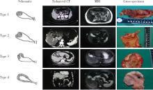
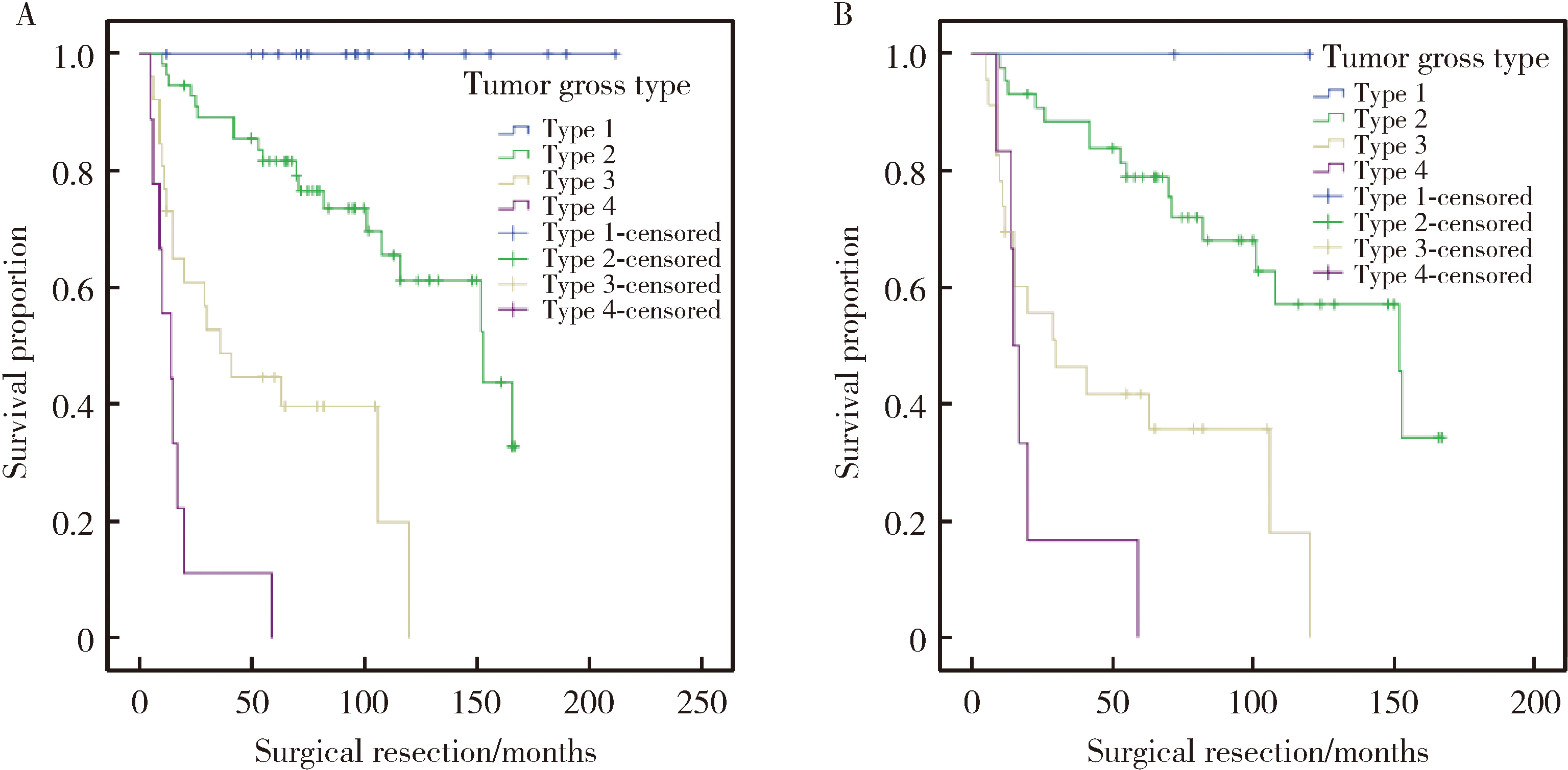
Gross classification of gallbladder cancer with primary lesion limited to the gallbladder wall and its correlation with prognosis and precancerous lesions

Lingfu ZHANG1, Ming CHEN2, Xiaoyu ZHAO1, Gang WANG1, Long CUI1, Xiaofeng LING1, Lixin WANG1, Zhi XU1, Limei GUO3,*(), Chunsheng HOU1,*()   

  1. 1. Department of General Surgery, Peking University Third Hospital, Beijing 100191, China
    2. Department of Radiology, Peking University Third Hospital, Beijing 100191, China
    3. Department of Pathology, Peking University Third Hospital, Beijing 100191, China
  • Received:2025-06-10 Online:2026-02-18 Published:2025-10-10
  • Contact: Limei GUO, Chunsheng HOU

RICH HTML


Abstract:

Objective: To explore the gross classification of gallbladder cancer with primary lesion confined within the gallbladder wall, and its correlation with prognosis and precancerous lesions. Methods: A retrospective study was conducted on 123 patients who were admitted to Peking University Third Hospital from January 2006 to December 2020. These patients had preoperative imaging findings suggesting that the primary lesion was confined within the gallbladder wall and had postoperative pathology of adenocarcinoma. Based on CT, MRI, or gross specimens, they were divided into the following four types: Type 1, simple intraluminal lesion: Intraluminal lesions without focal thickening of the gallbladder wall; Type 2, complex intraluminal lesion: Intraluminal lesions associated with focal thickening of the gallbladder wall and/or outer surface dimpling at the tumor base; Type 3, focal wall thickening: Circumferential focal wall thickening with heterogeneous enhancement within 2 continuous parts of the gallbladder; Type 4, diffuse wall thickening: Circumferential diffuse wall thickening extending more than 2 continuous parts of the gallbladder with heterogeneous enhancement. The clinical pathological characteristics, types of precancerous lesions, and survival status were compared among the different types. Results: Both preoperative CT/MRI and intraoperative gross specimens could serve as the basis for gross classification, with gross specimens demonstrating the highest accuracy rate. Among the 123 patients, 13 could not be classified, while the remaining 110 underwent gross classification. The gross classification of gallbladder cancer was strongly or moderately correlated with histopathological parameters such as T-stage (P < 0.001, rs=0.682), lymph node metastasis (P < 0.001, rs=0.478), tissue differentiation degree (P < 0.001, rs=0.484), nerve infiltration (P < 0.001, rs=0.490), and vascular invasion (P < 0.001, rs=0.334). The higher the classification, the more adverse histopathological parameters were observed. Additionally, the gross classification of gallbladder cancer was moderately strongly and highly strongly correlated with residual lesions after surgical treatment (P < 0.001, rs=0.328) and postoperative recurrence (P < 0.001, rs=0.619) in the patients. Survival analysis revealed that the higher the classification, the shorter the median survival time of the patients (Type 1: 96 months, Type 2: 73 months, Type 3: 30 months, Type 4: 14 months, P < 0.001). Multivariate Cox regression indicated that the gross classification of gallbladder cancer was an independent prognostic factor (HR=3.609, 95%CI: 2.177-5.983, P < 0.001). In the patients with the most heterogeneous biological behavior in stage T2, the gross classification of gallbladder cancer was also closely associated with prognosis (median survival times were 72, 70, 29, and 16 months, respectively, P < 0.001). Multivariate Cox regression further demonstrated that the gross classification of gallbladder cancer was an independent prognostic factor (HR=2.723, 95%CI: 1.566-4.736, P < 0.001). In terms of tumor origin, the gross classification of gallbladder cancer was significantly correlated with the type of precancerous lesions: Type 1 mainly originated from intracholecystic papillary neoplasm of the gallbladder, while Types 3 and 4 were mostly high-grade biliary intraepithelial neoplasm or without precancerous lesions. Analysis of the natural history of the tumor suggested that Type 1 progressed slowly, Type 4 progressed rapidly, and Type 2 exhibited greater heterogeneity. Conclusion: The gallbladder cancer with primary lesion confined within the gallbladder wall is closely related to prognosis and precancerous lesions, and can serve as an important reference for surgical decision-making and stratified management.

Key words: Gallbladder cancer, Gross classification, Precancerous lesions, Prognosis, Biological heterogeneity

CLC Number: 

  • R735.8

Figure 1

Each type of gallbladder cancer gross appearance Type 1, simple intraluminal lesion: Intraluminal lesions without focal thickening of the gallbladder wall; Type 2, complex intraluminal lesion: Intraluminal lesions associated with focal thickening of the gallbladder wall and/or outer surface dimpling at the tumor base; Type 3, focal wall thickening: Circumferential focal wall thickening with heterogeneous enhancement within 2 continuous parts of the gallbladder; Type 4, diffuse wall thickening: Circumferential diffuse wall thickening extending more than 2 continuous parts of the gallbladder with heterogeneous enhancement. The gallbladder is composed of 4 parts: fundus, body, neck and cystic duct."

Table 1

Comparison of clinical pathological characteristics among patients with different gross tumor types"

Parameters Type 1 (n=19) Type 2 (n=56) Type 3 (n=26) Type 4 (n=9) rs value P value
Baseline demographics
  Age/years, $\bar x \pm s$ 59.8±12.9 65.9±10.7 63.6±10.6 62.9±12.8 0.221
  Male/female, n 2/17 19/37 9/17 4/5 0.157
Clinicopathological characteristics
  Pancreaticobiliary maljunction, n 3 13 3 2 0.630
  Gallstone, n 6 19 9 5 0.628
  CA199 elevation*, n 4 10 5 4 0.089 0.371
  CEA elevation*, n 0 6 3 2 0.167 0.090
  T stage, n 0.682 <0.001
    Tis 12 2 0 0
    T1 5 10 0 0
    T2 2 44 23 6
    T3 0 0 3 3
  Lymph node metastasis, n (%) 0 (0) 12 (21) 15 (58) 5 (56) 0.478 <0.001
  Histology grade, n 0.484 <0.001
    Well 16 20 3 1
    Moderate 2 26 14 3
    Poor 1 10 9 5
  Neural invasion, n (%) 0 (0) 5 (9) 13 (50) 5 (56) 0.490 <0.001
  Lymphovascular invasion, n (%) 1 (5) 10 (18) 10 (19) 5 (56) 0.334 <0.001
  Residual disease, n (%) 0 (0) 3 (5) 6 (23) 3 (33) 0.328 <0.001
  Recurrence, n (%) 0 (0) 12 (21) 18 (69) 9 (100) 0.619 <0.001

Figure 2

Comparing cumulative survival based on different tumour gross type A, Kaplan-Meier survival curves comparing cumulative survival of cases among all 110 gallbladder cancers based on tumour gross type (Log-rank test, P < 0.001); B, Kaplan-Meier survival curves comparing cumulative survival of cases among 75 T2 gallbladder cancers based on tumour gross type (Log-rank test, P < 0.001)."

Table 2

Correlation between gross classification of gallbladder cancer and precancerous lesions"

Gross classification and precancerous lesions Type 1 (n=19) Type 2 (n=56) Type 3 (n=26)* Type 4 (n=9)
ICPN 18 23 0 0
High-grade BilIN 1 26 12 2
No precancerous lesion 0 6 12 7

Table 3

Natural course of the tumor based on gallbladder cancer gross classification"

Cases Tumor gross type Precancerous lesions Fist CT scan charateristics Interval of CTscan/months Second CT scan charateristics Operation date T stage
Case 1 Type 2 High-grade BilIN No polyps or localized thickening of the gallbladder wall 24 Intraluminal single polyp without focal thickening of the gallbladder wall 2010-11-5 Tis
Case 2 Type 3 ICPN Intraluminal single polyp without focal thickening of the gallbladder wall 12 Lesion diameter no change 2019-3-19 Tis
Case 3 Type 2 ICPN No polyps or localized thickening of the gallbladder wall 48 Intraluminal single polyp with focal thickening of the gallbladder wall 2013-7-18 T1b
Case 4 Type 2 ICPN No polyps or localized thickening of the gallbladder wall 12 Intraluminal single polyp with focal thickening of the gallbladder wall 2013-1-15 T2
Case 5 Type 4 No precancerous lesion No polyps or localized thickening of the gallbladder wall 9 Diffuse thickening of the gallbladder wall 2015-11-24 T3
1
Roa JC , García P , Kapoor VK , et al. Gallbladder cancer[J]. Nat Rev Dis Primers, 2022, 8, 69.

doi: 10.1038/s41572-022-00398-y
2
张铃福, 侯纯升, 徐智, 等. 腹腔镜胆囊切除术中或术后意外胆囊癌腹腔镜手术治疗: 单中心10年回顾性分析[J]. 中华外科杂志, 2019, 57 (4): 277- 281.
3
Zhang L , Hou C , Chen M , et al. Tumour radiological appearance evaluated by enhanced CT correlates with tumour progression and survival in curable gallbladder cancer[J]. Eur J Surg Oncol, 2020, 46 (11): 2099- 2105.

doi: 10.1016/j.ejso.2020.07.006
4
Mochidome N , Koga Y , Ohishi Y , et al. Prognostic implications of the coexisting precursor lesion types in invasive gallbladder cancer[J]. Hum Pathol, 2021, 114, 44- 53.

doi: 10.1016/j.humpath.2021.05.001
5
Nakanuma Y , Sugino T , Nomura Y , et al. Association of precursors with invasive adenocarcinoma of the gallbladder: A clinicopathological study[J]. Ann Diagn Pathol, 2022, 58, 151911.

doi: 10.1016/j.anndiagpath.2022.151911
6
Wakai T , Shirai Y , Yokoyama N , et al. Early gallbladder carcinoma does not warrant radical resection[J]. Br J Surg, 2001, 88 (5): 675- 678.

doi: 10.1046/j.1365-2168.2001.01749.x
7
张铃福, 侯纯升, 郭丽梅, 等. 术中冰冻或术后石蜡病理报告T1b期胆囊癌的外科治疗策略[J]. 北京大学学报(医学版), 2017, 49 (6): 1034- 1037.

doi: 10.3969/j.issn.1671-167X.2017.06.017
8
Kohya N , Kitahara K , Miyazaki K . Rational therapeutic strategy for T2 gallbladder carcinoma based on tumor spread[J]. World J Gastroenterol, 2010, 16 (28): 3567- 3572.

doi: 10.3748/wjg.v16.i28.3567
9
Okada KI , Kijima H , Imaizumi T , et al. Clinical significance of wall invasion pattern of subserosa-invasive gallbladder carcinoma[J]. Oncol Rep, 2012, 28 (5): 1531- 1536.

doi: 10.3892/or.2012.1971
10
Sung MK , Lee W , Lee JH , et al. Comparing survival rate and appropriate surgery methods according to tumor location in T2 gallbladder cancer[J]. Surg Oncol, 2022, 40, 101693.

doi: 10.1016/j.suronc.2021.101693
[1] Boda GUO, Min LU, Guoliang WANG, Hongxian ZHANG, Lei LIU, Xiaofei HOU, Lei ZHAO, Xiaojun TIAN, Shudong ZHANG. Clinicopathological and prognostic differences between clear cell and non-clear cell renal cell carcinoma with venous tumor thrombus [J]. Journal of Peking University (Health Sciences), 2025, 57(4): 644-649.
[2] Weihao LI, Jing LI, Xuemin ZHANG, Wei LI, Qingle LI, Xiaoming ZHANG. Effect of intraoperative blood salvage autotransfusion on the prognosis of patients after carotid body tumor resection [J]. Journal of Peking University (Health Sciences), 2025, 57(2): 272-276.
[3] Yaqing MAO, Zhen CHEN, Yao YU, Wenbo ZHANG, Yang LIU, Xin PENG. Impact of type 2 diabetes mellitus on the prognosis of patients with oral squamous cell carcinoma [J]. Journal of Peking University (Health Sciences), 2024, 56(6): 1089-1096.
[4] Junyong OU,Kunming NI,Lulin MA,Guoliang WANG,Ye YAN,Bin YANG,Gengwu LI,Haodong SONG,Min LU,Jianfei YE,Shudong ZHANG. Prognostic factors of patients with muscle invasive bladder cancer with intermediate-to-high risk prostate cancer [J]. Journal of Peking University (Health Sciences), 2024, 56(4): 582-588.
[5] Shuai LIU,Lei LIU,Zhuo LIU,Fan ZHANG,Lulin MA,Xiaojun TIAN,Xiaofei HOU,Guoliang WANG,Lei ZHAO,Shudong ZHANG. Clinical treatment and prognosis of adrenocortical carcinoma with venous tumor thrombus [J]. Journal of Peking University (Health Sciences), 2024, 56(4): 624-630.
[6] Le YU,Shaohui DENG,Fan ZHANG,Ye YAN,Jianfei YE,Shudong ZHANG. Clinicopathological characteristics and prognosis of multilocular cystic renal neoplasm of low malignant potential [J]. Journal of Peking University (Health Sciences), 2024, 56(4): 661-666.
[7] Zezhen ZHOU,Shaohui DENG,Ye YAN,Fan ZHANG,Yichang HAO,Liyuan GE,Hongxian ZHANG,Guoliang WANG,Shudong ZHANG. Predicting the 3-year tumor-specific survival in patients with T3a non-metastatic renal cell carcinoma [J]. Journal of Peking University (Health Sciences), 2024, 56(4): 673-679.
[8] Yangyi FANG,Qiang LI,Zhigao HUANG,Min LU,Kai HONG,Shudong ZHANG. Well-differentiated papillary mesothelial tumour of the tunica vaginalis: A case report [J]. Journal of Peking University (Health Sciences), 2024, 56(4): 741-744.
[9] Yuanyuan ZENG,Yun XIE,Daonan CHEN,Ruilan WANG. Related factors of euthyroid sick syndrome in patients with sepsis [J]. Journal of Peking University (Health Sciences), 2024, 56(3): 526-532.
[10] Jian-bin LI,Meng-na LYU,Qiang CHI,Yi-lin PENG,Peng-cheng LIU,Rui WU. Early prediction of severe COVID-19 in patients with Sjögren’s syndrome [J]. Journal of Peking University (Health Sciences), 2023, 55(6): 1007-1012.
[11] Huan-rui LIU,Xiang PENG,Sen-lin LI,Xin GOU. Risk modeling based on HER-2 related genes for bladder cancer survival prognosis assessment [J]. Journal of Peking University (Health Sciences), 2023, 55(5): 793-801.
[12] Zi-xuan XUE,Shi-ying TANG,Min QIU,Cheng LIU,Xiao-jun TIAN,Min LU,Jing-han DONG,Lu-lin MA,Shu-dong ZHANG. Clinicopathologic features and prognosis of young renal tumors with tumor thrombus [J]. Journal of Peking University (Health Sciences), 2023, 55(5): 802-811.
[13] Han LU,Jian-yun ZHANG,Rong YANG,Le XU,Qing-xiang LI,Yu-xing GUO,Chuan-bin GUO. Clinical factors affecting the prognosis of lower gingival squamous cell carcinoma [J]. Journal of Peking University (Health Sciences), 2023, 55(4): 702-707.
[14] Yun-fei SHI,Hao-jie WANG,Wei-ping LIU,Lan MI,Meng-ping LONG,Yan-fei LIU,Yu-mei LAI,Li-xin ZHOU,Xin-ting DIAO,Xiang-hong LI. Analysis of clinicopathological and molecular abnormalities of angioimmunoblastic T-cell lymphoma [J]. Journal of Peking University (Health Sciences), 2023, 55(3): 521-529.
[15] Xiao-juan ZHU,Hong ZHANG,Shuang ZHANG,Dong LI,Xin LI,Ling XU,Ting LI. Clinicopathological features and prognosis of breast cancer with human epidermal growth factor receptor 2 low expression [J]. Journal of Peking University (Health Sciences), 2023, 55(2): 243-253.
Viewed
Full text


Abstract

Cited

  Shared   
  Discussed   
No Suggested Reading articles found!