Journal of Peking University (Health Sciences) ›› 2020, Vol. 52 ›› Issue (4): 730-737. doi: 10.19723/j.issn.1671-167X.2020.04.026

Previous Articles     Next Articles

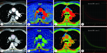
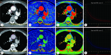
Comparative imaging study of mediastinal lymph node from pre-surgery dual energy CT versus post-surgeron verifications in non-small cell lung cancer patients

Qiao ZHU,Cui REN,Yan ZHANG,Mei-jiao LI,Xiao-hua WANG()   

  1. Department of Radiology, Peking University Third Hospital, Beijing 100191, China
  • Received:2019-12-19 Online:2020-08-18 Published:2020-08-06
  • Contact: Xiao-hua WANG E-mail:medwxh@126.com
  • Supported by:
    National Natural Foundation of China(81871326)

Abstract:

Objective: To validate the value of dual energy CT (DECT) in the differentiation of mediastinal metastatic lymph nodes from non-metastatic lymph nodes in non-small cell lung cancer (NSCLC). Methods: In the study, 57 surgically confirmed NSCLC patients who underwent enhanced DECT scan within 2 weeks before operation were enrolled. Two radiologists analyzed the CT images before operation. All mediastinal lymph nodes with short diameter≥5 mm on axial images were included in this study. The morphological parameters [long-axis diameter (L), short-axis diameter (S) and S/L of lymph nodes] and the DECT parameters [iodine concentration (IC), normalized iodine concentration (NIC), slope of spectral hounsfield unit curve (λHU) and effective atomic number (Zeff) in arterial and venous phase] were measured. The differences of morphological parameters and DECT parameters between metastatic and non-metastatic lymph nodes were compared. The parameters with significant difference were analyzed by the Logistic regression model, then a new predictive variable was established. Receiver operator characteristic (ROC) analyses were performed for S, NIC in venous phase and the new predictive variable. Results: In 57 patients, 49 metastatic lymph nodes and 938 non-metastatic lymph nodes were confirmed by surgical pathology. A total of 163 mediastinal lymph nodes (49 metastatic, 114 non-metastatic) with S≥5 mm were detected on axial CT images. The S, L and S/L of metastatic lymph nodes were significantly higher than those of non-metastatic lymph nodes (P<0.05). The DECT parameters of metastatic lymph nodes were significantly lower than those of non-metastatic lymph nodes (P<0.05). The best single morphological parameter for differentiation between metastatic and nonmetastatic lymph nodes was S (AUC, 0.752; threshold, 8.5 mm; sensitivity, 67.4%; specificity, 73.7%; accuracy, 71.8%). The best single DECT parameter for differentiation between metastatic and nonmetastatic lymph nodes was NIC in venous phase (AUC, 0.861; threshold, 0.53; sensitivity, 95.9%; specificity, 70.2%; accuracy, 77.9%). Multivariate analysis showed that S and NIC were independent predictors of lymph node metastasis. The AUC of combined S and NIC in the venous phase was 0.895(sensitivity, 79.6%; specificity, 87.7%; accuracy, 85.3%), which were significantly higher than that of S (P<0.001) and NIC (P=0.037). Conclusion: The ability of quantitative DECT parameters to distinguish mediastinal lymph node metastasis in NSCLC patients is better than that of morphological parameters. Combined S and NIC in venous phase can be used to improve preoperative diagnostic accuracy of metastatic lymph nodes.

Key words: Non-small cell lung cancer, Lymph node metastasis, Tomography, X-ray computed

CLC Number: 

  • R734.2

Figure 1

A 63-year-old female patient with right lower lobe adenocarcinoma, clinical stage was T2bN2M0, and the right fourth group of lymph nodes were metastatic A, 70 kV monochromatic contrast-enhancement image in arterial phase shows the lymph node with a short-axis diameter of 7.32 mm, and the ROI with an area of 33.18 mm2; B, iodine based material-decomposition image in arterial phase shows that iodine concentration (IC) of the lymph node is 13.59×100 μg/cm3 and IC of the thoracic aorta in the same slice is 164.38×100 μg/cm3; C, effective atomic number CT image in arterial phase shows the effective atomic number (Zeff) of the lymph node is 8.44; D, spectral attenuation curve of lymph node in arterial phase, and the slope of attenuation curve (λHU) is 1.61; E, venous phase 70 kV monochromatic contrast-enhanced CT image; F, iodine based material-decomposition image in venous phase, and IC of the lymph node is 27.35×100 μg/cm3 and IC of the thoracic aorta in the same slice is 52.24×100 μg/cm3; G, effective atomic number CT image in venous phase, and Zeff of the lymph node is 9.12; H, spectral attenuation curve of lymph node in venous phase. λHU is 3.24."

Figure 2

A 71-year-old male patient with right upper lobe adenocarcinoma, clinical stage was T1N0M0,and the right fourth group of lymph nodes were non-metastatic A, 70 kV monochromatic contrast-enhancement image in arterial phase (AP) shows the lymph node with a short-axis diameter of 9.98 mm, and the ROI with an area of 71.03 mm2; B, iodine based material-decomposition image in arterial phase shows that iodine concentration (IC) of the lymph node is 21.20×100 μg/cm3 and IC of the thoracic aorta in the same slice is 135.24×100 μg/cm3; C, effective atomic number CT image in arterial phase shows the effective atomic number (Zeff) of the lymph node is 8.81; D, spectral attenuation curve of lymph node in arterial phase, and the slope of attenuation curve (λHU) is 2.51; E, venous phase (VP) 70 kV monochromatic contrast-enhanced CT image; F, iodine based material-decomposition image in venous phase, and IC of the lymph node is 28.81×100 μg/cm3 and IC of the thoracic aorta in the same slice is 37.49×100 μg/cm3; G, effective atomic number CT image in venous phase, and Zeff of the lymph node is 9.14; H, spectral attenuation curve of lymph node in venous phase. λHU is 3.35."

Table 1

Distribution of metastatic and non-metastatic lymph nodes in the regions of mediastinum"

Group 2R 2L 3A 3P 4R 4L 5 6 7 8R 8L 9R 9L 10R 10L
Metastatic (n=49) 2 1 2 2 4 1 4 2 15 1 1 2 3 5 4
Non-metastatic (n=114) 1 0 0 1 14 19 8 7 28 4 0 5 1 15 11
Total 3 1 2 3 18 20 12 9 43 5 1 7 4 20 15

Table 2

Intra-observer and inter-observer agreement on morphological parameters and dual-energy parameters measurement"

Items Doctor A Doctor A and B
ICC 95%CI ICC 95%CI
S 0.872 0.566-0.947 0.849 0.743-0.912
L 0.891 0.643-0.954 0.932 0.873-0.963
Arterial phase
IC 0.930 0.303-0.980 0.897 0.818-0.942
NIC 0.880 0.798-0.930 0.853 0.730-0.919
λHU 0.891 0.690-0.952 0.839 0.614-0.923
Zeff 0.949 0.723-0.982 0.920 0.864-0.954
Venous phase
IC 0.833 0.484-0.929 0.901 0.420-0.967
NIC 0.891 0.690-0.952 0.839 0.614-0.923
λHU 0.903 0.335-0.970 0.812 0.340-0.927
Zeff 0.869 0.150-0.960 0.927 0.874-0.958

Table 3

Comparison of morphologic indexes between metastatic and non-metastatic mediastinal lymph nodes in non-small cell lung cancer (x?±s)"

Group L/mm S/mm S/L
Metastatic (n=49) 13.10±2.99 9.90±2.55 0.76±0.09
Non-metastatic (n=114) 11.38±2.91 7.72±1.96 0.69±0.08
t 3.432 5.899 5.075
P 0.001 <0.001 <0.001

Table 4

Comparison of quantitative dual-energy CT parameters between metastatic and non-metastatic mediastinal lymph nodes in non-small cell lung cancer ($\bar{x}±s$)"

Characteristic Metastatic (n=49) Non-metastatic (n=114) t P
Arterial phase
IC/(100 μg/cm3) 22.14±5.11 24.94±3.79 -3.448 <0.001
NIC 0.25±0.04 0.36±0.09 -10.300 <0.001
λHU 2.13±0.21 2.33±0.23 -5.194 <0.001
Zeff 8.07±0.46 8.34±0.51 -3.213 0.002
Venous phase
IC/(100 μg/cm3) 27.24±5.68 30.26±5.60 -3.139 0.002
NIC 0.43±0.07 0.60±0.14 -10.875 <0.001
λHU 2.46±0.31 2.84±0.29 -7.405 <0.001
Zeff 8.25±0.37 8.55±0.45 -4.052 <0.001

Table 5

Results of ROC analysis of morphologic indexes and dual-energy CT parameters in differential diagnosis of mediastinal metastatic and non-metastatic lymph nodes in non-small cell lung cancer (NSCLC)"

Items AUC Sensitivity/% Specivicity/% Positive predictive
value/%
Negative predictive
value/%
Accuracy/%
Parameter threshold
S 8.50 0.752(0.672-0.832) 67.4(52.5-80.1) 73.7(64.6-81.5) 52.4(43.3-61.3) 84.0(77.6-88.8) 71.8(64.2 -78.5)
L 10.94 0.654(0.564-0.745) 79.6(65.7-89.8) 50.0(40.5-59.5) 40.6(35.2-46.3) 85.1(76.1-91.1) 58.9(50.9 -66.5)
S/L 0.76 0.734(0.644-0.825) 57.1(42.2-71.2) 84.2(76.2-90.4) 60.9(48.8-71.7) 82.1(76.6-86.4) 76.1(68.8-82.4)
Arterial phase
IC 22.23 0.660(0.561-0.759) 61.2(46.2-74.8) 70.2(60.9-78.4) 46.9(38.1-55.8) 80.8(74.4-85.9) 67.5(59.7-74.6)
NIC 0.31 0.850(0.793-0.908) 91.8(80.4-97.7) 69.3(60.0-77.6) 56.3(49.1-63.2) 95.2(88.5-98.1) 76.1(68.8-82.4)
λHU 2.11 0.754(0.674-0.834) 57.1(42.2-71.2) 84.2(76.2-90.4) 60.9(48.8-71.7) 82.1(76.6-86.4) 76.1(68.8-82.4)
Zeff 8.34 0.657(0.569-0.744) 73.5(58.9-85.1) 56.1(46.5-65.4) 41.9(35.5-48.5) 83.1(75.0-89.0) 61.4(53.4-68.9)
Venous phase
IC 25.56 0.643(0.544-0.742) 44.9(30.7-59.8) 83.3(75.2-89.7) 53.7(40.9-66.0) 77.9(73.0-82.1) 71.8(64.2-78.5)
NIC 0.53 0.861(0.806-0.915) 95.9(86.0-99.5) 70.2(60.9-78.4) 58.0(50.9-64.8) 97.6(91.1- 99.4) 77.9(70.8-84.0)
λHU 2.72 0.807(0.736-0.878) 81.6(68.0-91.2) 66.7(57.2-75.2) 51.3(44.0-58.5) 89.4(82.2-93.9) 71.2(63.6-78.0)
Zeff 8.74 0.701(0.624-0.779) 89.8(77.8-96.6) 52.6(43.1-62.1) 44.9(39.6-50.3) 92.3(83.7-96.6) 63.8(55.9-71.2)
LogitP 0.56 0.895(0.846-0.943) 79.6(65.7-89.8) 87.7(80.3-93.1) 73.6(62.6-82.3) 90.9(85.1- 94.6) 85.3(78.9-90.3)

Figure 3

ROC curves of short-axis diameter (S), normalized iodine concentration (NIC) in venous phase and combined S and NIC in venous phase (LogitP) for differentiating metastatic and non-metastatic mediastinal lymph nodes in patients with NSCLC T, threshold."

[1] Siegel RL, Miller KD, Jemal A. Cancer statistics, 2018[J]. CA Cancer J Clin, 2018,68(1):7-30.
[2] Ferlay J, Colombet M, Soerjomataram I, et al. Estimating the global cancer incidence and mortality in 2018: GLOBOCAN sources and methods[J]. Int J Cancer, 2019,144(8):1941-1953.
doi: 10.1002/ijc.31937 pmid: 30350310
[3] 中华医学会, 中华医学会肿瘤学分会, 中华医学会杂志社. 中华医学会肺癌临床诊疗指南(2018版)[J]. 中华肿瘤杂志, 2018,40(12):935-964.
[4] Torabi M, Aquino SL, Harisinghani MG. Current concepts in lymph node imaging[J]. J Nucl Med, 2004,45(9):1509-1518.
pmid: 15347718
[5] Vansteenkiste J, Dooms C, De Leyn P. Early stage non-small cell lung cancer: challenges in staging and adjuvant treatment: evidence-based staging[J]. Ann Oncol, 2010, 21(Suppl 7): vii189-vii195.
[6] 宁先英, 李浩, 杨明, 等. CT能谱定量分析对肺腺癌与鳞癌的鉴别诊断价值[J]. 放射学实践, 2017,32(3):237-241.
[7] Lv P, Lin XZ, Li J, et al. Differentiation of small hepatic hemangioma from small hepatocellular carcinoma: recently introduced spectral CT method[J]. Radiology, 2011,259(3):720-729.
doi: 10.1148/radiol.11101425 pmid: 21357524
[8] Martin SS, Weidinger S, Czwikla R, et al. Iodine and fat quantification for differentiation of adrenal gland adenomas from metas-tases using third-generation dual-source dual-energy computed tomography[J]. Invest Radiol, 2018,53(3):173-178.
pmid: 28990974
[9] Muenzel D, Lo GC, Yu HS, et al. Material density iodine images in dual-energy CT: detection and characterization of hypervascular liver lesions compared to magnetic resonance imaging[J]. Eur J Radiol, 2017,95(10):300-306.
doi: 10.1016/j.ejrad.2017.08.035
[10] Liu X, Ouyang D, Li H, et al. Papillary thyroid cancer: dual-energy spectral CT quantitative parameters for preoperative diagnosis of metastasis to the cervical lymph nodes[J]. Radiology, 2015,275(1):167-176.
pmid: 25521777
[11] De Leyn P, Vansteenkiste J, Cuypers P, et al. Role of cervical mediastinoscopy in staging of non-small cell lung cancer without enlarged mediastinal lymph nodes on CT scan[J]. Eur J Cardiothorac Surg, 1997,12(5):706-712.
doi: 10.1016/s1010-7940(97)00253-4 pmid: 9458140
[12] Fukuya T, Honda H, Hayashi T, et al. Lymph-node metastases: efficacy for detection with helical CT in patients with gastric cancer[J]. Radiology, 1995,197(3):705-711.
pmid: 7480743
[13] Yoshimura G, Sakurai T, Oura S, et al. Evaluation of axillary lymph node status in breast cancer with MRI[J]. Breast Cancer, 1999,6(3):249-258.
pmid: 11091725
[14] Li X, Meng X, Ye Z. Iodine quantification to characterize primary lesions, metastatic and non-metastatic lymph nodes in lung cancers by dual energy computed tomography: an initial experience[J]. Eur J Radiol, 2016,85(6):1219-1223.
doi: 10.1016/j.ejrad.2016.03.030 pmid: 27161073
[15] Rizzo S, Radice D, Femia M, et al. Metastatic and non-metastatic lymph nodes: quantification and different distribution of iodine uptake assessed by dual-energy CT[J]. Eur Radiol, 2018,28(2):760-769.
doi: 10.1007/s00330-017-5015-5 pmid: 28835993
[16] Yang Z, Zhang X, Fang M, et al. Preoperative diagnosis of regional lymph node metastasis of colorectal cancer with quantitative parameters from dual-energy CT[J]. AJR Am J Roentgenol, 2019,213(6):1-9.
[17] Zhang X, Zheng C, Yang Z, et al. Axillary sentinel lymph nodes in breast cancer: quantitative evaluation at dual-energy CT[J]. Radiology, 2018,289(2):337-346.
doi: 10.1148/radiol.2018180544 pmid: 30152748
[18] Yang F, Dong J, Wang X, et al. Non-small cell lung cancer: spectral computed tomography quantitative parameters for preoperative diagnosis of metastatic lymph nodes[J]. Eur J Radiol, 2017,89(4):129-135.
[19] Lin LY, Zhang Y, Suo ST, et al. Correlation between dual-energy spectral CT imaging parameters and pathological grades of non-small cell lung cancer[J]. Clin Radiol, 2018, 73(4): 412.e1-412.e7.
doi: 10.1016/j.crad.2017.10.020 pmid: 29195660
[20] 崔元龙, 许毛荣, 文智. 能谱CT定量参数对非小细胞肺癌纵隔淋巴结转移中的应用价值[J]. 临床放射学杂志, 2019,38(5):825-829.
[21] 叶亚君, 莫淑琼. CT能谱成像鉴别诊断纵隔淋巴结良恶性的临床意义[J]. 现代医用影像学, 2018,27(8):2702-2703.
[22] Pan Z, Pang L, Ding B, et al. Gastric cancer staging with dual energy spectral CT imaging[J]. PLoS One, 2013,8(2):e53651.
doi: 10.1371/journal.pone.0053651 pmid: 23424614
[23] Yang L, Luo D, Li L, et al. Differentiation of malignant cervical lymphadenopathy by dual-energy CT: a preliminary analysis[J]. Sci Rep, 2016,6(1):31020.
[1] Xiaotong LING,Liuyang QU,Danni ZHENG,Jing YANG,Xuebing YAN,Denggao LIU,Yan GAO. Three-dimensional radiographic features of calcifying odontogenic cyst and calcifying epithelial odontogenic tumor [J]. Journal of Peking University (Health Sciences), 2024, 56(1): 131-137.
[2] Deng-hui DUAN,Hom-Lay WANG,En-bo WANG. Role of collagen membrane in modified guided bone regeneration surgery using buccal punch flap approach: A retrospective and radiographical cohort study [J]. Journal of Peking University (Health Sciences), 2023, 55(6): 1097-1104.
[3] Xiang LIU,Hui-hui XIE,Yu-feng XU,Xiao-dong ZHANG,Xiao-feng TAO,Lin LIU,Xiao-ying WANG. Value of artificial intelligence in the improvement of diagnostic consistency of radiology residents [J]. Journal of Peking University (Health Sciences), 2023, 55(4): 670-675.
[4] Jin-hua ZHANG,Jie PAN,Zhi-peng SUN,Xiao WANG. Effect of various intracanal materials on the diagnostic accuracy of cone-beam computed tomography in vertical root fractures [J]. Journal of Peking University (Health Sciences), 2023, 55(2): 333-338.
[5] Jia-xue YE,Yu-hong LIANG. A prevalence survey of cone-beam computed tomography use among endodontic practitioners [J]. Journal of Peking University (Health Sciences), 2023, 55(1): 114-119.
[6] Meng-qiao PAN,Jian LIU,Li XU,Xiao XU,Jian-xia HOU,Xiao-tong LI,Xiao-xia WANG. A long-term evaluation of periodontal phenotypes before and after the periodontal-orthodontic-orthognathic combined treatment of lower anterior teeth in patients with skeletal Angle class Ⅲ malocclusion [J]. Journal of Peking University (Health Sciences), 2023, 55(1): 52-61.
[7] Yu FU,Xin-nong HU,Sheng-jie CUI,Jie SHI. Decompensation effectiveness and alveolar bone remodeling analysis of mandibular anterior teeth after preoperative orthodontic treatment in high-angle patients with skeletal class Ⅱ malocclusion [J]. Journal of Peking University (Health Sciences), 2023, 55(1): 62-69.
[8] Yu WANG,Hui-min ZHANG,Xue-rong DENG,Wei-wei LIU,Lu CHEN,Ning ZHAO,Xiao-hui ZHANG,Zhi-bo SONG,Yan GENG,Lan-lan JI,Yu WANG,Zhuo-li ZHANG. Diagnostic values of urinary citrate for kidney stones in patients with primary gout [J]. Journal of Peking University (Health Sciences), 2022, 54(6): 1134-1140.
[9] Juan GAO,Hang-miao LV,Hui-min MA,Yi-jiao ZHAO,Xiao-tong LI. Evaluation of root resorption after surgical orthodontic treatment of skeletal Class Ⅲ malocclusion by three-dimensional volumetric measurement with cone-beam CT [J]. Journal of Peking University (Health Sciences), 2022, 54(4): 719-726.
[10] WANG Shu-lei,GAO Yang-xu,ZHANG Hong-wu,YANG Hai-bo,LI Hui,LI Yu,SHEN Li-xue,YAO Hong-xin. Clinical analysis of 30 cases of basal ganglia germinoma in children [J]. Journal of Peking University (Health Sciences), 2022, 54(2): 222-226.
[11] LIU Wei-tao,WANG Yi-ran,WANG Xue-dong,ZHOU Yan-heng. A cone-beam computed tomography evaluation of three-dimensional changes of circummaxillary sutures following maxillary protraction with alternate rapid palatal expansions and constrictions [J]. Journal of Peking University (Health Sciences), 2022, 54(2): 346-355.
[12] Gang YANG,Wen-jie HU,Jie CAO,Deng-gao LIU. Three-dimensional morphology analysis of the supraosseous gingival profile of periodontally healthy maxillary anterior teeth [J]. Journal of Peking University (Health Sciences), 2021, 53(5): 990-994.
[13] LI Xin-fei, PENG Yi-ji, YU Xiao-teng, XIONG Sheng-wei, CHENG Si-da, DING Guang-pu, YANG Kun-lin, TANG Qi, MI Yue, WU Jing-yun, ZHANG Peng, XIE Jia-xin, HAO Han, WANG He, QIU Jian-xing, YANG Jian, LI Xue-song, ZHOU Li-qun. Three dimensional nephrometry system for partial nephrectomy: Our initial exploration [J]. Journal of Peking University (Health Sciences), 2021, 53(3): 613-622.
[14] LIAO Xu-he,WANG Rong-fu,LIU Meng,CHEN Xue-qi,XIONG Yan,NONG Lin,YIN Lei,ZHANG Bing-ye,DU Yu-jing. Semiquantitative parameters of 18F-FDG PET/CT, gene mutation states of epidermal growth factor receptor and anaplastic lymphoma kinase in prognosis evaluation of patients with lung adenocarcinoma [J]. Journal of Peking University (Health Sciences), 2021, 53(2): 246-254.
[15] MENG Yuan,ZHANG Li-qi,ZHAO Ya-ning,LIU Deng-gao,ZHANG Zu-yan,GAO Yan. Three-dimentional radiographic features of 67 maxillary radicular cysts [J]. Journal of Peking University (Health Sciences), 2021, 53(2): 396-401.
Viewed
Full text


Abstract

Cited

  Shared   
  Discussed   
[1] . [J]. Journal of Peking University(Health Sciences), 2007, 39(3): 225 -328 .
[2] . [J]. Journal of Peking University(Health Sciences), 2001, 33(6): 565 .
[3] . [J]. Journal of Peking University(Health Sciences), 2008, 40(1): 39 -42 .
[4] . [J]. Journal of Peking University(Health Sciences), 2008, 40(6): 600 -602 .
[5] . [J]. Journal of Peking University(Health Sciences), 2009, 41(1): 90 -94 .
[6] . [J]. Journal of Peking University(Health Sciences), 2007, 39(5): 498 -502 .
[7] . [J]. Journal of Peking University(Health Sciences), 2001, 33(6): 559 -561 .
[8] . [J]. Journal of Peking University(Health Sciences), 2003, 35(5): 499 -502 .
[9] . [J]. Journal of Peking University(Health Sciences), 2005, 37(6): 599 -602 .
[10] . [J]. Journal of Peking University(Health Sciences), 2006, 38(2): 197 -200 .