Journal of Peking University (Health Sciences) ›› 2024, Vol. 56 ›› Issue (4): 735-740. doi: 10.19723/j.issn.1671-167X.2024.04.030
Previous Articles Next Articles
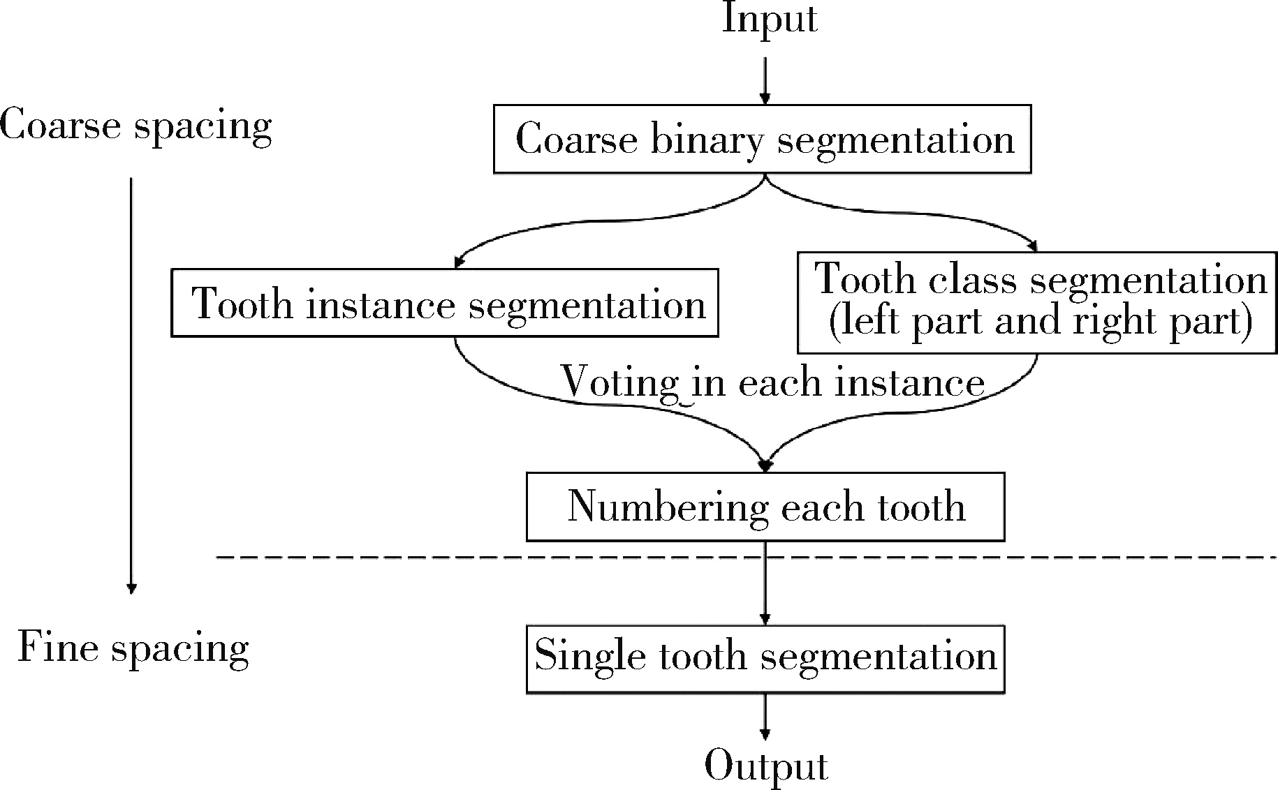
Tooth segmentation and identification on cone-beam computed tomography with convolutional neural network based on spatial embedding information
Shishi BO1,2,Chengzhi GAO2,*( )
)
- 1. Department of General Dentistry Ⅱ, Peking University School and Hospital of Stomatology & National Center for Stomatology & National Clinical Research Center for Oral Diseases & National Engineering Research Center of Oral Biomaterials and Digital Medical Devices, Beijing 100081, China
2. Department of Dentistry, Peking University People' s Hospital, Beijing 100044, China
CLC Number:
- R780.1


| 1 |
Liao FZ , Liang M , Li Z , et al. Evaluate the malignancy of pulmonary nodules using the 3-D deep leaky noisy-OR network[J]. IEEE Trans Neural Netw Learn Syst, 2019, 30 (11): 3484- 3495.
doi: 10.1109/TNNLS.2019.2892409 |
| 2 |
Long J , Shelhamer E , Darrell T . Fully convolutional networks for semantic segmentation[J]. IEEE Trans Pattern Anal Mach Intell, 2017, 39 (4): 640- 651.
doi: 10.1109/TPAMI.2016.2572683 |
| 3 | Ronneberger O, Fischer P, Brox T. U-Net: Convolutional networks for biomedical image segmentation: International conference on medical image computing and computer-assisted intervention[C]. Cham: Springer, 2015. |
| 4 | Neven D, Brabandere BD, Proesmans M, et al. Instance segmentation by jointly optimizing spatial embeddings and clustering bandwidth: 2019 IEEE/CVF conference on computer vision and pattern recognition (CVPR)[C]. Long Beach, CA: IEEE, 2019. |
| 5 |
Pei YR , Ai XS , Zha HB , et al. 3D exemplar-based random walks for tooth segmentation from cone-beam computed tomography images[J]. Med Phys, 2016, 43 (9): 5040- 5050.
doi: 10.1118/1.4960364 |
| 6 | Patil S , Kulkarni V , Bhise A . Algorithmic analysis for dental caries detection using an adaptive neural network architecture[J]. Heliyon, 2019, 5 (5): e1579. |
| 7 |
Zhang KL , Wu J , Chen H , et al. An effective teeth recognition method using label tree with cascade network structure[J]. Comput Med Imaging Graph, 2018, 68, 61- 70.
doi: 10.1016/j.compmedimag.2018.07.001 |
| 8 |
Hosntalab M , Aghaeizadeh ZR , Abbaspour TA , et al. Classification and numbering of teeth in multi-slice CT images using wavelet-Fourier descriptor[J]. Int J Comput Assist Radiol Surg, 2010, 5 (3): 237- 249.
doi: 10.1007/s11548-009-0389-8 |
| 9 |
Hwang JJ , Jung YH , Cho BH , et al. An overview of deep learning in the field of dentistry[J]. Imaging Sci Dent, 2019, 49 (1): 1- 7.
doi: 10.5624/isd.2019.49.1.1 |
| 10 |
Chen H , Zhang KL , Lyu PJ , et al. A deep learning approach to automatic teeth detection and numbering based on object detection in dental periapical films[J]. Sci Rep, 2019, 9 (1): 3840.
doi: 10.1038/s41598-019-40414-y |
| 11 |
Miki Y , Muramatsu C , Hayashi T , et al. Classification of teeth in cone-beam CT using deep convolutional neural network[J]. Comput Biol Med, 2017, 80, 24- 29.
doi: 10.1016/j.compbiomed.2016.11.003 |
| 12 | Cui ZM, Li CJ, Wang WP. ToothNet: Automatic tooth instance segmentation and identification from cone beam CT images: 2019 IEEE/CVF conference on computer vision and pattern recognition (CVPR)[C]. Long Beach, CA: IEEE, 2019. |
| 13 | He KM, Zhang XY, Ren SQ, et al. Deep residual learning for image recognition: 2016 IEEE conference on computer vision and pattern recognition (CVPR)[C]. Las Vegas: IEEE, 2016. |
| 14 | Dentistry-designation system for teeth and areas of the oral cavity: ISO 3950: 2009[S/OL]. [2016-03-01]. https://www.iso.org/standard/68292.html. |
| 15 |
Dice LR . Measures of the amount of ecologic association between species[J]. Ecology, 1945, 26 (3): 297- 302.
doi: 10.2307/1932409 |
| [1] | Yue WANG, Yuhong LIANG. Florid cemento-osseous dysplasia: A case report [J]. Journal of Peking University (Health Sciences), 2026, 58(1): 220-224. |
| [2] | Jie LIU, Mingwei MA, Qing'an WANG, Ming SHI, Jinpeng YIN, Zhanping WANG, Jingtao SHEN, Xianshu GAO. Comparison of setup errors between two immobilization methods in prostate cancer radiotherapy based on cone-beam computed tomography [J]. Journal of Peking University (Health Sciences), 2025, 57(4): 692-697. |
| [3] | Xiaotong LING,Liuyang QU,Danni ZHENG,Jing YANG,Xuebing YAN,Denggao LIU,Yan GAO. Three-dimensional radiographic features of calcifying odontogenic cyst and calcifying epithelial odontogenic tumor [J]. Journal of Peking University (Health Sciences), 2024, 56(1): 131-137. |
| [4] | Deng-hui DUAN,Hom-Lay WANG,En-bo WANG. Role of collagen membrane in modified guided bone regeneration surgery using buccal punch flap approach: A retrospective and radiographical cohort study [J]. Journal of Peking University (Health Sciences), 2023, 55(6): 1097-1104. |
| [5] | Jin-hua ZHANG,Jie PAN,Zhi-peng SUN,Xiao WANG. Effect of various intracanal materials on the diagnostic accuracy of cone-beam computed tomography in vertical root fractures [J]. Journal of Peking University (Health Sciences), 2023, 55(2): 333-338. |
| [6] | Jia-xue YE,Yu-hong LIANG. A prevalence survey of cone-beam computed tomography use among endodontic practitioners [J]. Journal of Peking University (Health Sciences), 2023, 55(1): 114-119. |
| [7] | Meng-qiao PAN,Jian LIU,Li XU,Xiao XU,Jian-xia HOU,Xiao-tong LI,Xiao-xia WANG. A long-term evaluation of periodontal phenotypes before and after the periodontal-orthodontic-orthognathic combined treatment of lower anterior teeth in patients with skeletal Angle class Ⅲ malocclusion [J]. Journal of Peking University (Health Sciences), 2023, 55(1): 52-61. |
| [8] | Yu FU,Xin-nong HU,Sheng-jie CUI,Jie SHI. Decompensation effectiveness and alveolar bone remodeling analysis of mandibular anterior teeth after preoperative orthodontic treatment in high-angle patients with skeletal class Ⅱ malocclusion [J]. Journal of Peking University (Health Sciences), 2023, 55(1): 62-69. |
| [9] | Juan GAO,Hang-miao LV,Hui-min MA,Yi-jiao ZHAO,Xiao-tong LI. Evaluation of root resorption after surgical orthodontic treatment of skeletal Class Ⅲ malocclusion by three-dimensional volumetric measurement with cone-beam CT [J]. Journal of Peking University (Health Sciences), 2022, 54(4): 719-726. |
| [10] | LIU Wei-tao,WANG Yi-ran,WANG Xue-dong,ZHOU Yan-heng. A cone-beam computed tomography evaluation of three-dimensional changes of circummaxillary sutures following maxillary protraction with alternate rapid palatal expansions and constrictions [J]. Journal of Peking University (Health Sciences), 2022, 54(2): 346-355. |
| [11] | Gang YANG,Wen-jie HU,Jie CAO,Deng-gao LIU. Three-dimensional morphology analysis of the supraosseous gingival profile of periodontally healthy maxillary anterior teeth [J]. Journal of Peking University (Health Sciences), 2021, 53(5): 990-994. |
| [12] | MENG Yuan,ZHANG Li-qi,ZHAO Ya-ning,LIU Deng-gao,ZHANG Zu-yan,GAO Yan. Three-dimentional radiographic features of 67 maxillary radicular cysts [J]. Journal of Peking University (Health Sciences), 2021, 53(2): 396-401. |
| [13] | ZHOU Jing,LIU Yi. Cone-beam CT evaluation of temporomandibular joint in skeletal class Ⅱ female adolescents with different vertical patterns [J]. Journal of Peking University (Health Sciences), 2021, 53(1): 109-119. |
| [14] | GAO Lu,GU Yan. Chinese morphological stages of midpalatal suture and its correlation with Demirjian dental age [J]. Journal of Peking University (Health Sciences), 2021, 53(1): 133-138. |
| [15] | Ye YAN,Hai-zhui XIA,Xu-sheng LI,Wei HE,Xue-hua ZHU,Zhi-ying ZHANG,Chun-lei XIAO,Yu-qing LIU,Hua HUANG,Liang-hua HE,Jian LU. Application of U-shaped convolutional neural network in auto segmentation and reconstruction of 3D prostate model in laparoscopic prostatectomy navigation [J]. Journal of Peking University(Health Sciences), 2019, 51(3): 596-601. |
|
||