Journal of Peking University (Health Sciences) ›› 2024, Vol. 56 ›› Issue (1): 131-137. doi: 10.19723/j.issn.1671-167X.2024.01.020

Previous Articles     Next Articles

Three-dimensional radiographic features of calcifying odontogenic cyst and calcifying epithelial odontogenic tumor

Xiaotong LING1,Liuyang QU1,Danni ZHENG1,Jing YANG1,Xuebing YAN2,Denggao LIU1,*(),Yan GAO3   

  1. 1. Department of Oral and Maxillofacial Radiology, Peking University School and Hospital of Stomatology & National Center for Stomatology & National Clinical Research Center for Oral Diseases & National Engineering Research Center of Oral Biomaterials and Digital Medical Devices & Beijing Key Laboratory of Digital Stomatology & NHC Research Center of Engineering and Technology for Computerized Dentistry & NMPA Key Laboratory for Dental Materials, Beijing 100081, China
    2. Special Dental Department, The First Clinical Division, Peking University School and Hospital of Stomatology & National Center for Stomatology & National Clinical Research Center for Oral Diseases & National Engineering Research Center of Oral Biomaterials and Digital Medical Devices & Beijing Key Laboratory of Digital Stomatology & NHC Research Center of Engineering and Technology for Computerized Dentistry & NMPA Key Laboratory for Dental Materials, Beijing 100081, China
    3. Department of Oral Pathology, Peking University School and Hospital of Stomatology & National Center for Stomatology & National Clinical Research Center for Oral Diseases & National Engineering Research Center of Oral Biomaterials and Digital Medical Devices & Beijing Key Laboratory of Digital Stomatology & NHC Research Center of Engineering and Technology for Computerized Dentistry & NMPA Key Laboratory for Dental Materials, Beijing 100081, China
  • Received:2023-06-27 Online:2024-02-18 Published:2024-02-06
  • Contact: Denggao LIU E-mail:kqldg@bjmu.edu.cn

RICH HTML


Abstract:

Objective: To analyze the three-dimensional radiographic characteristics of calcifying odontogenic cyst and calcifying epithelial odontogenic tumor using spiral computed tomography (CT) and cone-beam computed tomography (CBCT). Methods: Clinical records, histopathological reports, and CBCT or non-enhanced spiral CT images of 19 consecutive patients with calcifying odontogenic cyst (COC) and 16 consecutive patients with calcifying epithelial odontogenic tumor (CEOT) were retrospectively acquired, and radiographic features, including location, size, expansion, internal structure and calcification, were analyzed. Results: Among the 19 COC cases (12 males and 7 females, with an average age of 27 years), 89.5% (17/19) of the lesions originated from the anterior and premolar areas, 100.0% of them exhibited cortex expansion, and 78.9% had discontinued cortex. Among the 16 CEOT cases (3 males and 13 females, with an average age of 36 years), 81.3% (13/16) of the lesions were in the premolar and molar areas, 56.3% of them exhibited cortex expansion, and 96.8% had discontinued cortex. According to the distribution of internal calcifications, these lesions were divided into: Ⅰ (non-calcification type): absence of calcification; Ⅱ (eccentric marginal type): multiple calcifications scattered along one side of the lesion; Ⅲ (diffused type): numerous calcifications diffusely distributed into the lesion; Ⅳ (plaque type): with a ≥ 5 mm calcified patch; Ⅴ (peri-coronal type): multiple calcifications clustered around impacted teeth. Calcifications were present in 73.7% of COC lesions, including 9 type Ⅱ, 3 type Ⅲ and 2 type Ⅳ lesions, and 42.8% of CEOT lesions had calcification images, including 2 type Ⅲ and 5 type Ⅴ lesions. Six COC lesions had odontoma-like images. Moreover, 8 of 9 type Ⅰ CEOTs were histologically Langerhans cell-rich subtype, which had a smaller size (with an average mesiodistal diameter of 17.8 mm) and were not associated with impacted teeth. Conclusion: COC lesions tended to originate from the anterior part of the jaw and exhibit cortex expansion, and were sometimes associated with odontoma. CEOT commonly occurred in the posterior jaw and had discontinued cortex. Two lesions had significantly different calcification map. Over 70% of COC lesions had calcification images, which were mostly scattered along one side of the cysts, far from the impacted teeth. Approximately 60% of CEOT lesions exhibited smaller size and non-calcification, and the remaining CEOT cases often had calcification images clustered around the impacted teeth.

Key words: Calcifying odontogenic cyst, Calcifying epithelial odontogenic tumor, Three-dimensional imaging, Cone-beam computed tomography

CLC Number: 

  • R739.8

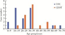
Figure 1

Age distribution of patients with COC or CEOT COC, calcifying odontogenic cyst; CEOT, calcifying epithelial odontogenic tumor."

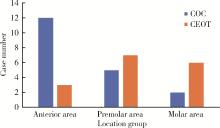
Figure 2

Location distribution of COC or CEOT COC, calcifying odontogenic cyst; CEOT, calcifying epithelial odontogenic tumor."

Table 1

Cortex expansion and discontinued cortex in COC and CEOT"

Lesions Cortex expansion Discontinued cortex
Presence, n Absence, n P value Presence, n Absence, n P value
COC 19 0 0.002 15 4 0.374
CEOT 9 7 15 1

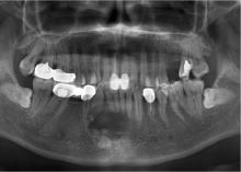
Figure 3

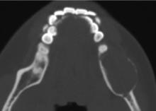
COC associated with an odontoma COC, calcifying odontogenic cyst."

Figure 4

A multilocular image of a COC COC, calcifying odontogenic cyst."

Figure 5

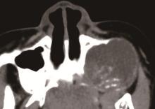
Non-calcification type: Absence of calcification (CEOT) CEOT, calcifying epithelial odontogenic tumor."

Figure 6

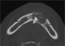
Eccentric marginal type of COC: Multiple calcifications scattered along one side of the lesion (COC) COC, calcifying odontogenic cyst."

Figure 7

Diffused type: Numerous calcifications diffusely distributed into the lesion (COC) COC, calcifying odontogenic cyst."

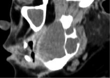
Figure 8

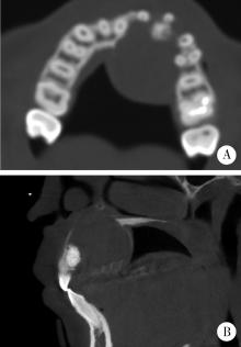
Plaque type: With a ≥5 mm calcified patch A, axial; B, sagital. CBCT images showed a expansive radiolucent lesion in the left maxilla. Note the calcified patch located at the margin of the cyst (COC). CBCT, cone-beam computed tomography; COC, calcifying odontogenic cyst."

Figure 9

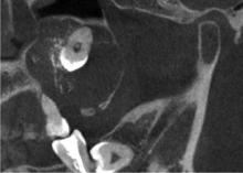
Peri-coronal type: Multiple calcifications clustered around an impacted tooth (CEOT) CEOT, calcifying epithelial odontogenic tumor."

Table 2

Calcification features of COC and CEOT"

Lesions Calcification features, n P value
Type Ⅱ Type Ⅲ Type Ⅳ Type Ⅴ
COC 9 3 2 0 < 0.001
CEOT 0 2 0 5

Figure 10

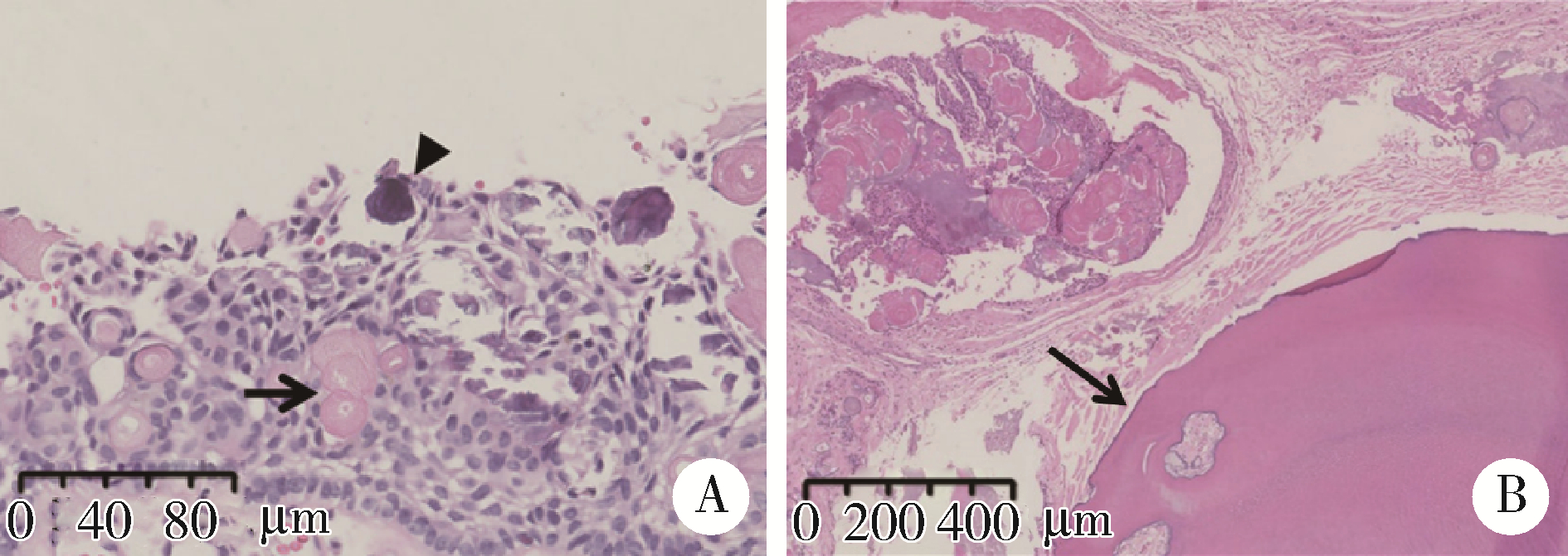
Photomicrographs of COC (HE staining) A, cystic epithelium is keratinized and produces ghost cells (arrow) and calcifications (arrowhead); B, the dental hard tissues resembling an odontoma (arrow) were laid adjacent to the cyst wall. COC, calcifying odontogenic cyst."

Figure 11

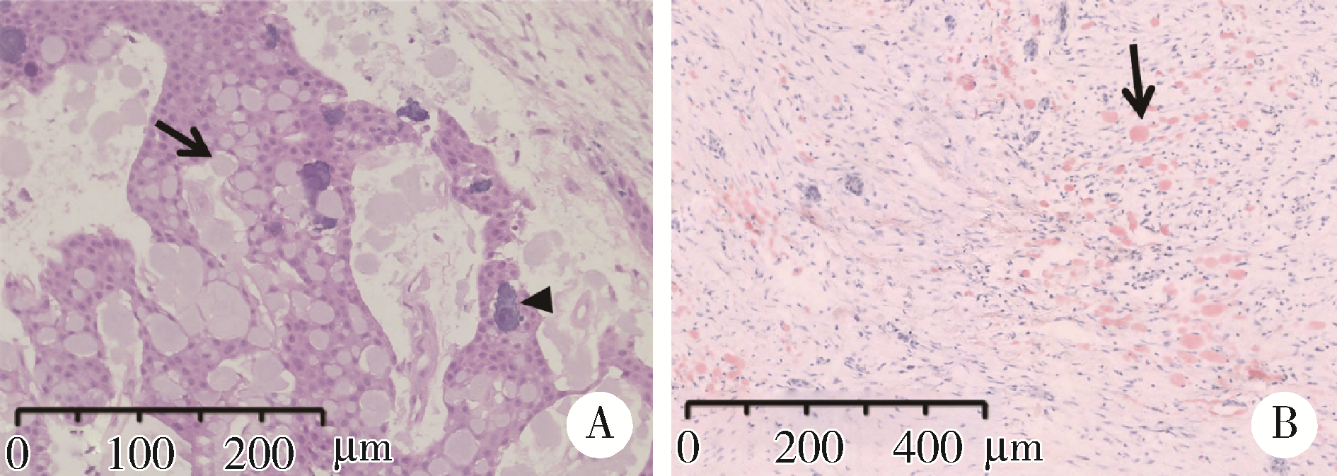
Photomicrographs of CEOT A, HE staining showed proliferative epithelial islands with amyloid-like materials (arrow) and concentric calcifications (arrowhead); B, Congo red staining showed the brick-red amyloid-like materials distributed in the tumor cells (arrow). CEOT, calcifying epithelial odontogenic tumor."

1 Lee SK , Kim YS . Current concepts and occurrence of epithelial odontogenic tumors: Ⅱ. Calcifying epithelial odontogenic tumor versus ghost cell odontogenic tumors derived from calcifying odontogenic cyst[J]. Korean J Pathol,, 2014, 48 (3): 175- 187.
doi: 10.4132/KoreanJPathol.2014.48.3.175
2 马绪臣. 口腔颌面医学影像学[M]. 北京: 北京大学医学出版社, 2006.
3 Philipsen HP , Reichart PA . Adenomatoid odontogenic tumour: Facts and figures[J]. Oral Oncol, 1999, 35 (2): 125- 131.
doi: 10.1016/S1368-8375(98)00111-0
4 Bansal SP , Shaikh S , Arvandekar AS , et al. Analysis of 55 cases of adenomatoid odontogenic tumor in an Indian population and review of literature[J]. Med Oral Patol Oral Cir Bucal, 2022, 27 (1): e85- e93.
5 陈菲, 张庆庆, 陆东辉, 等. 牙源性钙化囊性瘤临床病理研究[J]. 临床与实验病理学杂志, 2012, 28 (8): 891- 894.
doi: 10.3969/j.issn.1001-7399.2012.08.014
6 付文荣, 程正江. 牙源性钙化上皮瘤3例临床病理观察[J]. 诊断病理学杂志, 2014, 21 (4): 231- 233.
doi: 10.3969/j.issn.1007-8096.2014.04.012
7 Sonone A , Sabane VS , Desai R . Calcifying ghost cell odontogenic cyst: Report of a case and review of literature[J]. Case Rep Dent, 2011, 2011, 328743.
8 刘梅, 孙国文, 唐恩溢, 等. 牙源性钙化囊肿的临床病理分析[J]. 实用口腔医学杂志, 2020, 36 (1): 96- 99.
doi: 10.3969/j.issn.1001-3733.2020.01.021
9 陶谦, 梁培盛. 2017版WHO牙源性肿瘤新分类之述评[J]. 口腔疾病防治, 2017, 25 (12): 749- 754.
doi: 10.12016/j.issn.2096-1456.2017.12.001
10 Soluk-Tekkesin M , Wright JM . The World Health Organization classification of odontogenic lesions: A summary of the changes of the 2022 (5th) edition[J]. Turk Patoloji Derg, 2022, 38 (2): 168- 184.
11 Buchner A . The central (intraosseous) calcifying odontogenic cyst: An analysis of 215 cases[J]. J Oral Maxillofac Surg, 1991, 49 (4): 330- 339.
doi: 10.1016/0278-2391(91)90365-S
12 Ahmad SA , Popli DB , Sircar K , et al. Calcifying odontogenic cyst: Report of an uncommon entity with a brief literature review[J]. J Oral Maxillofac Pathol, 2022, 26 (1): 131.
13 Chandran A , Nachiappan S , Selvakumar R , et al. Calcifying epithelial odontogenic cyst of maxilla: Report of a case and review and discussion on the terminology and classification[J]. J Microsc Ultrastruct, 2020, 9 (2): 98- 102.
14 de Arruda JAA , Schuch LF , Abreu LG , et al. A multicenter study of 268 cases of calcifying odontogenic cysts and a literature review[J]. Oral Dis, 2018, 24 (7): 1282- 1293.
doi: 10.1111/odi.12906
15 Rojo R , Prados-Frutos JC , Gutierrez Lázaro I , et al. Calcifying odontogenic cysts[J]. J Stomatol Oral Maxillofac Surg, 2017, 118 (2): 122- 124.
doi: 10.1016/j.jormas.2016.10.007
16 Chindasombatjaroen J , Poomsawat S , Boonsiriseth K . Two unique cases of calcifying cystic odontogenic tumor in the maxillary posterior region[J]. Oral Surg Oral Med Oral Pathol Oral Radiol, 2014, 118 (4): 497- 504.
doi: 10.1016/j.oooo.2014.06.006
17 Santos HBP , de Morais EF , Moreira DGL , et al. Calcifying odontogenic cyst with extensive areas of dentinoid: uncommon case report and update of main findings[J]. Case Rep Pathol, 2018, 2018, 8323215.
18 Gamoh S , Akiyama H , Furukawa C , et al. Calcifying cystic odontogenic tumor accompanied by a dentigerous cyst: A case report[J]. Oncol Lett, 2017, 14 (5): 5785- 5790.
19 Hirshberg A , Kaplan I , Buchner A . Calcifying odontogenic cyst associated with odontoma: A possible separate entity (odontocalcifying odontogenic cyst)[J]. J Oral Maxillofac Surg, 1994, 52 (6): 555- 558.
doi: 10.1016/0278-2391(94)90087-6
20 Vizuete-Bolaños MX , Salgado-Chavarria F , Ramírez-Martínez CM , et al. Compound odontoma associated with a calcifying odontogenic cyst. Case report and systematic review[J]. J Stomatol Oral Maxillofac Surg, 2022, 123 (3): e97- e105.
doi: 10.1016/j.jormas.2021.10.008
21 张艳宁, 侯亚丽, 于美清, 等. 牙源性钙化上皮瘤1例临床病理分析及文献复习[J]. 实用口腔医学杂志, 2021, 37 (3): 431- 433.
doi: 10.3969/j.issn.1001-3733.2021.03.032
22 Mujib BR , Kulkarni PG , Lingappa A , et al. An atypical presentation of Pindborg tumor in anterior maxilla[J]. Dent Res J (Isfahan), 2012, 9 (4): 495- 498.
23 Ibituruna ACH , Costa ARGF , Paulo LFB , et al. Multiple calcifying epithelial odontogenic tumor: Case report and review of the literature[J]. Oral Surg Oral Med Oral Pathol Oral Radiol, 2019, 128 (3): 268- 272.
doi: 10.1016/j.oooo.2019.03.018
24 Zhang A , Chaw SY , Talacko AA , et al. Central calcifying epithelial odontogenic tumour in the posterior maxilla: A case report[J]. Aust Dent J, 2016, 61 (3): 381- 385.
doi: 10.1111/adj.12384
25 王凯利, 郑广宁, 刘莉, 等. 牙源性钙化上皮瘤2例[J]. 华西口腔医学杂志, 2016, 34 (1): 104- 107.
26 Vered M , Wright JM . Update from the 5th edition of the World Health Organization classification of head and neck tumors: Odontogenic and maxillofacial bone tumours[J]. Head Neck Pathol, 2022, 16 (1): 63- 75.
doi: 10.1007/s12105-021-01404-7
27 王丽, 汪说之, 陈新明, 等. 牙源性钙化上皮瘤中朗格汉斯细胞的研究[J]. 口腔医学研究, 2005, 21 (6): 645- 648.
doi: 10.3969/j.issn.1671-7651.2005.06.016
28 Hong SP , Ellis GL , Hartman KS . Calcifying odontogenic cyst. A review of ninety-two cases with reevaluation of their nature as cysts or neoplasms, the nature of ghost cells, and subclassification[J]. Oral Surg Oral Med Oral Pathol, 1991, 72 (1): 56- 64.
doi: 10.1016/0030-4220(91)90190-N
29 刘轶芳. 245例颌骨良恶性肿瘤的CT影像学特征的对比研究[D]. 上海: 上海交通大学, 2020.
30 叶婷婷, 陈蔚华, 裴婧, 等. 青少年牙本质生成性影细胞瘤1例[J]. 口腔医学研究, 2022, 38 (10): 995- 996.
31 王予江, 李琳琳. 外周型牙源性钙化囊性瘤病例报道[C]//中华口腔医学会口腔颌面外科专业委员会. 第十四次中国口腔颌面外科学术会议论文汇编. 重庆: [出版者不详], 2018: 1.
[1] Shishi BO,Chengzhi GAO. Tooth segmentation and identification on cone-beam computed tomography with convolutional neural network based on spatial embedding information [J]. Journal of Peking University (Health Sciences), 2024, 56(4): 735-740.
[2] Deng-hui DUAN,Hom-Lay WANG,En-bo WANG. Role of collagen membrane in modified guided bone regeneration surgery using buccal punch flap approach: A retrospective and radiographical cohort study [J]. Journal of Peking University (Health Sciences), 2023, 55(6): 1097-1104.
[3] Wen ZHANG,Xiao-jing LIU,Zi-li LI,Yi ZHANG. Effect of alar base cinch suture based on anatomic landmarks on the morphology of nasolabial region in patients after orthognathic surgery [J]. Journal of Peking University (Health Sciences), 2023, 55(4): 736-742.
[4] Jin-hua ZHANG,Jie PAN,Zhi-peng SUN,Xiao WANG. Effect of various intracanal materials on the diagnostic accuracy of cone-beam computed tomography in vertical root fractures [J]. Journal of Peking University (Health Sciences), 2023, 55(2): 333-338.
[5] Ao-nan WEN,Wei LIU,Da-wei LIU,Yu-jia ZHU,Ning XIAO,Yong WANG,Yi-jiao ZHAO. Preliminary evaluation of the trueness of 5 chairside 3D facial scanning techniques [J]. Journal of Peking University (Health Sciences), 2023, 55(2): 343-350.
[6] Jia-xue YE,Yu-hong LIANG. A prevalence survey of cone-beam computed tomography use among endodontic practitioners [J]. Journal of Peking University (Health Sciences), 2023, 55(1): 114-119.
[7] Meng-qiao PAN,Jian LIU,Li XU,Xiao XU,Jian-xia HOU,Xiao-tong LI,Xiao-xia WANG. A long-term evaluation of periodontal phenotypes before and after the periodontal-orthodontic-orthognathic combined treatment of lower anterior teeth in patients with skeletal Angle class Ⅲ malocclusion [J]. Journal of Peking University (Health Sciences), 2023, 55(1): 52-61.
[8] Yu FU,Xin-nong HU,Sheng-jie CUI,Jie SHI. Decompensation effectiveness and alveolar bone remodeling analysis of mandibular anterior teeth after preoperative orthodontic treatment in high-angle patients with skeletal class Ⅱ malocclusion [J]. Journal of Peking University (Health Sciences), 2023, 55(1): 62-69.
[9] Juan GAO,Hang-miao LV,Hui-min MA,Yi-jiao ZHAO,Xiao-tong LI. Evaluation of root resorption after surgical orthodontic treatment of skeletal Class Ⅲ malocclusion by three-dimensional volumetric measurement with cone-beam CT [J]. Journal of Peking University (Health Sciences), 2022, 54(4): 719-726.
[10] LIU Wei-tao,WANG Yi-ran,WANG Xue-dong,ZHOU Yan-heng. A cone-beam computed tomography evaluation of three-dimensional changes of circummaxillary sutures following maxillary protraction with alternate rapid palatal expansions and constrictions [J]. Journal of Peking University (Health Sciences), 2022, 54(2): 346-355.
[11] Gang YANG,Wen-jie HU,Jie CAO,Deng-gao LIU. Three-dimensional morphology analysis of the supraosseous gingival profile of periodontally healthy maxillary anterior teeth [J]. Journal of Peking University (Health Sciences), 2021, 53(5): 990-994.
[12] MENG Yuan,ZHANG Li-qi,ZHAO Ya-ning,LIU Deng-gao,ZHANG Zu-yan,GAO Yan. Three-dimentional radiographic features of 67 maxillary radicular cysts [J]. Journal of Peking University (Health Sciences), 2021, 53(2): 396-401.
[13] ZHOU Jing,LIU Yi. Cone-beam CT evaluation of temporomandibular joint in skeletal class Ⅱ female adolescents with different vertical patterns [J]. Journal of Peking University (Health Sciences), 2021, 53(1): 109-119.
[14] GAO Lu,GU Yan. Chinese morphological stages of midpalatal suture and its correlation with Demirjian dental age [J]. Journal of Peking University (Health Sciences), 2021, 53(1): 133-138.
[15] WANG Yi-ran, ZHOU Yan-heng, WANG Xue-dong, WEI Song, LIU Wei-tao. Evaluation of maxillary three-dimensional changes in maxillary protraction with alternating rapid palatal expansion and constriction based on the cone-beam computed tomography [J]. Journal of Peking University(Health Sciences), 2018, 50(4): 685-693.
Viewed
Full text


Abstract

Cited

  Shared   
  Discussed   
[1] . [J]. Journal of Peking University(Health Sciences), 2007, 39(4): 434 -436 .
[2] . [J]. Journal of Peking University(Health Sciences), 2001, 33(1): 50 -53 .
[3] . [J]. Journal of Peking University(Health Sciences), 2001, 33(1): 45 .
[4] . [J]. Journal of Peking University(Health Sciences), 2001, 33(6): 540 -544 .
[5] . [J]. Journal of Peking University(Health Sciences), 2002, 34(1): 33 -35 .
[6] . [J]. Journal of Peking University(Health Sciences), 2002, 34(2): 97 -98 .
[7] . [J]. Journal of Peking University(Health Sciences), 2002, 34(2): 140 -143 .
[8] . [J]. Journal of Peking University(Health Sciences), 2010, 42(4): 476 -479 .
[9] . [J]. Journal of Peking University(Health Sciences), 2008, 40(2): 208 -210 .
[10] . [J]. Journal of Peking University(Health Sciences), 2010, 42(5): 520 -525 .