北京大学学报(医学版) ›› 2019, Vol. 51 ›› Issue (3): 530-535. doi: 10.19723/j.issn.1671-167X.2019.03.023
弥散张量成像联合虚拟现实三维重建在功能区胶质瘤手术中的应用
陈素华1,杨军1△( ),韩鸿宾2,3,崔德华2,孙建军1,马长城1,和清源2,3,林国中1,韩芸峰1,吴超1,马凯明1,张一博1
- 1. 北京大学第三医院神经外科,北京 100191
2. 磁共振成像设备与技术北京市重点实验室,北京 100191
3. 北京大学第三医院放射科,北京 100191
Application of diffusion tensor imaging combined with virtual reality three-dimensional reconstruction in the operation of gliomas involved eloquent regions
Su-hua CHEN1,Jun YANG1△(),Hong-bin HAN2,3,De-hua CUI2,Jian-jun SUN1,Chang-cheng MA1,Qing-yuan HE2,3,Guo-zhong LIN1,Yun-feng HAN1,Chao WU1,Kai-ming MA1,Yi-bo ZHANG1
- 1. Department of Neurosurgery, Peking University Third Hospital, Beijing 100191, China
2. Beijing Key Lab of Magnetic Resonance Imaging Device and Technique, Beijing 100191, China
3. Department of Radiology, Peking University Third Hospital, Beijing 100191, China
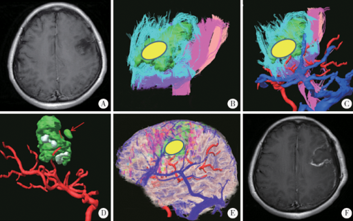
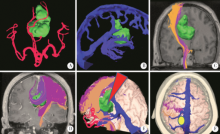
摘要: 目的 探讨弥散张量成像(diffusion tensor imaging,DTI)联合Dextroscope虚拟现实技术(virtual reality,VR)在功能区胶质瘤患者手术的作用。方法 回顾性纳入2015年1月至2019年1月北京大学第三医院经手术治疗的35例累及语言区及运动区的胶质瘤患者,术前将患者的磁共振成像(magnetic resonance imaging,MRI)、DTI、磁共振动脉成像(magnetic resonance arteriography,MRA)等数据输入Dextroscope虚拟现实系统中进行影像融合,重建神经纤维束、肿瘤、血管等重要结构,模拟操作并设计个体化手术方案,从而指导手术治疗,术后1周、1个月及3个月评估神经功能。结果 通过进行肿瘤及周围神经纤维束、血管、脑组织等结构的三维虚拟现实影像重建,可清晰判断纤维束位移与破坏,显示肿瘤与重要纤维束、动脉、静脉等的解剖关系。所有患者均成功完成虚拟现实的手术模拟与手术设计,所得三维影像与术中所见基本吻合。35例患者中,累及运动区10例,累及语言区14例,同时累及语言区和运动区11例,肿瘤影像学全部切除30例(85.7%),次全部切除5例(14.3%),术后神经功能改善34例(97.1%),1例较术前无改善(2.9%)。13例术前无神经功能缺失患者,术后出现一过性神经功能缺失,术后10 d左右恢复;22例术前存在神经功能缺失患者,其中12例患者术后1周评估时神经功能改善,9例患者术后1个月随访改善,1例运动区复发胶质母细胞瘤患者术后肢体活动障碍加重,术后2个月肿瘤再次复发因脑疝死亡。结论 应用Dextroscope虚拟现实系统三维重建肿瘤及周围神经纤维束、血管,通过解剖研究以及手术模拟,有助于个体化设计最佳手术方案,提高手术疗效。
中图分类号:
- R651.1


| [1] |
Lacroix M, Abisaid D, Fourney DR , et al. A multivariate analysis of 416 patients with glioblastoma multiforme: prognosis, extent of resection, and survival[J]. J Neurosurg, 2001,95(2):190-198.
doi: 10.3171/jns.2001.95.2.0190 |
| [2] |
Sawaya R, Hammoud M, Schoppa D , et al. Neurosurgical outcomes in a modern series of 400 craniotomies for treatment of parenchymal tumors[J]. Neurosurgery, 1998,42(5):1044-1055.
doi: 10.1097/00006123-199805000-00054 |
| [3] |
Schucht P, Beck J, Abu-Isa J , et al. Gross total resection rates in contemporary glioblastoma surgery: results of an institutional protocol combining 5-aminolevulinic acid intraoperative fluorescence imaging and brain mapping[J]. Neurosurgery, 2012,71(5):927-935.
doi: 10.1227/NEU.0b013e31826d1e6b |
| [4] |
Della Puppa A, de Pelleqrin S, d’Avella E , et al. 5-aminolevulinic acid (5-ALA) fluorescence guided surgery of high-grade gliomas in eloquent areas assisted by functional mapping. Our experience and review of the literature[J]. Acta Neurochir(Wien), 2013,155(6):965-972.
doi: 10.1007/s00701-013-1660-x |
| [5] | Wölfer J, Stummer W . Brain tumor imaging[M]. Berlin: Sprin-ger, 2014: 143-154. |
| [6] |
de Witt Hamer PC, Robles SG, Zwinderman AH , et al. Impact of intraoperative stimulation brain mapping on glioma surgery outcome: a meta analysis[J]. J Clin Oncol, 2012,30(20):2559-2565.
doi: 10.1200/JCO.2011.38.4818 |
| [7] |
McGirt MJ, Mukherjee D, Chaichana KL , et al. Association of surgically acquired motor and language deficits on overall survival after resection of glioblastoma multiforme[J]. Neurosurgery, 2009,65(3):463-470.
doi: 10.1227/01.NEU.0000349763.42238.E9 |
| [8] | 陈晓雷, 许百男, 王飞 , 等. 功能神经导航及术中磁共振成像在语言区胶质瘤手术中的应用[J]. 中华外科杂志, 2011(8):688-692. |
| [9] |
Senft C, Bink A, Franz K , et al. Intraoperative MRI guidance and extent of resection in glioma surgery: a randomised, controlled trial.[J]. Lancet Oncol, 2011,12(11):997-1003.
doi: 10.1016/S1470-2045(11)70196-6 |
| [10] |
Guyotat J, Pallud J, Armoiry X , et al. 5-aminolevulinic acid-protoporphyrin Ⅸ fluorescence-guided surgery of high-grade gliomas: a systematic review[J]. Adv Tech Stand Neurosurg, 2016,43(43):61-83.
doi: 10.1007/978-3-319-21359-0 |
| [11] |
Signorelli F . The value of cortical stimulation applied to the sur-gery of malignant gliomas in language areas[J]. Neurol Sci, 2001,22(3):217-218.
doi: 10.1007/s100720100016 |
| [12] |
Pereira LC, Oliveira KM , L’Abbate GL, et al. Outcome of fully awake craniotomy for lesions near the eloquent cortex: analysis of a prospective surgical series of 79 supratentorial primary brain tumors with long follow-up[J]. Acta Neurochir (Wien), 2009,151(10):1215-1230.
doi: 10.1007/s00701-009-0363-9 |
| [13] | 漆松涛, 李志勇, 方陆雄 , 等. 功能区胶质瘤手术的基本策略与方法[J]. 中华神经外科杂志, 2013,29(11):1083-1086. |
| [14] |
Kockro RA, Serra L, Tseng-Tsai Y , et al. Planning and simulation of neurosurgery in a virtual reality environment[J]. Neurosurgery, 2000,46(1):118-137.
doi: 10.1093/neurosurgery/46.1.118 |
| [15] |
Stadie AT, Kockro RA, Reisch R , et al. Virtual reality system for planning minimally invasive neurosurgery[J]. J Neurosurg, 2008,108(2):382-394.
doi: 10.3171/JNS/2008/108/2/0382 |
| [16] | 陈素华, 杨军, 马顺昌 , 等. 虚拟现实技术在颅颈交界区病变手术中的应用[J]. 中华神经外科杂志, 2018,34(6):591-595. |
| [17] |
Shi J, Xia J, Wei Y , et al. Three-dimensional virtual reality simulation of periarticular tumors using Dextroscope reconstruction and simulated surgery: a preliminary 10-case study[J]. Med Sci Monit, 2014,20:1043-1050.
doi: 10.12659/MSM.889770 |
| [18] |
Ng I, Hwang PY, Kumar D , et al. Surgical planning for micro-surgical excision of cerebral arterio-venous malformations using virtual reality technology[J]. Acta Neurochir (Wien), 2009,151(5):453-463.
doi: 10.1007/s00701-009-0278-5 |
| [19] |
Hattingen E, Rathert J, Jurcoane A , et al. A standardised evaluation of pre-surgical imaging of the corticospinal tract: where to place the seed ROI[J]. Neurosurg Rev, 2009,32(4):445-456.
doi: 10.1007/s10143-009-0197-1 |
| [20] |
Kim Y, Kim H, Kim YO . Virtual reality and augmented reality in plastic surgery: a review[J]. Arch Plast Surg, 2017,44(3):179-187.
doi: 10.5999/aps.2017.44.3.179 |
| [21] |
Guan XP, Wang W, Wang AB , et al. Brain interstitial fluid drainage alterations in glioma-bearing rats[J]. Aging Dis, 2018,9(2):228-234.
doi: 10.14336/AD.2017.0415 |
| [1] | 胡迪,张苗,康惠颖,彭芸. 0~2岁婴幼儿磁共振脑白质模板的建立及验证[J]. 北京大学学报(医学版), 2021, 53(2): 341-347. |
| [2] | 赵思铭,赵晓含,张杰,王党校,王晓燕. 虚拟现实技术用于龋坏识别教学[J]. 北京大学学报(医学版), 2021, 53(1): 139-142. |
| [3] | 唐祖南,Hui Yuh Soh,胡耒豪,于尧,章文博,彭歆. 混合现实技术在口腔颌面部肿瘤手术中的应用[J]. 北京大学学报(医学版), 2020, 52(6): 1124-1129. |
| [4] | 洪洪,钱宇婷,付磊,王武,李成辉,尹毅青. 困难气道中运用CT 三维重建技术指导硬质纤维气管镜行气管插管[J]. 北京大学学报(医学版), 2019, 51(5): 870-874. |
| [5] | 于尧, 章文博, 王洋, 刘筱菁, 郭传瑸, 俞光岩, 彭歆. iPlan CMF软件辅助下增强CT三维重建在头颈部肿瘤治疗中的应用[J]. 北京大学学报(医学版), 2017, 49(5): 878-882. |
| [6] | 赵一姣,王斯维,刘怡,王勇. 基于影像学牙周膜解剖特征快速提取活体牙三维牙根形态的方法[J]. 北京大学学报(医学版), 2017, 49(1): 54-059. |
| [7] | 肖宗宇, 陈晓娟, 杨艺, 徐如祥. 肿瘤干细胞样细胞RNA致敏树突状细胞治疗大鼠9L脑肿瘤[J]. 北京大学学报(医学版), 2015, 47(4): 661-666. |
| [8] | 孙洪赞, 范国光, 王桂, 郭启勇. 脑胶质瘤致癫痫的化学突触机制研究进展[J]. 北京大学学报(医学版), 2008, 40(4): 446-封3. |
| [9] | 孙建军, 王振宇, 刘彬, 钟延丰, 杜娟, 陈英玉, 马长城, 陈晓东. 治疗性协同给药抑制裸鼠脑胶质瘤的增生浸润[J]. 北京大学学报(医学版), 2006, 38(3): 252-256. |
| [10] | 刘波, 梁冶矢, 石祥恩, 张庆俊. 视神经胶质瘤7例的诊断与治疗[J]. 北京大学学报(医学版), 2005, 37(6): 645-647. |
| [11] | 孙建军, 王振宇, 刘彬, 马长城, 陈晓东, 于涛, 杜娟, 钟延丰. U251MG人脑胶质瘤模型的建立和成瘤率测定[J]. 北京大学学报(医学版), 2005, 37(5): 553-554. |
| [12] | 刘波, 李运海, 栾文忠, 梁冶矢, 虞有智, 郑爱萍. P16蛋白和Ki-67抗原在人脑胶质瘤中的表达及其意义[J]. 北京大学学报(医学版), 2003, 35(2): 223-224. |
|
||
