北京大学学报(医学版) ›› 2026, Vol. 58 ›› Issue (3): 641-649. doi: 10.19723/j.issn.1671-167X.2026.03.026

• 论著 • 上一篇    下一篇

动态牵张促进人骨髓间充质干细胞三维培养的成骨分化

白晓强1,2, 袁芷若2, 周永胜1,2,*(), 吕珑薇2,3,*()   

  1. 1. 北京大学医学部医学技术研究院, 北京 100191
    2. 北京大学口腔医学院·口腔医院修复科, 国家口腔医学中心, 国家口腔疾病临床医学研究中心, 口腔生物材料和数字诊疗装备国家工程研究中心, 颅颌面组织生物智造与修复再生北京市重点实验室, 国家卫生健康委口腔数字医学重点实验室, 北京 100081
    3. 北京大学口腔医院三亚医院(三亚口腔医学中心), 海南三亚 572013
  • 收稿日期:2024-01-16 出版日期:2026-06-18 发布日期:2026-04-09
  • 通讯作者: 周永胜, 吕珑薇
  • 基金资助:
    国家自然科学基金(82271032); 国家自然科学基金(82470943); 海南省自然科学基金优秀青年基金(825YXQN602); 北京大学临床科学家培养计划(BMU2025PYJH011)

Dynamic stretching promotes osteogenic differentiation of human bone marrow mesenchymal stem cells in three-dimensional culture

Xiaoqiang BAI1,2, Zhiruo YUAN2, Yongsheng ZHOU1,2,*(), Longwei LV2,3,*()   

  1. 1. Institute of Medical Technology, Peking University Health Science Center, Beijing 100191, China
    2. Department of Prosthodontics, Peking University School and Hospital of Stomatology & National Center for Stomatology & National Clinical Research Center for Oral Diseases & National Engineering Research Center of Oral Biomaterials and Digital Medical Devices & Beijing Key Laboratory for Intelligent Biomanufacturing and Regeneration of Craniofacial Tissues & NHC Key Laboratory of Digital Stomatology, Beijing 100081, China
    3. Peking University Hospital of Stomatology Sanya Division(Sanya Stomatology Center), Sanya 572013, Hainan, China
  • Received:2024-01-16 Online:2026-06-18 Published:2026-04-09
  • Contact: Yongsheng ZHOU, Longwei LV
  • Supported by:
    the National Natural Science Foundation of China(82271032); the National Natural Science Foundation of China(82470943); the Hainan Provincial Natural Science Foundation of China(825YXQN602); the Peking University Clinical Scientist Training Program(BMU2025PYJH011)

RICH HTML


摘要:

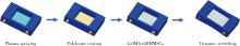
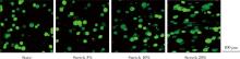
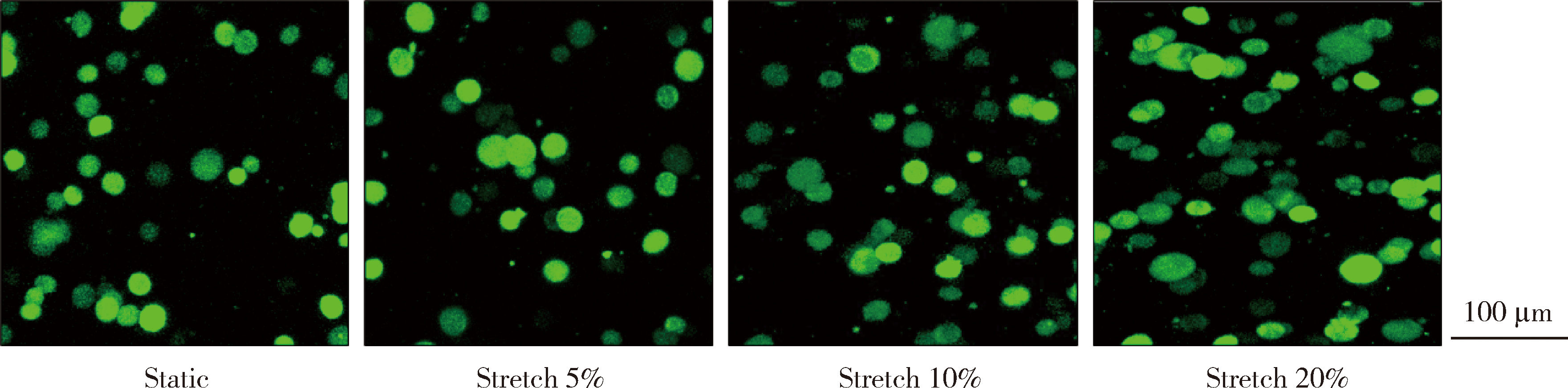
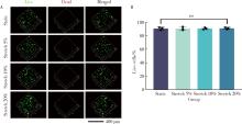
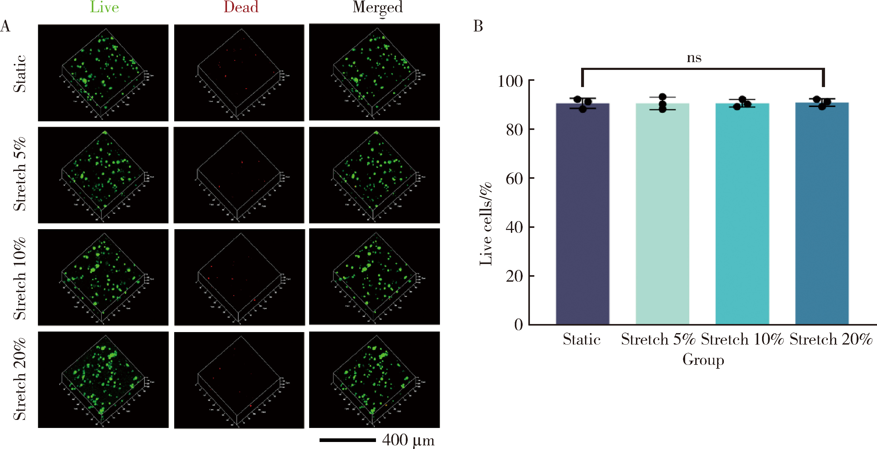
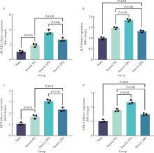
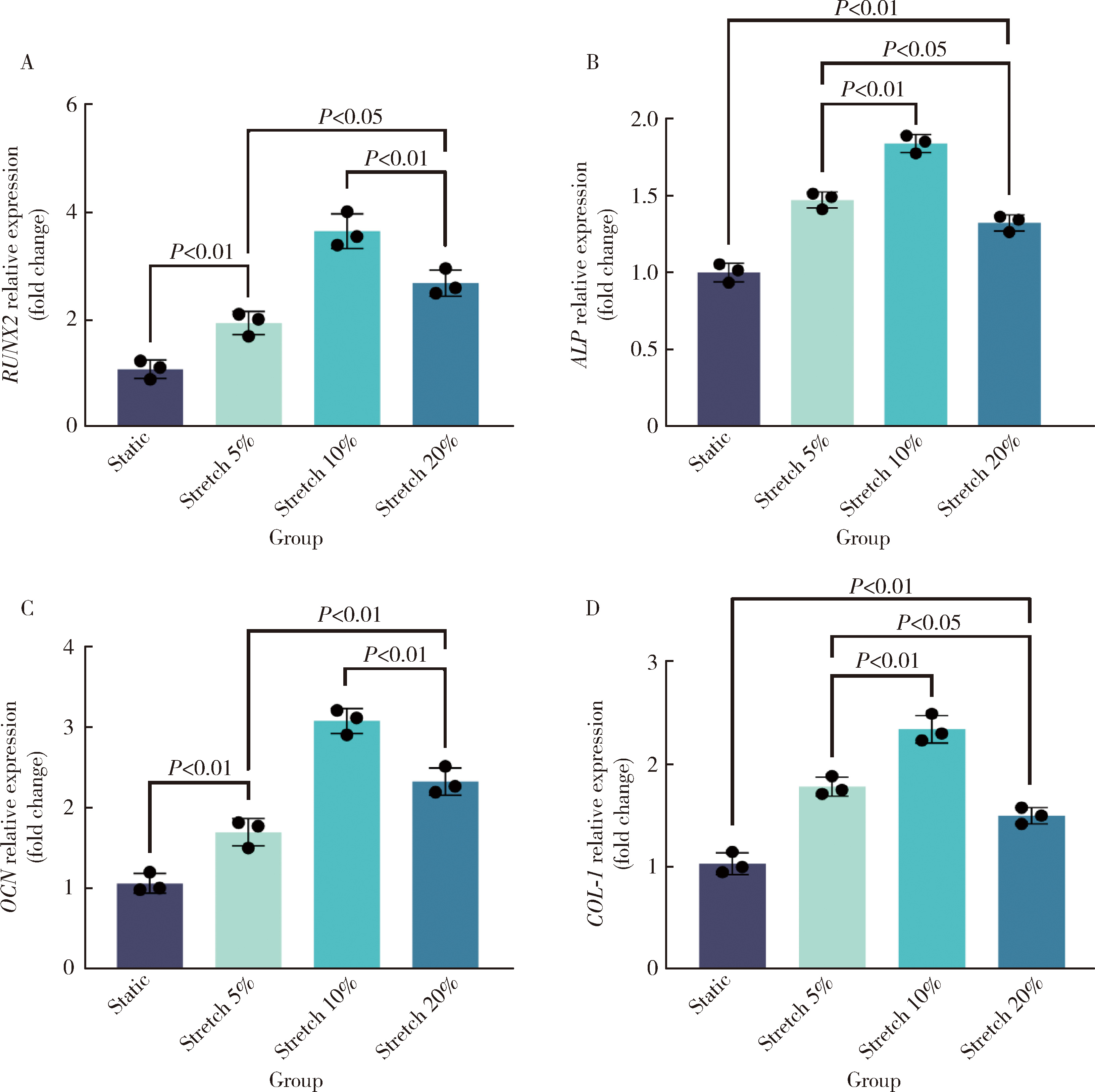
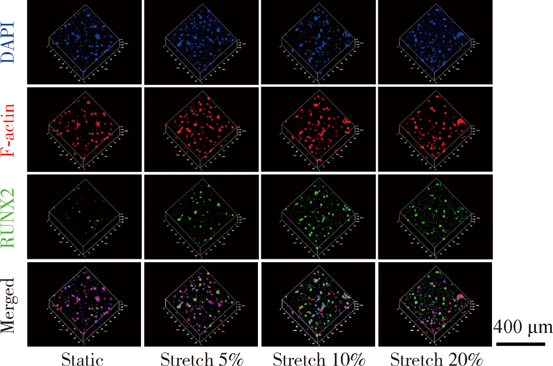
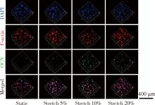
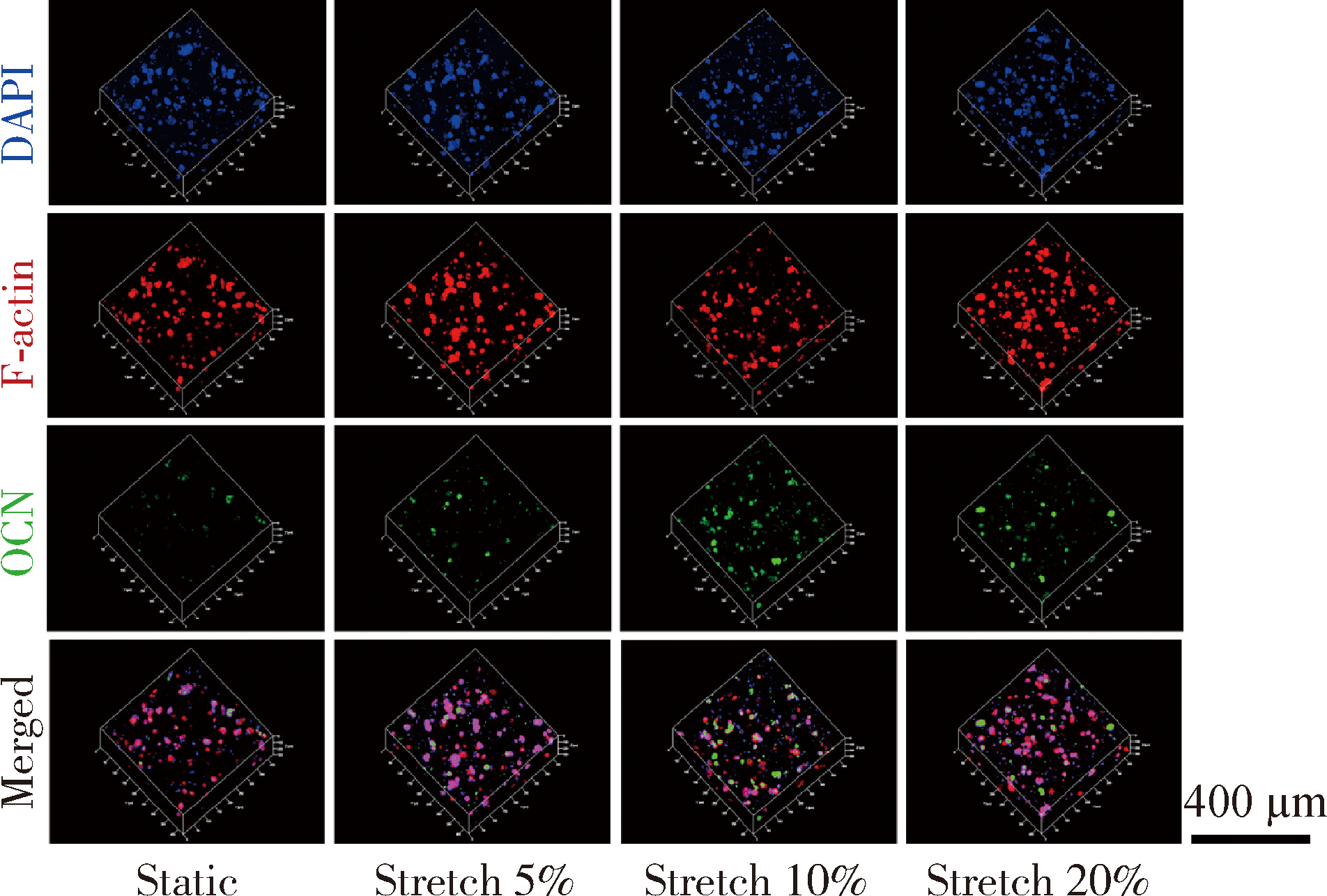
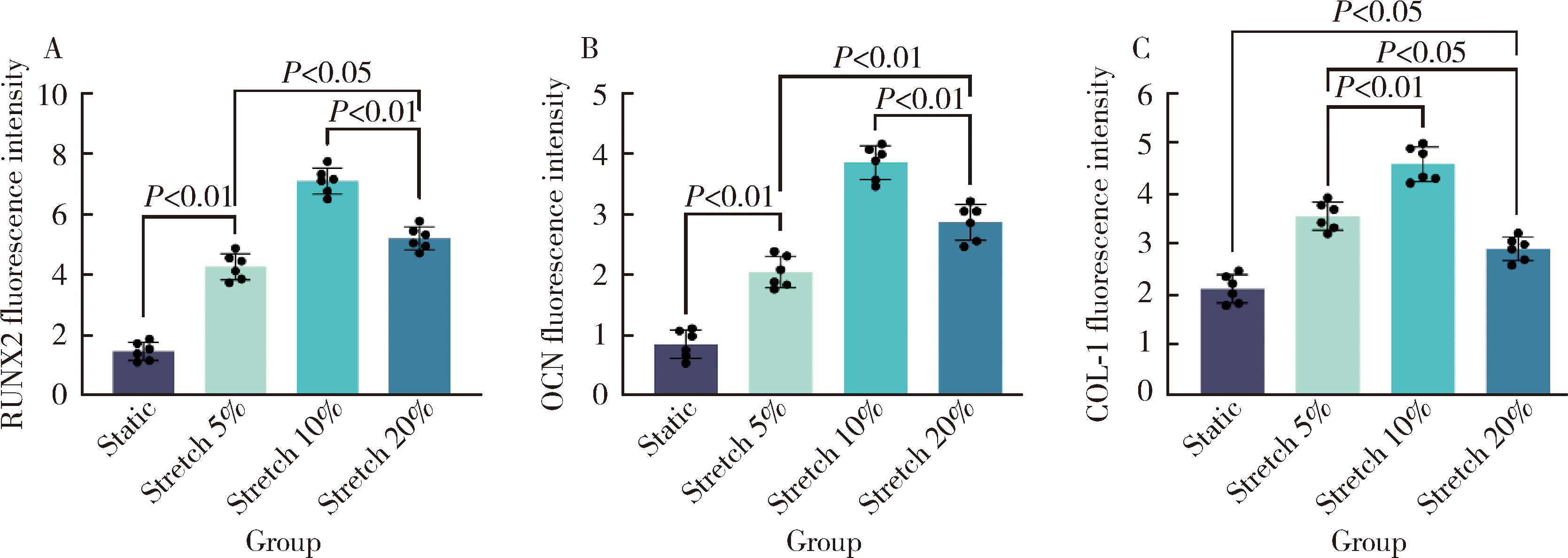
目的: 探究动态牵张对人骨髓间充质干细胞(human bone marrow mesenchymal stem cells,hBMMSCs)三维培养中成骨分化的促进作用。方法: 对载有hBMMSCs的三维培养体系进行动态牵张,设置动态牵张的比例(5%、10%、20%)、频率(0.5 Hz)和时间(2 h/d),以静态培养为对照组;通过胞质荧光染色观察动态牵张引起的hBMMSCs形变;动态牵张培养7 d后,通过细胞活死染色观察动态牵张对hBMMSCs活性的影响,通过碱性磷酸酶(alkaline phosphatase,ALP)染色及定量实验,成骨相关基因和蛋白的表达观察动态牵张对hBMMSCs成骨分化的影响。结果: 成功构建动态牵张三维培养hBMMSCs,可实现不同持续时间、频率和动态牵张的比例;动态牵张使得hBMMSCs发生形变,牵张比例越大,细胞形变越明显,从圆形向扁椭圆形变化;动态牵张培养7 d后,静态对照组和动态牵张各实验组中hBMMSCs基本为绿染的活细胞,仅有个别红染的死细胞,活细胞比例各组之间的差异无统计学意义(P>0.05);动态牵张各组较静态对照组ALP大体染色深,显微镜下ALP染色阳性细胞数量多,成骨相关基因和蛋白的表达增加,差异具有统计学意义(P<0.05),其中动态牵张10%组ALP大体染色最深,阳性细胞数量最多,成骨相关基因和蛋白的表达较静态对照组增加最为显著。结论: 动态牵张使得hBMMSCs发生形变,对细胞活性无明显影响,能够有效促进hBMMSCs成骨向分化。

关键词: 动态牵张, 三维培养, 人骨髓间充质干细胞, 成骨分化

Abstract:

Objective: To explore the promotion of dynamic distraction on osteogenic differentiation of human bone marrow mesenchymal stem cells (hBMMSCs) in three-dimensional culture. Methods: Dynamic stretching in a three-dimensional culture for hBMMSCs was achieved with proportions set at 5%, 10%, and 20%, a frequency of 0.5 Hz, and a dynamic stretching duration of 2 hours per day. Static culture was used as the control group. Deformation of hBMMSCs induced by dynamic stretching was observed through cytoplasmic fluorescence staining. After 7 days of dynamic stretching culture, the impact of dynamic stretching on the viability of hBMMSCs was observed through cell live/dead staining. The effect of dynamic stretching on the osteogenic differentiation of hBMMSCs was observed through alkaline phosphatase (ALP) staining and the expression of osteogenic related genes and proteins after 7 days of dynamic stretching culture. Results: Dynamic stretching in a three-dimensional culture for hBMMSCs was successfully constructed, which could achieve different durations, frequencies, and ratios of dynamic stretching. Dynamic stretching led to deformation of hBMMSCs, and the greater the stretching ratio, the more pronounced the cell deformation, transitioning from a circular to a flat oval shape. After 7 days of dynamic stretching culture, hBMMSCs in the static control group and dynamic stretching groups were mostly green stained live cells, with only a few red stained dead cells. The difference in the proportion of live cells between the groups was not statistically significant (P>0.05). The ALP staining in the dynamic stretching group was deeper than that in the static control group, and the number of ALP staining positive cells observed under the microscope was higher. The expression of osteogenic related genes and proteins increased after 7 days of dynamic stretching culture, and the difference was statistically significant (P < 0.05). Among them, the dynamic stretching group with 10% had the deepest ALP staining, the highest number of positive cells, and the most significant increase in the expression of osteogenic related genes and proteins compared with the static control group. Conclusion: Dynamic stretching caused deformation of hBMMSCs without a significant impact on cell viability, and it could effectively promote the osteogenic differentiation of hBMMSCs.

Key words: Dynamic stretching, Three-dimensional culture, Human bone marrow mesenchymal stem cells, Osteogenic differentiation

中图分类号: 

  • R78

表1

qRT-PCR引物序列"

Gene Forward primer(5′to 3′) Reverse prime(5′to 3′)
GAPDH GAAGGTGAAGGTCGGAGTC GAAGATGGTGATGGGATTTC
RUNX2 GAACCACAAGTGCGGTGCAA ACTGCTTGCAGCCTTAAATGACT
ALP ATGGGATGGGTGTCTCCACA CCACGAAGGGGAACTTGTC
OCN CATGAGAGCCCTCACACTCCTC CCTGCTTGGACACAAAGGCTGC
COL-1 ACAGGGCTCTAATGATGTTGA AGGCGTGATGGCTTATTTGT

图1

动态牵张三维培养的构建"

图2

不同比例动态牵张下hBMMSCs的形变"

图3

不同比例动态牵张下hBMMSCs活死染色(A)和活细胞百分比(B)"

图4

不同比例动态牵张下hBMMSCs ALP染色大体(A)、显微镜下(B)和活性相对定量(C)"

图5

不同比例动态牵张下hBMMSCs成骨相关基因的表达"

图6

不同比例动态牵张下hBMMSCs成骨相关蛋白RUNX2的表达"

图7

不同比例动态牵张下hBMMSCs成骨相关蛋白OCN的表达"

图8

不同比例动态牵张下hBMMSCs成骨相关蛋白COL-1的表达"

图9

不同比例动态牵张下hBMMSCs成骨相关蛋白RUNX2(A)、OCN(B)、COL-1(C)表达的半定量分析"

1
Hartmann A , Hildebrandt H , Younan Z , et al. Long-term results in three-dimensional, complex bone augmentation procedures with customized titanium meshes[J]. Clin Oral Implants Res, 2022, 33 (11): 1171- 1181.

doi: 10.1111/clr.14000
2
Khachatryan L , Khachatryan G , Hakobyan G . The treatment of lower jaw defects using vascularized fibula graft and dental implants[J]. J Craniofacial Surg, 2018, 29 (8): 2214- 2217.

doi: 10.1097/SCS.0000000000005015
3
Fernandez de Grado G , Keller L , Idoux-Gillet Y , et al. Bone substitutes: A review of their characteristics, clinical use, and perspectives for large bone defects management[J]. J Tissue Eng, 2018, 9, 2041731418776819.

doi: 10.1177/2041731418776819
4
Tang XY , Wu S , Wang D , et al. Human organoids in basic research and clinical applications[J]. Sig Transduct Target Ther, 2022, 7, 168.

doi: 10.1038/s41392-022-01024-9
5
Kratochvil MJ , Seymour AJ , Li TL , et al. Engineered materials for organoid systems[J]. Nat Rev Mater, 2019, 4 (9): 606- 622.

doi: 10.1038/s41578-019-0129-9
6
Zhang J , Griesbach J , Ganeyev M , et al. Long-term mechanical loading is required for the formation of 3D bioprinted functional osteocyte bone organoids[J]. Biofabrication, 2022, 14 (3): 035018.

doi: 10.1088/1758-5090/ac73b9
7
Boccafoschi F , Mosca C , Ramella M , et al. The effect of mechanical strain on soft (cardiovascular) and hard (bone) tissues: Common pathways for different biological outcomes[J]. Cell Adhes Migr, 2013, 7 (2): 165- 173.

doi: 10.4161/cam.23020
8
Chen X , Yan J , He F , et al. Mechanical stretch induces antioxidant responses and osteogenic differentiation in human mesenchymal stem cells through activation of the AMPK-SIRT1 signaling pathway[J]. Free Radic Biol Med, 2018, 126, 187- 201.

doi: 10.1016/j.freeradbiomed.2018.08.001
9
Wang L , You X , Lotinun S , et al. Mechanical sensing protein PIEZO1 regulates bone homeostasis via osteoblast-osteoclast crosstalk[J]. Nat Commun, 2020, 11, 282.

doi: 10.1038/s41467-019-14146-6
10
Li W , Zhao J , Sun W , et al. Osteocytes promote osteoclastogenesis via autophagy-mediated RANKL secretion under mechanical compressive force[J]. Arch Biochem Biophys, 2020, 694, 108594.

doi: 10.1016/j.abb.2020.108594
11
Xu H , Xia M , Sun L , et al. Osteocytes enhance osteogenesis by autophagy-mediated FGF23 secretion under mechanical tension[J]. Front Cell Dev Biol, 2022, 9, 782736.

doi: 10.3389/fcell.2021.782736
12
Xie C , Liang R , Ye J , et al. High-efficient engineering of osteo-callus organoids for rapid bone regeneration within one month[J]. Biomaterials, 2022, 288, 121741.

doi: 10.1016/j.biomaterials.2022.121741
13
Sun J , Chan YT , Ho KWK , et al. "Slow walk" mimetic tensile loading maintains human meniscus tissue resident progenitor cells homeostasis in photocrosslinked gelatin hydrogel[J]. Bioact Mater, 2023, 25, 256- 272.
14
Wu S , Wu X , Wang X , et al. Hydrogels for bone organoid construction: From a materiobiological perspective[J]. J Mater Sci Technol, 2023, 136, 21- 31.
15
Skerry TM . The response of bone to mechanical loading and disuse: Fundamental principles and influences on osteoblast/osteocyte homeostasis[J]. Arch Biochem Biophys, 2008, 473 (2): 117- 123.

doi: 10.1016/j.abb.2008.02.028
16
Williams D , Kuipers A , Mukai C , et al. Acclimation during space flight: Effects on human physiology[J]. Can Med Assoc J, 2009, 180 (13): 1317- 1323.

doi: 10.1503/cmaj.090628
17
Man J , Graham T , Squires-Donelly G , et al. The effects of microgravity on bone structure and function[J]. NPJ Microgravity, 2022, 8, 9.

doi: 10.1038/s41526-022-00194-8
18
Cooper AN , Cunningham CB , Morris JS , et al. Musculoskeletal mass and shape are correlated with competitive ability in male house mice (Mus musculus)[J]. J Exp Biol, 2020, 223 (Pt 3): jeb213389.
19
Fahy N , Alini M , Stoddart MJ . Mechanical stimulation of mesenchymal stem cells: Implications for cartilage tissue engineering[J]. J Orthop Res, 2018, 36 (1): 52- 63.

doi: 10.1002/jor.23670
20
Zhang Y , Habibovic P . Delivering mechanical stimulation to cells: State of the art in materials and devices design[J]. Adv Mater, 2022, 34 (32): 2110267.

doi: 10.1002/adma.202110267
21
Chen S , Chen X , Geng Z , et al. The horizon of bone organoid: A perspective on construction and application[J]. Bioact Mater, 2022, 18, 15- 25.
22
Weivoda MM , Bradley EW . Macrophages and bone remodeling[J]. J Bone & Mineral Res, 2023, 38 (3): 359- 369.
23
Yang C , Liu Y , Wang Z , et al. Controlled mechanical loading improves bone regeneration by regulating type H vessels in aS1Pr1-dependent manner[J]. FASEB J, 2022, 36 (10): e22530.

doi: 10.1096/fj.202200339RRR
24
Wu T , Yin F , Wang N , et al. Involvement of mechanosensitive ion channels in the effects of mechanical stretch induces osteogenic differentiation in mouse bone marrow mesenchymal stem cells[J]. J Cell Physiol, 2021, 236 (1): 284- 293.

doi: 10.1002/jcp.29841
25
Wang D , Cai J , Zeng Z , et al. The interactions between mTOR and NF-κB: A novel mechanism mediating mechanical stretch-stimulated osteoblast differentiation[J]. J Cell Physiol, 2021, 236 (6): 4592- 4603.

doi: 10.1002/jcp.30184
26
Wang H , Yu H , Huang T , et al. Hippo-YAP/TAZ signaling in osteogenesis and macrophage polarization: Therapeutic implications in bone defect repair[J]. Genes Dis, 2023, 10 (6): 2528- 2539.
27
Qin L , He T , Chen S , et al. Roles of mechanosensitive channel Piezo1/2 proteins in skeleton and other tissues[J]. Bone Res, 2021, 9, 44.
28
Fischer C , Milting H , Fein E , et al. Long-term functional and structural preservation of precision-cut human myocardium under continuous electromechanical stimulation in vitro[J]. Nat Commun, 2019, 10, 117.
29
Mihic A , Li J , Miyagi Y , et al. The effect of cyclic stretch on maturation and 3D tissue formation of human embryonic stem cell-derived cardiomyocytes[J]. Biomaterials, 2014, 35 (9): 2798- 2808.
30
Abdel Fattah AR , Daza B , Rustandi G , et al. Actuation enhances patterning in human neural tube organoids[J]. Nat Commun, 2021, 12, 3192.
31
Qian X , Nguyen HN , Song MM , et al. Brain-region-specific organoids using mini-bioreactors for modeling ZIKV exposure[J]. Cell, 2016, 165 (5): 1238- 1254.
[1] 曾立婷, 程凯远, 刘中宁, 李健, 杨静文, 姜婷. miR-488-5p促进大鼠骨髓间充质干细胞成神经、成骨分化及神经化骨再生[J]. 北京大学学报(医学版), 2026, 58(1): 10-21.
[2] 盛春辉, 张晓, 吕珑薇, 周永胜. 人脂肪间充质干细胞外泌体对去势小鼠骨质疏松的预防[J]. 北京大学学报(医学版), 2025, 57(2): 217-226.
[3] 胡轶博, 吕伟佳, 夏炜, 刘亦洪. 基于细胞生长与成骨分化的不同孔径生物支架流体力学有限元分析[J]. 北京大学学报(医学版), 2025, 57(1): 97-105.
[4] 帅婷, 郭艳艳, 林春平, 侯晓玫, 金婵媛. 敲减NPTX1促进人骨髓间充质干细胞成骨分化[J]. 北京大学学报(医学版), 2025, 57(1): 7-12.
[5] 尤鹏越,刘玉华,王新知,王思雯,唐琳. 脱细胞猪心包膜生物相容性及成骨性能的体内外评价[J]. 北京大学学报(医学版), 2021, 53(4): 776-784.
[6] 刘霞,李英妮,孙晓麟,彭清林,卢昕,王国春. 去整合素金属蛋白酶对成骨分化的影响[J]. 北京大学学报(医学版), 2018, 50(6): 962-967.
[7] 朱云艳,李倩,张怡美,周彦恒. MAPK和AKT磷酸化下调参与Toll样受体抑制的人牙周膜干细胞的成骨分化[J]. 北京大学学报(医学版), 2018, 50(1): 33-41.
[8] 常嘉, 马绪臣, 魏明洁, 王晶, 焦岩涛. 兔髁状突软骨细胞藻酸盐凝胶三维培养体系的建立[J]. 北京大学学报(医学版), 2002, 34(2): 103-107.
Viewed
Full text


Abstract

Cited

  Shared   
  Discussed   
No Suggested Reading articles found!