北京大学学报(医学版) ›› 2021, Vol. 53 ›› Issue (1): 69-75. doi: 10.19723/j.issn.1671-167X.2021.01.011
数字化评估CAD/CAM个性化基台与成品基台影响粘接剂残留的体外研究
岳兆国1,张海东1,杨静文2,侯建霞1,Δ( )
)
- 1.北京大学口腔医学院·口腔医院,牙周科 国家口腔疾病临床医学研究中心 口腔数字化医疗技术和材料国家工程实验室 口腔数字医学北京市重点实验室,北京 100081
2.北京大学口腔医学院·口腔医院,修复科 国家口腔疾病临床医学研究中心 口腔数字化医疗技术和材料国家工程实验室 口腔数字医学北京市重点实验室,北京 100081
Comparison of residual cement between CAD/CAM customized abutments and stock abutments via digital measurement in vitro
YUE Zhao-guo1,ZHANG Hai-dong1,YANG Jing-wen2,HOU Jian-xia1,Δ()
- 1. Department of Periodontology, Peking University School and Hospital of Stomatology & National Clinical Research Center for Oral Diseases & National Engineering Laboratory for Digital and Material Technology of Stomatology & Beijing Key Laboratory of Digital Stomatology, Beijing 100081, China
2. Department of Prosthetics, Peking University School and Hospital of Stomatology & National Clinical Research Center for Oral Diseases & National Engineering Laboratory for Digital and Material Technology of Stomatology & Beijing Key Laboratory of Digital Stomatology, Beijing 100081, China
摘要:
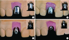
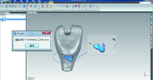
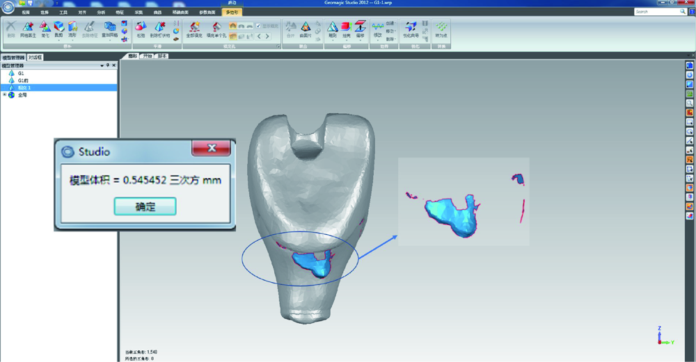
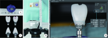
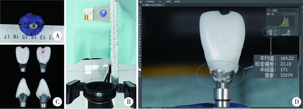
目的: 通过体外模型比较计算机辅助设计/计算机辅助制造个性化基台(computer aided design/computer aided manufacturing customized abutments,CCA)与成品基台(stock abutments,SA)对粘接剂残留的影响,同时初步评价数字化三维扫描技术定量评估残留粘接剂的可行性。方法: 本研究所需20个工作模型皆取自同一例已在北京大学口腔医院牙周科接受了右上中切牙种植手术的患者。通过个性化愈合基台成形植体周软组织后,测得植体平台位于颊侧黏膜下5 mm。利用个性化转移杆取模后灌制20副工作模型,并根据基台种类及粘接边缘位置将工作模型分为四组,每组5个:CCA1(穿黏膜高度5 mm,即平齐黏膜粘接边缘)、CCA2(穿黏膜高度4 mm,即黏膜下1 mm粘接边缘)、SA1(穿黏膜高度3 mm,即黏膜下2 mm粘接边缘)和SA2(穿黏膜高度1 mm,即黏膜下4 mm粘接边缘)。在工作模型上模拟临床粘接过程并清除多余粘接剂后,利用三维扫描技术获得残留粘接剂的体积,利用数码相机拍摄二维图像获得残留粘接剂面积百分比,利用称量的方式获得残留粘接剂的质量,并分析三维扫描方法获取的体积与传统评价方法所得的面积百分比及质量的相关性。结果: 所有冠-基台复合体粘接边缘均有粘接剂残留。其中,CCA组残留粘接剂的体积明显小于SA组[(0.635 3±0.535 4) mm3 vs. (2.293 8±0.943 8) mm 3,P<0.001], 面积百分比及质量也显著低于SA组[面积百分比:7.57%±2.99% vs. 22.68%±10.06%,P<0.001;质量:(0.001 5±0.001 0) g vs. (0.003 7±0.001 4) g,P<0.001],而三者在CCA组及SA组内差异均无统计学意义(P>0.05)。三维扫描所得残留粘接剂的体积与传统评价方法所得残留粘接剂的面积百分比及残留粘接剂的质量间均具强相关性(r>0.75,P<0.001)。结论: 与SA相比,CCA能更有效地减少粘接剂的残留。基于三维扫描技术数字化评估残留粘接剂的方法切实可行,但其效度和信度还需进一步研究。
中图分类号:
- R783


| [1] |
Jung RE, Pjetursson BE, Glauser R, et al. A systematic review of the 5-year survival and complication rates of implant-supported single crowns[J]. Clin Oral Implants Res, 2008,19(2):119-130.
doi: 10.1111/j.1600-0501.2007.01453.x pmid: 18067597 |
| [2] |
Chee W, Felton DA, Johnson PF, et al. Cemented versus screw-retained implant prostheses: Which is better?[J]. Int J Oral Maxillofac Implants, 1999,14(1):137-141.
pmid: 10074764 |
| [3] |
Linkevicius T, Vindasiute E, Puisys A, et al. The influence of margin location on the amount of undetected cement excess after delivery of cement-retained implant restorations[J]. Clin Oral Implants Res, 2011,22(12):1379-1384.
doi: 10.1111/j.1600-0501.2010.02119.x pmid: 21382089 |
| [4] |
Staubli N, Walter C, Schmidt JC, et al. Excess cement and the risk of peri-implant disease: A systematic review[J]. Clin Oral Implants Res, 2017,28(10):1278-1290.
doi: 10.1111/clr.12954 pmid: 27647536 |
| [5] |
Korsch M, Obst U, Walther W. Cement-associated peri-implantitis: A retrospective clinical observational study of fixed implant-supported restorations using a methacrylate cement[J]. Clin Oral Implants Res, 2014,25(7):797-802.
doi: 10.1111/clr.12173 pmid: 23600620 |
| [6] |
Wilson TG Jr. The positive relationship between excess cement and peri-implant disease: A prospective clinical endoscopic study[J]. J Periodontol, 2009,80(9):1388-1392.
doi: 10.1902/jop.2009.090115 pmid: 19722787 |
| [7] |
Linkevicius T, Puisys A, Vindasiute E, et al. Does residual cement around implant-supported restorations cause peri-implant disease? A retrospective case analysis[J]. Clin Oral Implants Res, 2013,24(11):1179-1184.
doi: 10.1111/j.1600-0501.2012.02570.x pmid: 22882700 |
| [8] |
Ichikawa T, Ishida O, Watanabe M, et al. A new retrieval system for cement-retained implant superstructures: A technical report[J]. J Prosthodont, 2008,17(6):487-489.
doi: 10.1111/j.1532-849X.2008.00329.x pmid: 18544129 |
| [9] |
Galván G, Kois JC, Chaiyabutr Y, et al. Cemented implant restoration: A technique for minimizing adverse biologic consequences[J]. J Prosthet Dent, 2015,114(4):482-485.
doi: 10.1016/j.prosdent.2014.10.017 pmid: 26119018 |
| [10] |
Seo CW, Seo JM. A technique for minimizing subgingival residual cement by using rubber dam for cement-retained implant crowns[J]. J Prosthet Dent, 2017,117(2):327-328.
doi: 10.1016/j.prosdent.2016.08.024 pmid: 27771147 |
| [11] | Linkevicius T. Zero bone loss concepts [M]. Illinois: Quintessence Publishing Co, 2019. |
| [12] |
Lewis S, Beumer J 3rd, Hornburg W, et al. The “UCLA” abutment[J]. Int J Oral Maxillofac Implants, 1988,3(3):183-189.
pmid: 3074050 |
| [13] | 戴文雍, 汤春波. 种植体修复个性化基台研究现状及展望[J]. 口腔医学, 2012,32(11):685-687. |
| [14] | 宿玉成. 口腔种植学[M]. 2版. 北京: 人民卫生出版社, 2014: 403-404. |
| [15] |
Shapoff CA, Lahey BJ. Crestal bone loss and the consequences of retained excess cement around dental implants[J]. Compend Contin Educ Dent, 2012,33(2):94-101.
pmid: 22545427 |
| [16] |
Schwarz F, Derks J, Monje A, et al. Peri-implantitis[J]. J Clin Periodontol, 2018,45(Suppl 20):S246-S266.
doi: 10.1111/jcpe.2018.45.issue-S20 |
| [17] |
Andersson B, Odman P, Lindvall AM, et al. Cemented single crowns on osseointegrated implants after 5 years: Results from a prospective study on CeraOne[J]. Int J Prosthodont, 1998,11(3):212-218.
pmid: 9728114 |
| [18] |
Linkevicius T, Vindasiute E, Puisys A, et al. The influence of the cementation margin position on the amount of undetected cement. A prospective clinical study[J]. Clin Oral Implants Res, 2013,24(1):71-76.
doi: 10.1111/j.1600-0501.2012.02453.x pmid: 22487018 |
| [19] |
Kappel S, Eiffler C, Lorenzo-Bermejo J, et al. Undetected resi-dual cement on standard or individualized all-ceramic abutments with cemented zirconia single crowns: A prospective randomized pilot trial[J]. Clin Oral Implants Res, 2016,27(9):1065-1071.
doi: 10.1111/clr.12691 pmid: 26381392 |
| [20] |
Kotsakis GA, Zhang L, Gaillard P, et al. Investigation of the association between cement retention and prevalent peri-implant diseases: A cross-sectional study[J]. J Periodontol, 2016,87(3):212-220.
doi: 10.1902/jop.2015.150450 pmid: 26537368 |
| [21] |
Daubert DM, Weinstein BF, Bordin S, et al. Prevalence and predictive factors for peri-implant disease and implant failure: A cross-sectional analysis[J]. J Periodontol, 2015,86(3):337-347.
doi: 10.1902/jop.2014.140438 pmid: 25415249 |
| [22] | Fuchigami K, Munakata M, Kitazume T, et al. A diversity of peri-implant mucosal thickness by site[J]. Clin Oral Impl Res, 2017,28(2):171-176. |
| [23] | 张众, 孟焕新, 韩劼, 等. 软组织垂直厚度对牙周炎患者种植修复临床效果的影响[J]. 北京大学学报(医学版), 2020,52(2):332-338. |
| [24] |
Dumbrigue HB, Abanomi AA, Cheng LL. Techniques to minimize excess luting agent in cement-retained implant restorations[J]. J Prosthet Dent, 2002,87(1):112-114.
doi: 10.1067/mpr.2002.119418 pmid: 11807495 |
| [25] |
Vindasiute E, Puisys A, Maslova N, et al. Clinical factors influencing removal of the cement excess in implant-supported restorations[J]. Clin Implant Dent Relat Res, 2015,17(4):771-778.
doi: 10.1111/cid.12170 pmid: 24224895 |
| [26] |
Andersson B, Odman P, Lindvall AM, et al. Single-tooth restorations supported by osseointegrated implants: results and experiences from a prospective study after 2 to 3 years[J]. Int J Oral Maxillofac Implants, 1995,10(6):702-711.
pmid: 8530173 |
| [27] | Higginbottom F, Belser U, Jones JD, et al. Prosthetic management of implants in the esthetic zone[J]. Int J Oral Maxillofac Implants, 2004,19(Suppl.):62-72. |
| [28] |
Berglundh T, Lindhe J, Marinello C, et al. Soft tissue reaction to de novo plaque formation on implants and teeth. An experimental study in the dog[J]. Clin Oral Implants Res, 1992,3(1):1-8.
doi: 10.1034/j.1600-0501.1992.030101.x pmid: 1420721 |
| [29] | 高鹏程, 谢理哲, 严斌. 牙颌模型三维数字化技术及其在口腔正畸学中的应用进展[J]. 口腔生物医学, 2014,5(3):152-157. |
| [1] | 白晓强, 袁芷若, 周永胜, 吕珑薇. 动态牵张促进人骨髓间充质干细胞三维培养的成骨分化[J]. 北京大学学报(医学版), 2026, 58(3): 641-649. |
| [2] | 温奥楠, 张晓会, 杨咏涛, 高梓翔, 李文博, 单珅瑶, 商相宜, 田淯文, 郭殊玮, 王艺蓁, 王勇, 赵一姣. 基于非刚性配准构建三维颜面微笑仿真序列数据的方法[J]. 北京大学学报(医学版), 2026, 58(1): 139-144. |
| [3] | 于录, 吴灵, 刘筱菁, 李自力. 基于数据库相似性检索的正颌外科手术规划技术流程可行性研究: 随机对照试验[J]. 北京大学学报(医学版), 2026, 58(1): 145-152. |
| [4] | 邵梁, 马雯洁, 陈莹, 丁茜, 张磊. 上颌切牙前伸和正中咬合接触解剖特征的数字化测量与分析[J]. 北京大学学报(医学版), 2026, 58(1): 99-106. |
| [5] | 王翠萍, 陈哲, 程永静. 超微血流成像评估与膝骨关节炎临床症状的关联研究[J]. 北京大学学报(医学版), 2025, 57(6): 1096-1100. |
| [6] | 宋凤岐, 徐心雨, 刘筱菁, 李自力. 上颌骨前部和整体顺时针旋转改善骨性Ⅲ类牙颌面畸形患者鼻旁凹陷的对比[J]. 北京大学学报(医学版), 2025, 57(5): 980-988. |
| [7] | 肖宇嘉, 毛渤淳, 周彦恒. 姿势性微笑的三维形态学研究[J]. 北京大学学报(医学版), 2025, 57(5): 989-995. |
| [8] | 王泽远, 于栓宝, 郑浩轲, 陶金, 范雅峰, 张雪培. 基于临床特征和多参数MRI的前列腺癌盆腔淋巴结转移的术前预测模型[J]. 北京大学学报(医学版), 2025, 57(4): 684-691. |
| [9] | 宁圆, 张晓盈, 李雪, 李原, 何菁, 金月波. 干燥综合征并发乳腺淋巴瘤1例[J]. 北京大学学报(医学版), 2025, 57(4): 808-811. |
| [10] | 孙建军, 马千权, 尹晓亮, 杨辰龙, 张嘉, 陈素华, 吴超, 谢京城, 韩芸峰, 林国中, 司雨, 杨军, 邬海博, 赵强. 任意维度重建磁共振对骶管囊肿进行精准分型对于指导微创手术和康复的意义[J]. 北京大学学报(医学版), 2025, 57(2): 303-308. |
| [11] | 仇师禹, 练洋, 康一帆, 张雷, 蔡义望, 单小峰, 蔡志刚. 基于下颌骨数据库和全连接神经网络的三维检索模型辅助下的下颌骨个性化重建[J]. 北京大学学报(医学版), 2025, 57(2): 360-368. |
| [12] | 马丽娟, 腾雍辉, 王勇, 赵一姣, 张馨月, 秦庆钊, 尹东. 乳牙缺失数字化丝圈间隙保持器的三维有限元分析[J]. 北京大学学报(医学版), 2025, 57(2): 376-383. |
| [13] | 朱玉佳, 沈华, 温奥楠, 高梓翔, 秦庆钊, 单珅瑶, 李文博, 傅湘玲, 赵一姣, 王勇. 三维颌面对称参考平面智能构建的深度学习算法[J]. 北京大学学报(医学版), 2025, 57(1): 113-120. |
| [14] | 徐昕恺, 赵建江, 田素坤, 刘中宁, 赵晓一, 赵晓波, 江腾飞, 陈晓军, 马超, 孙玉春. 集成压缩气流系统扫描头辅助获取液体干扰表面三维数据精度评价[J]. 北京大学学报(医学版), 2025, 57(1): 121-127. |
| [15] | 吴灵, 方嘉琨, 刘筱菁, 李自力, 李阳, 王晓霞. 基于牙颌面畸形患者三维颅面特征相似性度量模型的建立及评估[J]. 北京大学学报(医学版), 2025, 57(1): 128-135. |
|
||
