北京大学学报(医学版) ›› 2021, Vol. 53 ›› Issue (1): 76-82. doi: 10.19723/j.issn.1671-167X.2021.01.012
数字化辅助确定再定位牙合垫颌位方法的探索和精度评价
房硕博1,2,杨广聚1,2,康艳凤1,2,孙玉春1,谢秋菲1,2,Δ( )
)
- 1.北京大学口腔医学院·口腔医院,修复科 国家口腔疾病临床医学研究中心 口腔数字化医疗技术和材料国家工程实验室 口腔数字医学北京市重点实验室,北京 100081
2.北京大学口腔医学院·口腔医院,口颌功能诊疗研究中心 国家口腔疾病临床医学研究中心 口腔数字化医疗技术和材料国家工程实验室 口腔数字医学北京市重点实验室,北京 100081
Method and accuracy of determining the jaw position of repositioning splint with the aid of digital technique
FANG Shuo-bo1,2,YANG Guang-ju1,2,KANG Yan-feng1,2,SUN Yu-chun1,XIE Qiu-fei1,2,Δ()
- 1. Department of Prosthodontics, Peking University School and Hospital of Stomatology & National Clinical Research Center for Oral Diseases & National Engineering Laboratory for Digital and Material Technology of Stomatology & Beijing Key Laboratory of Digital Stomatology, Beijing 100081, China
2. Center for Oral and Jaw Functional Diagnosis, Peking University School and Hospital of Stomatology & National Clinical Research Center for Oral Diseases & National Engineering Laboratory for Digital and Material Technology of Stomatology & Beijing Key Laboratory of Digital Stomatology, Beijing 100081, China
摘要:
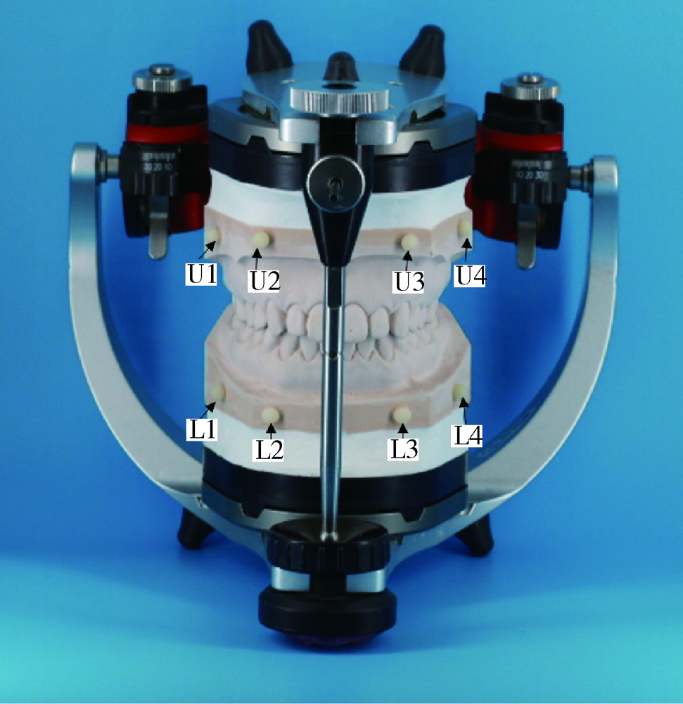
目的: 建立数字化辅助确定再定位牙合垫颌位的方法,并在体外评价此方法的精度以及垂直距离升高量对精度的影响。方法: 招募受试者1例,获取上下颌光学牙列模型、锥形束CT(cone beam computed tomography,CBCT)牙颌图像、下颌运动数据,融合光学牙列模型与重建的CBCT模型,并将融合后的模型配准到下颌运动数据的参考系。根据受试者前牙开口3 mm和矢状位上髁突位于关节窝中部确定再定位牙合垫的颌位,进行计算机辅助设计并3D打印制作再定位牙合垫。受试者试戴牙合垫后拍摄CBCT,观察比较戴入后的髁突位置与设计时的髁突位置。体外模型实验中,灌制一副标准全牙列石膏模型,在底座上粘固标志球体,模型于牙尖交错位上全可调牙合架。扫描模型建立数字化牙列模型,使用超声下颌运动轨迹描记仪记录牙合架上模拟的下颌运动并重复3次。融合牙列模型与下颌运动数据,基于下颌运动数据辅助确定3个颌位,分别为切点沿运动轨迹升高4 mm、5 mm、6 mm且下颌均前伸2 mm,保存设计颌位为STL格式数据,设计并3D打印制作再定位牙合垫。将再定位牙合垫戴入石膏牙列模型进行 3次颌位扫描,获得扫描颌位的STL格式数据。以下颌模型为共同区域配准设计颌位与扫描颌位,比较上颌模型的均方根误差。拟合设计颌位与扫描颌位上颌标志球的球心坐标,计算球心在X、Y、Z轴方向上的差值绝对值。采用SPSS 18.0软件进行单因素方差分析,双侧检验,显著性水平α取0.05。结果: 利用多源数据融合和个体下颌运动,建立数字化辅助确定再定位牙合垫颌位的方法,右侧髁突实现了预期的调整,左侧髁突较预期位置偏前下。体外模型实验中,扫描颌位与设计颌位上颌模型的整体偏差(均方根误差)为(0.25±0.04) mm,不同垂直距离升高量的偏差大小差异无统计学意义(P>0.05)。标志球球心在X、Y、Z轴方向的偏差(差值绝对值)分别为(0.08±0.01) mm、(0.30±0.02) mm、(0.21±0.04) mm,不同垂直距离升高量的偏差大小差异无统计学意义(P>0.05)。结论: 建立了数字化辅助确定再定位牙合垫颌位的新方法,实现多源数据融合、数字化设计与制作并完成个体试戴,证明此方法可行。体外模型研究表明此方法可以实现较为精确的颌位调整,其应用效果尚需进一步临床研究评价。
中图分类号:
- R782.6


| [1] |
Clark GT. The TMJ repositioning appliance: a technique for construction, insertion, and adjustment[J]. Cranio, 1986,4(1):37-46.
doi: 10.1080/08869634.1986.11678129 pmid: 3456393 |
| [2] |
Aslanidou K, Kau CH, Vlachos C, et al. The fabrication of a customized occlusal splint based on the merging of dynamic jaw tracking records, cone beam computed tomography, and CAD-CAM digital impression[J]. J Orthod Sci, 2017,6(3):104-109.
doi: 10.4103/jos.JOS_61_16 pmid: 28717635 |
| [3] |
Kühnöl C, Kordaß B. Digital workflow for TMD diagnostics and bite alteration: description of a case treated using Sicat Function[J]. Int J Comput Dent, 2019,22(3):283-292.
pmid: 31463492 |
| [4] | 杨德圣, 韩科, 周书敏, 等. 临床弹响消失牙合位作为调位牙合垫治疗牙合位的研究[J]. 中华口腔医学杂志, 2000,35(6):465-466. |
| [5] | 陈慧敏, 傅开元, 李优伟, 等. 再定位牙合垫戴入前后颞下颌关节盘和髁突的位置改变[J]. 华西口腔医学杂志, 2009,27(4):408-412. |
| [6] |
Dao TT, Lavigne G. Oral splints: the crutches for temporomandi-bular disorders and bruxism?[J]. Crit Rev Oral Biol Med, 1998,9(3):345-361.
doi: 10.1177/10454411980090030701 pmid: 9715371 |
| [7] | 佘文珺, 谢俊良, 翁维民. 下颌运动轨迹描记在无牙颌颌位记录中的应用[J]. 临床口腔医学杂志, 2018,34(1):22-24. |
| [8] |
Li W, Xie Q, Wang Y, et al. A pilot study of digital recording of edentulous jaw relations using a handheld scanner and specially designed headgear[J]. Sci Rep, 2018,8(1):8975.
doi: 10.1038/s41598-018-27277-5 pmid: 29895978 |
| [9] | 吴冰, 吴国锋. 口腔修复CAD/CAM系统中的虚拟牙合架[J]. 实用口腔医学杂志, 2016,32(2):293-297. |
| [10] |
Gärtner C, Kordaß B. The virtual articulator: development and evaluation[J]. Int J Comput Dent, 2003,6(1):11-24.
pmid: 12838585 |
| [11] |
Bohner L, Gamba DD, Hanisch M, et al. Accuracy of digital technologies for the scanning of facial, skeletal, and intraoral tissues: A systematic review[J]. J Prosthet Dent, 2019,121(2):246-251.
doi: 10.1016/j.prosdent.2018.01.015 pmid: 30017156 |
| [12] | 宋杨, 孙玉春, 赵一姣, 等. 牙颌模型三维扫描仪精度定量评价[J]. 北京大学学报(医学版), 2013,45(1):140-144. |
| [13] |
Andreiotelli M, Kamposiora P, Papavasiliou G. Digital data management for CAD/CAM technology. An update of current systems[J]. Eur J Prosthodont Restor Dent, 2013,21(1):9-15.
pmid: 23682504 |
| [14] | Fang JJ, Kuo TH. Tracked motion-based dental occlusion surface estimation for crown restoration[J]. Computer Aided Design, 2009,41(4):315-323. |
| [15] | Li L, Sun Y, Wang Y, et al. Accuracy of a novel virtual articulator for recording three-dimensional dentition[J]. Int J Pros-thodont, 2020,33(4):441-451. |
| [16] | 陈磊, 张豪, 冯海兰, 等. 颅颌运动仿真系统牙合接触精度初探[J]. 中华口腔医学杂志, 2010,45(2):98-101. |
| [17] |
Uechi J, Okayama M, Shibata T, et al. A novel method for the 3-dimensional simulation of orthognathic surgery by using a multimodal image-fusion technique[J]. Am J Orthod Dentofacial Orthop, 2006,130(6):786-798.
doi: 10.1016/j.ajodo.2006.03.025 pmid: 17169742 |
| [18] |
Swennen GR, Mommaerts MY, Abeloos J, et al. The use of a wax bite wafer and a double computed tomography scan procedure to obtain a three-dimensional augmented virtual skull model[J]. J Craniofac Surg, 2007,18(3):533-539.
doi: 10.1097/scs.0b013e31805343df pmid: 17538314 |
| [19] |
Nkenke E, Zachow S, Benz M, et al. Fusion of computed tomography data and optical 3D images of the dentition for streak artefact correction in the simulation of orthognathic surgery[J]. Dentomaxillofac Radiol, 2004,33(4):226-232.
doi: 10.1259/dmfr/27071199 pmid: 15533975 |
| [20] | 赵一姣, 原福松, 谢晓艳, 等. 牙颌模型激光扫描数据与锥形束CT数据配准方法的精度比较[J]. 中华口腔医学杂志, 2013,48(3):173-176. |
| [21] | 谢秋菲. 牙合垫在口腔疾病治疗中的应用现状及未来发展趋势[J]. 中华口腔医学杂志, 2019,58(8):515-521. |
| [22] |
Dedem P, Türp JC. Digital Michigan splint: from intraoral scanning to plasterless manufacturing[J]. Int J Comput Dent, 2016,19(1):63-76.
pmid: 27027103 |
| [23] |
Berntsen C, Kleven M, Heian M, et al. Clinical comparison of conventional and additive manufactured stabilization splints[J]. Acta Biomater Odontol Scand, 2018,4(1):81-89.
doi: 10.1080/23337931.2018.1497491 pmid: 30128331 |
| [24] | 王时敏, 李峥, 王冠博, 等. 全程数字化夜磨牙保护牙合垫的制作和初步应用[J]. 北京大学学报(医学版), 2019,51(1):111-116. |
| [25] |
Li Z, Wang S, Ye H, et al. Preliminary clinical application of complete workflow of digitally designed and manufactured sports mouthguards[J]. Int J Prosthodont, 2020,33(1):99-104.
doi: 10.11607/ijp.6348 pmid: 31860919 |
| [1] | 单珅瑶, 杨咏涛, 李文博, 温奥楠, 高梓翔, 商相宜, 王勇, 赵一姣. 基于下颌运动轨迹的𬌗架关键参数计算方法[J]. 北京大学学报(医学版), 2026, 58(1): 115-125. |
| [2] | 于录, 吴灵, 刘筱菁, 李自力. 基于数据库相似性检索的正颌外科手术规划技术流程可行性研究: 随机对照试验[J]. 北京大学学报(医学版), 2026, 58(1): 145-152. |
| [3] | 马丽娟, 腾雍辉, 王勇, 赵一姣, 张馨月, 秦庆钊, 尹东. 乳牙缺失数字化丝圈间隙保持器的三维有限元分析[J]. 北京大学学报(医学版), 2025, 57(2): 376-383. |
| [4] | 徐心雨,吴灵,宋凤岐,李自力,张益,刘筱菁. 基于下颌运动轨迹的正颌外科术中下颌骨髁突定位方法及初步精度验证[J]. 北京大学学报(医学版), 2024, 56(1): 57-65. |
| [5] | 李穗,马雯洁,王时敏,丁茜,孙瑶,张磊. 上前牙种植单冠修复体切导的数字化设计正确度[J]. 北京大学学报(医学版), 2024, 56(1): 81-87. |
| [6] | 罗昊,田福聪,王晓燕. 不同椅旁可切削修复材料序列抛光时间及表面粗糙度与光泽度的比较[J]. 北京大学学报(医学版), 2022, 54(3): 565-571. |
| [7] | 冯莎蔚,国慧,王勇,赵一姣,刘鹤. 乳牙数字化参考牙冠模型的初步构建[J]. 北京大学学报(医学版), 2022, 54(2): 327-334. |
| [8] | 李怡,王丽瑜,刘晓强,周倜,吕季喆,谭建国. 不同材料及厚度椅旁CAD/CAM瓷贴面的边缘特征[J]. 北京大学学报(医学版), 2022, 54(1): 140-145. |
| [9] | 邱淑婷,朱玉佳,王时敏,王飞龙,叶红强,赵一姣,刘云松,王勇,周永胜. 姿势微笑位口唇对称参考平面的数字化构建及初步应用验证[J]. 北京大学学报(医学版), 2022, 54(1): 193-199. |
| [10] | 徐啸翔,曹烨,赵一姣,贾璐,谢秋菲. 数字化个齿托盘制取下颌全牙列全冠预备体印模的体外评价[J]. 北京大学学报(医学版), 2021, 53(1): 54-61. |
| [11] | 岳兆国,张海东,杨静文,侯建霞. 数字化评估CAD/CAM个性化基台与成品基台影响粘接剂残留的体外研究[J]. 北京大学学报(医学版), 2021, 53(1): 69-75. |
| [12] | 李峥,柳玉树,王时敏,张瑞,贾璐,叶红强,胡文杰,赵文艳,刘云松,周永胜. 数字化方法复制暂时修复体牙合面形态在重度磨耗病例中的应用[J]. 北京大学学报(医学版), 2021, 53(1): 62-68. |
| [13] | 罗佳,张宇,崔宏燕,祝宁,沈惠丹,邸萍,林野. 锥度固位结合数字化技术在后牙连续多牙种植即刻修复中的应用[J]. 北京大学学报(医学版), 2020, 52(5): 964-970. |
| [14] | 魏菱,邹东,陈虎,潘韶霞,孙玉春,周永胜. 一种数字化全口义齿的临床疗效评价[J]. 北京大学学报(医学版), 2020, 52(4): 762-770. |
| [15] | 刘洋,胡菳洳,赵一姣,王勇,孙玉春,潘韶霞,冯海兰. 一种新的基于全口义齿测量的牙列缺失修复用颌位记录托盘[J]. 北京大学学报(医学版), 2020, 52(2): 368-372. |
|
||
