北京大学学报(医学版) ›› 2026, Vol. 58 ›› Issue (4): 754-761. doi: 10.19723/j.issn.1671-167X.2026.04.011

• 论著 • 上一篇    下一篇

重复肾输尿管畸形合并上尿路结石内镜手术的策略

杨新纪1,*, 林秀石1,*, 谢志飞2, 刘余庆1, 肖春雷1, 张树栋1, 侯小飞1,*(), 邱敏1,*()   

  1. 1. 北京大学第三医院泌尿外科,北京 100191
    2. 天津市武清区第二人民医院泌尿外科,天津 301700
  • 收稿日期:2026-02-22 出版日期:2026-08-18 发布日期:2026-05-19
  • 通讯作者: 侯小飞, 邱敏
  • 作者简介:

    * These authors contributed equally to this work

  • 基金资助:
    吴阶平医学基金会临床科研专项资助基金(320.6750.2022-03-45)

Analysis of endoscopic surgical strategies for duplex kidney and ureteral malformation complicated with upper urinary tract calculi

Xinji YANG1, Xiushi LIN1, Zhifei XIE2, Yuqing LIU1, Chunlei XIAO1, Shudong ZHANG1, Xiaofei HOU1,*(), Min QIU1,*()   

  1. 1. Department of Urology, Peking University Third Hospital, Beijing 100191, China
    2. Department of Urology, Tianjin Wuqing District Second People' s Hospital, Tianjin 301700, China
  • Received:2026-02-22 Online:2026-08-18 Published:2026-05-19
  • Contact: Xiaofei HOU, Min QIU
  • Supported by:
    the Wu Jieping Medical Foundation Special Fund for Clinical Research(320.6750.2022-03-45)

RICH HTML


摘要:

目的: 探讨重复肾输尿管畸形合并上尿路结石的内镜手术治疗策略,总结不同畸形亚型的个体化诊疗经验,为该疾病的临床规范化处置提供参考。方法: 回顾性分析2012年11月至2025年12月于北京大学第三医院泌尿外科接受内镜手术治疗的重复肾输尿管畸形合并上尿路结石患者的临床资料,收集患者基线特征、影像学检查结果、围术期指标、并发症及肾功能变化等数据,总结不同畸形亚型的内镜手术技巧与核心诊疗要点,采用SPSS 22.0及R 3.6.1软件进行统计学分析。结果: 共纳入38例患者,其中男性16例,女性22例,平均年龄(49.92±14.27)岁,中位随访时间17.80(1.60~41.90)个月。临床症状以腰痛为主(32例,84.21%),畸形分型以单侧不完全性重复肾输尿管畸形占比最高(24例,63.16%)。根据畸形分型与结石特征,所有患者均依据术前计算机断层扫描尿路成像等影像学精准评估,个体化选择手术方案:15例患者行输尿管硬镜碎石取石术(ureteroscopic lithotripsy, URL),15例行输尿管软镜碎石取石术(flexible ureteroscopic lithotripsy, FURL),7例行经皮肾镜碎石取石术(percutaneous nephrolithotomy, PCNL),1例行PCNL联合FURL治疗。患者中位手术时间74.00(64.75~92.50) min,中位术中出血量2.00(1.00~4.25) mL,中位术后住院时间3.00(2.00~5.75) d,术后整体清石率达94.74%。围术期无严重并发症发生,14例(36.84%)患者出现术后发热,经对症治疗后均痊愈。术后患者血肌酐水平较术前显著降低(P<0.05),肾功能显著改善。结论: 内镜手术治疗重复肾输尿管畸形合并上尿路结石安全、有效,具有创伤小、恢复快、清石率高的优势;通过“精准诊断-个体化术式选择”的策略,可针对不同畸形亚型制定个性化方案,值得临床推广应用。

关键词: 重复肾输尿管畸形, 上尿路结石, 输尿管镜, 经皮肾镜, 内镜手术

Abstract:

Objective: To investigate the endoscopic surgical management strategies for duplex kidney and ureteral duplication anomalies complicated by upper urinary tract calculi, to summarize individualized diagnostic and therapeutic experiences for different anomaly subtypes, and to provide a reference for standardized clinical management of this condition. Methods: The clinical data of patients with duplex kidney and ureteral duplication anomalies and upper urinary tract calculi who underwent endoscopic surgery at the Department of Urology, Peking University Third Hospital from November 2012 to December 2025 were retrospectively analyzed. Baseline characteristics, imaging findings, perioperative parameters, complications, and changes in renal function were collected. Endoscopic surgical techniques and key diagnostic and therapeutic points for different anomaly subtypes were summarized. Statistical analysis was performed using SPSS 22.0 and R 3.6.1 software. Results: A total of 38 patients were enrolled, including 16 males and 22 females, with a mean age of (49.92±14.27) years. The median follow-up duration was 17.80 months (1.60-41.90). The predominant clinical symptom was flank pain (32 cases, 84.21%). The most common anomaly subtype was unilateral incomplete duplex kidney and ureteral duplication (24 cases, 63.16%). Based on the anomaly subtype and stone characteristics, all the patients underwent precise preoperative assessment using CT urography and other imaging modalities, and indivi-dualized surgical approaches were selected: In the study, 15 patients underwent ureteroscopic lithotripsy (URL), 15 underwent flexible ureteroscopic lithotripsy (FURL), 7 underwent percutaneous nephrolithotomy (PCNL), and 1 patient received PCNL combined with FURL. The median operative time was 74.00 (64.75-92.50) min, the median intraoperative blood loss was 2.00 (1.00-4.25) mL, and the median postoperative hospital stay was 3.00 (2.00-5.75) d. The overall postoperative stone-free rate was 94.74%. No severe perioperative complications occurred. Postoperative fever occurred in 14 patients (36.84%), all of whom recovered after symptomatic treatment. Postoperative serum creatinine levels were significantly lower than preoperative levels (P < 0.05), indicating significant improvement in renal function. Conclusion: Endoscopic surgery is safe and effective for the treatment of duplex kidney and ureteral duplication anomalies complicated by upper urinary tract calculi, offering advantages of minimal trauma, rapid recovery, and a high stone-free rate. The strategy of "precise diagnosis-individualized surgical approach selection" allows the formulation of tailored treatment plans for different anomaly subtypes and is worthy of clinical promotion and application.

Key words: Duplex kidney and ureteral duplication, Upper urinary tract calculi, Ureteroscopy, Percutaneous nephrolithotomy, Endoscopic surgery

中图分类号: 

  • R693.1

表1

重复肾输尿管畸形合并上尿路结石患者的人口学及基线临床特征"

Variables Patients (n=38)
Gender, n (%)
  Male 16 (42.11)
  Female 22 (57.89)
Age/years, $\bar x \pm s$ 49.92±14.27
BMI/(kg/m2), M (P25P75) 25.01 (22.63, 26.47)
Symptoms, n (%)
  Low back pain 32 (84.21)
  Lower urinary tract irritation symptoms 10 (26.32)
  Lower urinary tract obstruction symptoms 3 (7.89)
  Hematuria 12 (31.58)
  Fever 6 (15.79)
  Nausea and vomiting 7 (18.42)
  Detected during physical examination 3 (7.89)
Duration of symptoms/d, M (P25P75) 30.00 (21.50, 165.00)
Basic medical history, n (%)
  Hypertension 13 (34.21)
  Diabetes mellitus 6 (15.79)
  Coronary heart disease 4 (10.53)

表2

重复肾输尿管畸形类型、结石特征及肾积水情况的术前影像学评估"

Variables Patients (n=38)
Side of Ureteral duplication, n (%)
  Left 12 (31.58)
  Right 17 (44.74)
  Bilateral 9 (23.68)
Type of Ureteral duplication, n (%)
  Unilateral incompletely 24 (63.16)
  Unilateral completely 4 (10.53)
  Bilateral incompletely 1 (2.63)
  Bilateral completely 3 (7.89)
  Bilateral completely and contralateral incompletely 5 (13.16)
  Unilateral inverted Y 1 (2.63)
Stone location, n (%)
  Calices renales of the upper kidney 10 (26.32)
  Calices renales of the lower kidney 8 (21.05)
  Ureteropelvic junction 6 (15.79)
  Ureter 11 (28.95)
  Multiple locations 3 (7.89)
Number of stones, n (%)
  1 21 (55.26)
  2 2 (5.26)
  Multiple 15 (39.48)
Maximum stone diameter/cm, M (P25P75) 1.06 (0.84, 1.48)
Stone CT value/HU, $\bar x \pm s$ 897.82±348.62
Pelvic hydronephrosis, n (%) 23 (60.53)
Hydronephrosis size/cm, M (P25P75) 3.43 (2.96, 3.79)

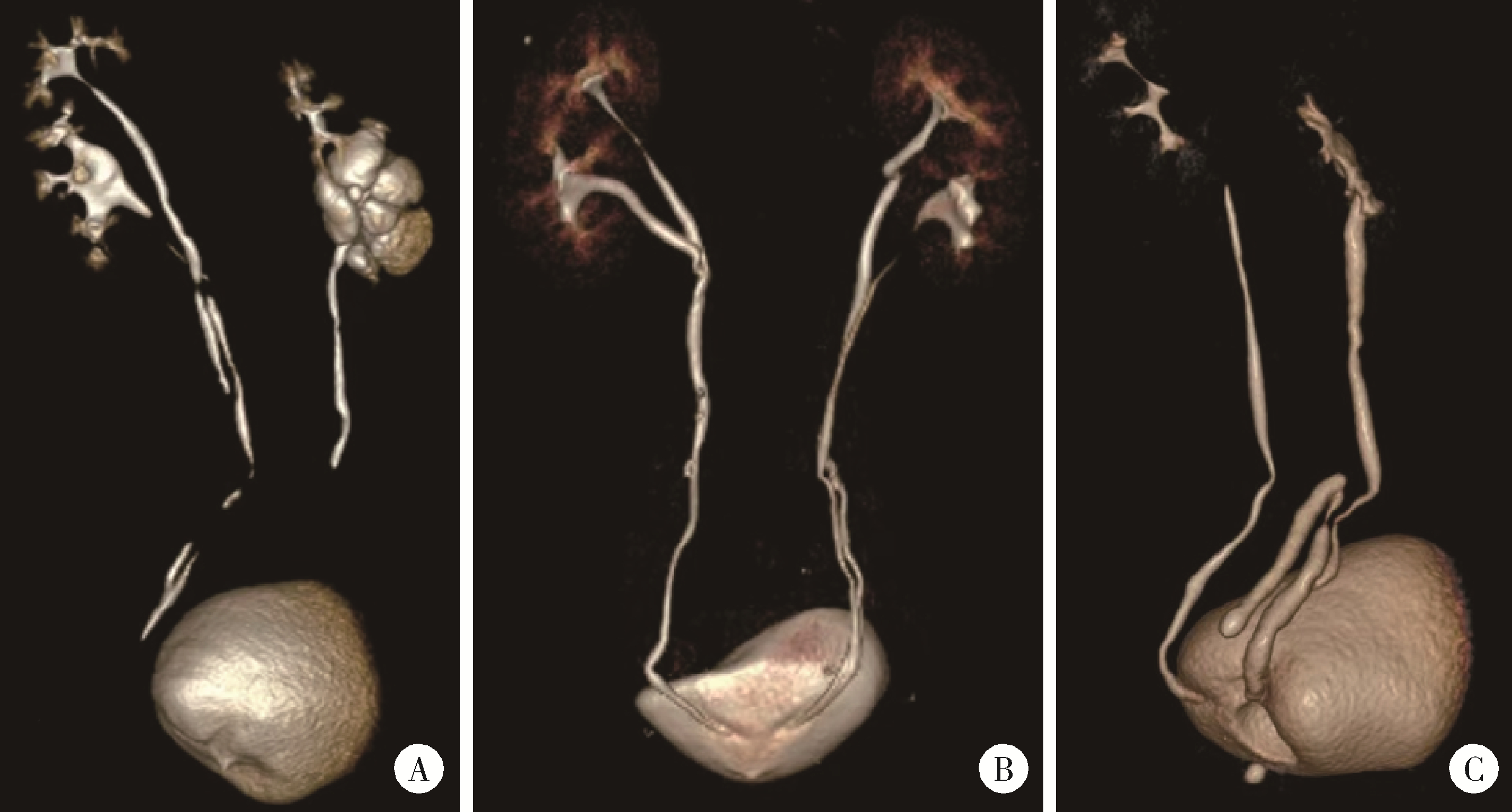
图1

不同类型肾盂输尿管重复畸形的CT三维重建表现"

表3

内镜手术方式、术中处理及特殊情况汇总"

Variables Patients (n=38)
ASA grade, n (%)
  Ⅰ 21 (55.26)
  Ⅱ 17 (44.74)
Anesthesia method, n (%)
  General anesthesia 25 (65.79)
  Spinal anesthesia 13 (34.21)
Surgical method, n (%)
  URL 15 (39.47)
  FURL 15 (39.47)
  PCNL 7 (18.43)
  PCNL+FURL 1 (2.63)
Intraoperative special conditions, n (%)
  Ureteral stricture 19 (50.00)
  Ureteral orifice cyst 3 (7.89)
  Complete impaction 2 (5.30)
  Stricture incision 3 (7.90)
  Cyst incision 1 (2.63)
  Balloon dilation 2 (5.26)
Surgical time/min, M (P25P75) 74.00 (64.75, 92.50)
Estimated blood loss/mL, M (P25P75) 2.0 (1.00, 4.25)

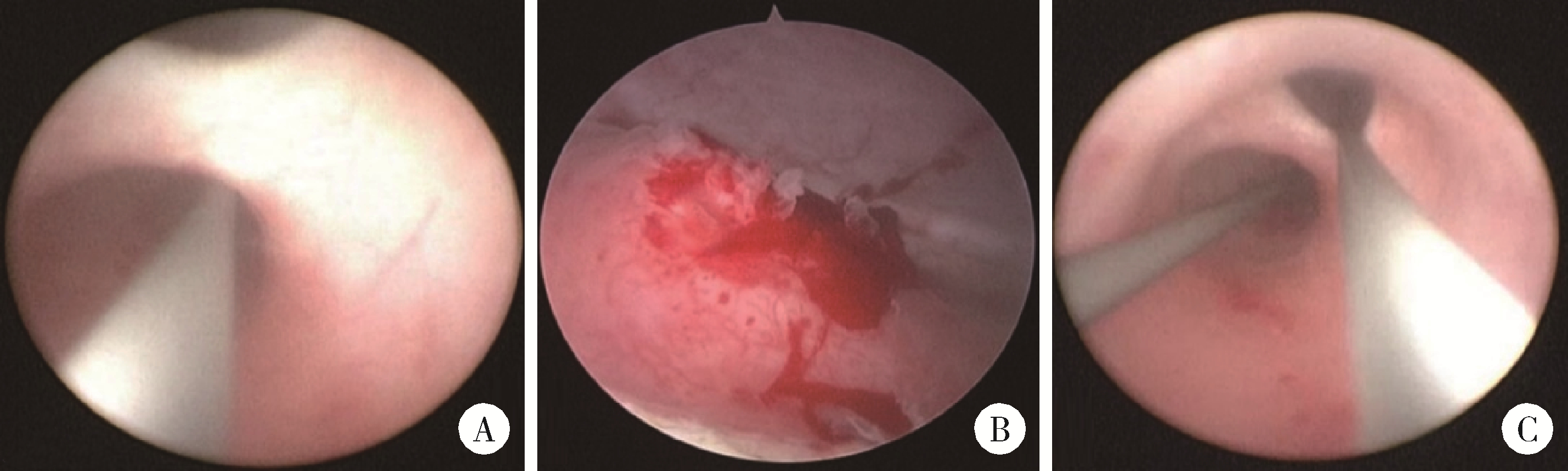
图2

重复肾输尿管畸形的术中镜下图像"

表4

术后清石效果、并发症及肾功能改善情况"

Variables Patients (n=38)
Postoperative fever, n (%) 14 (36.84)
Complete stone clearance, n (%) 36 (94.74)
Staged Surgery, n (%) 1 (2.63)
Follow-up time/months, M (P25P75) 17.80 (1.60, 41.90)
Postoperative extubation time/d, M (P25P75) 37.00 (31.25, 42.25)
Postoperative hospital stay/d, M (P25P75) 3.00 (2.00, 5.75)
Postoperative creatinine decline, n (%) 32 (84.21)

表5

手术前后肾功能相关实验室指标比较"

Variable Preoperative Postoperative V P value
Creatinine/(μmol/L), M (P25P75) 75.50 (65.50, 89.70) 70.00 (57.00, 80.00) 43 0.007
Glomerular filtration rate/[mL/(min·1.73 m2)], M (P25P75) 92.77 (82.79, 102.75) 100.00 (93.00, 113.00) 135 0.033
1
Horst M , Smith GHH . Pelvi-ureteric junction obstruction in duplex kidneys[J]. BJU Int, 2008, 101(12): 1580- 1584.

doi: 10.1111/j.1464-410X.2007.07386.x
2
Pioli Lamêgo de Faria C , Gomes Botelho L , Santos Vidal I , et al. Urinary incontinence due to complete ureteral duplication with an ectopic vulvar ureteral orifice: A case report[J]. Cureus, 2023, 15(7): e42114.
3
Lakshman G , Krishnamoorthy S , Kumaresan N . Duplex kidney with calculus obstruction of lower moiety lessons learnt[J]. J Med Pharm Allied Sci, 2022, 11(2): 4718- 4720.

doi: 10.55522/jmpas.V11I2.2335
4
Katims A , Rosen DC , Chandhoke R , et al. Extravesical ectopic ureteral calculus obstruction in a completely duplicated collecting system[J]. J Endourol Case Rep, 2020, 6(1): 42- 44.

doi: 10.1089/cren.2019.0108
5
Zheng Z , Hu W , Ji C , et al. A study of the difference in biochemical metabolism between patients with unilateral and bilateral upper urinary tract stones[J]. Sci Rep, 2024, 14(1): 30154.

doi: 10.1038/s41598-024-81454-3
6
梁月有, 郑克立, 戴宇平, 等. 先天性重复肾盂、输尿管畸形合并结石的诊断与治疗: 附32例报告[J]. 新医学, 2002, 33(12): 725- 726.
7
唐翠松, 汤光宇, 李伟, 等. 64排容积CT尿路成像在肾盂输尿管重复畸形诊断中的应用价值及优势[J]. 临床放射学杂志, 2010, 29(11): 1504- 1507.
8
冀竞辉, 林秀石, 潘大猛, 等. 输尿管硬镜联合勺状阻石篮激光碎石术治疗输尿管1.5 cm及以上结石的效果[J]. 北京大学学报(医学版), 2025, 57(4): 676- 683.

doi: 10.19723/j.issn.1671-167X.2025.04.008
9
余霄腾, 黄奕瑄, 李新飞, 等. 结石相关输尿管狭窄的上尿路修复手术技术与临床结局[J]. 北京大学学报(医学版), 2025, 57(4): 670- 675.

doi: 10.19723/j.issn.1671-167X.2025.04.007
10
李志军, 李勇, 钱坤, 等. 重复肾输尿管畸形合并上尿路结石的腔内外科治疗体会[J]. 中国内镜杂志, 2018, 24(5): 90- 94.
11
Pan Y , Chen G , Chen H , et al. The left ureterocele and stone of calyceal diverticulum in the patient with bilateral incomplete duplex kidneys managed by flexible ureteroscopy: A case report and literature review[J]. BMC Urol, 2020, 20(1): 35.

doi: 10.1186/s12894-020-00604-7
12
花梅免, 杨庆, 张振声, 等. 经尿道输尿管口囊肿电切术在重复肾输尿管畸形合并异位输尿管口囊肿患者中的应用(附9例报告)[J]. 海军军医大学学报, 2022, 43(12): 1481- 1484.
13
张韬, 赵贵成, 沈军, 等. 重复肾输尿管畸形12例诊治体会[J]. 实用医学杂志, 2015, 31(19): 3228- 3231.
14
Ye WX , Ren LG , Chen L . Inverted Y ureteral duplication with an ectopic ureter and multiple urinary calculi: A case report[J]. World J Clin Cases, 2022, 10(4): 1326- 1332.

doi: 10.12998/wjcc.v10.i4.1326
15
Falahatkar R , Falahatkar S , Khajavi Gaskarei MA , et al. The global, prevalence, and risk factors of postoperative fever after percutaneous nephrolithotomy: A systematic review and meta-analysis[J]. Asian J Urol, 2024, 11(2): 253- 260.

doi: 10.1016/j.ajur.2022.04.008
16
Bohare U , Gupta HL , Gupta M , et al. Evaluation of risk factors for fever and sepsis after ureteroscopic lithotripsy: A prospective observational study[J]. Int J Anat Radiol Surg, 2023, 12(6): 13- 16.
[1] 赖世聪, 王焕瑞, 倪润峰, 胡浩浦, 王辰龙, 徐涛, 胡浩. 免分腿侧卧位与截石位对输尿管软镜碎石术中肾盂内压及预后的影响[J]. 北京大学学报(医学版), 2026, 58(4): 748-753.
[2] 余霄腾, 黄奕瑄, 李新飞, 陈昶甫, 赵方舟, 应鸿刚, 陶子豪, 张一鸣, 徐丽清, 李志华, 杨昆霖, 周利群, 李学松, 赵峥. 结石相关输尿管狭窄的上尿路修复手术技术与临床结局[J]. 北京大学学报(医学版), 2025, 57(4): 670-675.
[3] 王焕瑞, 赖世聪, 胡浩浦, 丁泽华, 徐涛, 胡浩. 腹腔镜与输尿管软镜联合定位治疗复杂输尿管狭窄的疗效分析[J]. 北京大学学报(医学版), 2025, 57(4): 784-788.
[4] 乔佳佳,田聪,黄晓波,刘军. 肾结石合并系统性红斑狼疮行经皮肾镜碎石取石术的安全性和有效性评估[J]. 北京大学学报(医学版), 2024, 56(4): 745-749.
[5] 田聪,刘军,杨波,乔佳佳,黄晓波,许清泉. 经皮肾镜取石术中异常肾盂黏膜活检结果分析[J]. 北京大学学报(医学版), 2023, 55(5): 948-952.
[6] 张春龙,王明瑞,王起,许克新,徐涛,胡浩. 覆膜金属输尿管支架维持性治疗输尿管镜碎石术后难治性输尿管狭窄的远期疗效评价[J]. 北京大学学报(医学版), 2022, 54(4): 674-679.
[7] 戴翔,左美妮,张晓鹏,胡浩,徐涛. 经皮肾镜术中不同憩室颈部处理方式治疗肾盏憩室结石的长期预后[J]. 北京大学学报(医学版), 2021, 53(4): 704-709.
[8] 王明瑞,王起,胡浩,赖金惠,贺永新,熊杰,刘献辉,刘士军,许克新,徐涛. 标准通道经皮肾镜取石术治疗孤立肾肾结石的长期安全性和有效性[J]. 北京大学学报(医学版), 2020, 52(4): 663-666.
[9] 邱敏,郝一昌,肖春雷,马潞林. 肾盂癌保留肾脏的内镜手术策略[J]. 北京大学学报(医学版), 2020, 52(4): 610-613.
[10] 高健,胡立宝,陈尘,郅新,徐涛. 经皮肾镜去石术后出血的介入治疗[J]. 北京大学学报(医学版), 2020, 52(4): 667-671.
[11] 张军晖,蒋一航,蒋宇光,张际青,康宁. 双侧同步内镜手术治疗双侧上尿路结石的临床效果[J]. 北京大学学报(医学版), 2020, 52(4): 672-677.
[12] 马闰卓,夏海缀,陆敏,张智荧,张启鸣,卢剑,王国良,马潞林. 输尿管镜活体组织检查对上尿路尿路上皮癌根治性手术的影响[J]. 北京大学学报(医学版), 2019, 51(4): 665-672.
[13] 郝一昌,陈昆,刘余庆,卢剑,肖春雷,马潞林. 输尿管软镜下钬激光切除术治疗肾盂癌6例报道及文献复习[J]. 北京大学学报(医学版), 2018, 50(5): 816-821.
[14] 谌诚,张博,韩文科,林健,王刚,张晓春,宋毅,赵峥,张中元,金杰,虞巍. 超声造影技术引导下经皮肾穿刺应用于经皮肾镜取石术的单中心初步经验[J]. 北京大学学报(医学版), 2017, 49(6): 1071-1075.
[15] 王少刚, 余虓. 经皮肾镜碎石术——日间手术新探索[J]. 北京大学学报(医学版), 2017, 49(5): 753-755.
Viewed
Full text


Abstract

Cited

  Shared   
  Discussed   
No Suggested Reading articles found!