北京大学学报(医学版) ›› 2019, Vol. 51 ›› Issue (1): 86-92. doi: 10.19723/j.issn.1671-167X.2019.01.016

• 论著 • 上一篇    下一篇

骨性Ⅲ类错牙合畸形患者正畸-正颌联合治疗的稳定性

王秀婧1,张怡美1,周彦恒2,()   

  1. 1. 北京大学口腔医学院·口腔医院,门诊部 国家口腔疾病临床医学研究中心 口腔数字化医疗技术和材料国家工程实验室 口腔数字医学北京市重点实验室,北京 100081
    2. 北京大学口腔医学院?口腔医院正畸科,北京 100081
  • 收稿日期:2018-10-07 出版日期:2019-02-18 发布日期:2019-02-26
  • 通讯作者: 周彦恒 E-mail:yanhengzhou@vip.163.com
  • 基金资助:
    国家自然科学基金(81801014)

Orthodontic-orthognathic treatment stability in skeletal class Ⅲ malocclusion patients

Xiu-jing WANG1,Yi-mei ZHANG1,Yan-heng ZHOU2,()   

  1. 1. First Clinical Division, Peking University School and Hospital of Stomatology & National Clinical Research Center for Oral Diseases & National Engineering Laboratory for Digital and Material Technology of Stomatology & Beijing Key Laboratory of Digital Stomatology, Beijing 100081, China
    2. Department of Orthodontics, Peking University School and Hospital of Stomatology, Beijing 100081, China
  • Received:2018-10-07 Online:2019-02-18 Published:2019-02-26
  • Contact: Yan-heng ZHOU E-mail:yanhengzhou@vip.163.com
  • Supported by:
    Supported by the National Natural Science Foundation of China(81801014)

摘要:

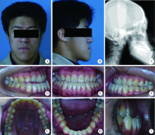
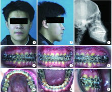
目的:测量骨性Ⅲ类错牙合畸形患者经过正畸-正颌联合治疗后及治疗结束3~12年软、硬组织的变化, 探讨正畸-正颌联合治疗后颌面部组织的长期稳定性。方法: 回顾2000年1月1日至2009年1月1日就诊于北京大学口腔医院行正畸-正颌联合治疗的骨性Ⅲ类患者22例,收集正畸-正颌联合治疗结束时及3~12年随访复诊的头颅侧位片,测量各牙性硬组织、骨性硬组织及软组织变化的项目。利用SPSS 17.0软件进行配对t检验,P<0.05为差异有统计学意义。结果:比较联合治疗术后3~12年和治疗结束时的牙性硬组织变化如下:上中切牙-SN角由110.98°±6.77°减少为109.21°±5.80°(P = 0.005),上中切牙-NA角由28.31°±6.80°减少为26.49°±6.18°(P = 0.002),上下切牙角由123.51°±8.14°增大为125.7°±10.01°(P = 0.035), 其余牙性硬组织项目变化均差异无统计学意义,说明联合治疗后3~12年相比联合治疗结束时,患者的上前牙有直立趋势。骨性硬组织变化中全面高由124.98°±11.98°减少为122.4°±11.05°(P = 0.024), 其余骨性硬组织项目变化均差异无统计学意义,提示联合治疗后3~12年骨性硬组织具有相对稳定性。比较联合治疗术后3~12年和治疗结束时的软组织测量值,上唇凸点至EP平面距离由(-2.78±2.20) mm减少为(-3.29±2.44) mm (P = 0.02), H角由8.27°±3.71°减少为7.32°±3.83° (P = 0.006),其余软组织项目变化均差异无统计学意义,上唇和颏部软组织变化表现为上唇少量回缩和颏部形态的少量改变。结论: 骨性Ⅲ类错牙合畸形正畸-正颌联合治疗后3~12年牙性硬组织、骨性硬组织及软组织改变基本稳定。

关键词: 错牙合畸形, 上颌后缩, 下颌前突, 正颌手术

Abstract:

Objective:To investigate stability of skeletal hard tissues, dental hard tissues and soft tissues after orthodontic-orthognathic treatment in a long term. This study reviewed longitudinal changes in orthodontic-orthognathic patients of skeletal class Ⅲ malocculsion, using lateral cephalometric radiographs in 3-12 years after treatment in comparison to treatment finishing. Methods: Twenty-two patients with skeletal Class Ⅲ malocclusion following orthodontic-orthognathic surgery in Peking University School and Hospital of Stomatology from January 1, 2000 to January 1, 2009 were observed. The lateral cephalometric radiographs of the following stages were collected: treatment finishing (T1), 3 to 12 years after treatment (T2). Statistical analyses of cephalometrics were evaluated. Paired student t test was performed by SPSS 17.0. Results: Data of all the 22 patients were studied in longitudinal timeline after treatment and 3-12 years after treatment. From T1 to T2, we evaluated 11-SN (angle between the upper incisors axis and SN plane), 11-NA angle (angle between the upper incisors axis and NA plane), 11-NA mm (perpendicular distance from upper incisors to NA plane), 11-41(angle between the upper incisors axis and lower incisors axis), 41-NB angle (angle between lower incisors and NB plane), 41-NB(perpendicular distance from lower incisors to NB plane), 41-MP angle (angle between lower incisors and GoGn plane), and IMPA [angle between lower incisor and mandibular plane (tangent line to submandibular border)]. Most hard tissues of the teeth remained stable but upper anterior teeth angulations decreased, indicating by significantly reducing 11-SN (T1:110.98°±6.77°; T2: 109.21°±5.80°; P=0.005); reducing 11-NA(T1: 28.31°±6.80°;T2: 26.49°±6.18°; P=0.002); increasing 11-41 (T1:123.51°±8.14°;T2:125.7°±10.01°;P=0.035). From T1 to T2, we also evaluated SNA (angle of sella-nasion-A-point ), SNB (angle of sella-nasion-B-point), ANB (angle of A-point-nasion-B-point), GoGn-SN(angle between GoGn and SN plane), GoGn-FH (angle between GoGn and Frankfort plane), Y axis(angel between Sella-Gn and Frankfort plane), N-ANS (distance from nasion point to ANS point), ANS-Me (distance from ANS point to Menton point), N-Me (distance from nasion point to Menton point), ANS-Me/N-Me% (proportion of ANS-Me to N-Me), and FMA (angle between Frankfort and mandibular plane), Wits appraisal (horizontal distance between points A and B on functional occlusal plane). Skeletal hard tissues also remained relatively stable, only N-Me value changed significantly with a decreasing facial height (T1:124.98°±11.98°; T2:122.4°±11.05°; P=0.024). From T1 to T2, we finally evaluated FH-NsPg angle (angle between NsPg and Frankfort plane), H angle (angel between H line and NB), FH-A’UL angle (angle between A’UL and Frankfort plane), FH-B’LL angle (angle between B’LL and Frankfort plane), UL-LL (angle between UL and LL), UL-EP (distance between UL and E line), LL-EP (distance between LL and E line), Sn-H(perpendicular distance between Sn point and H line), Nls-H (distance of nose-lip-sulcus to H line), Li-H (lower lip to H line), Si-H (lower lip sulcus to H line), and NLA (nasolabial angle, angle of Cm-Sn-UL-point).Soft tissues changes were observed in decreasing UL-EP [T1: (-2.78±2.20) mm; (-3.29±2.44) mm; P=0.02] and H angle(T1: 8.27°±3.71°; 7.32°±3.83°; P=0.006).Other soft tissues remained relatively stable by retruding upper lip position and chin changes with no statistical significance. Conclusion: Orthodontic-orthognathic treatment can improve esthetics and occlusal function in patients of skeletal class Ⅲ malocclusion with a stable long-term outcome.

Key words: Malocclusion, Maxillary retrognathism, Mandibular prognathism, Orthognathic surgery

中图分类号: 

  • R783.5

表1

骨性硬组织测量项目治疗结束时和治疗结束3~12年的比较"

Variables Tfinished Tfinished 3-12 t P
SNA/(°) 82.87±4.62 82.87±4.54 -0.008 0.990
SNB/(°) 81.76±4.80 81.91±4.33 -0.320 0.752
ANB/(°) 1.10±2.26 0.97±2.35 0.657 0.518
GoGn-SN/(°) 37.17±7.77 36.52±7.44 1.124 0.274
GoGn-FH/(°) 33.61±8.00 33.70±6.36 -0.096 0.925
Y axis/(°) 70.08±4.81 69.83±4.34 0.660 0.517
N-ANS/mm 55.88±5.22 54.84±5.49 2.003 0.058
ANS-Me/mm 69.34±7.91 67.76±6.93 1.981 0.061
N-Me/mm 124.98±11.98 122.40±11.05 2.437 0.024*
ANS-Me/N-Me/% 55.60±2.48 55.50±2.42 0.260 0.797
FMA/(°) 34.72±9.51 35.16±7.93 -0.432 0.671
Wits value/mm -4.71±2.48 -5.11±2.74 1.455 0.160

表2

牙性硬组织测量项目治疗结束时和治疗结束3~12年的比较"

Variables Tfinished Tfinished 3-12 t P
11-SN/(°) 110.98±6.77 109.21±5.80 3.085 0.005#
11-NA angle/(°) 28.31±6.80 26.49±6.18 3.471 0.002#
11-NA/mm 10.04±8.63 8.87±6.07 0.870 0.394
11-41/(°) 123.51±8.14 125.70±10.01 -2.251 0.035*
41-NB angle/(°) 27.13±6.95 26.80±8.15 0.425 0.675
41-NB/(mm) 7.56±5.74 6.94±4.59 0.782 0.442
41-MP/(°) 88.24±9.04 88.33±10.18 -0.097 0.923
IMPA/(°) 87.38±8.90 87.45±9.92 -0.078 0.928

表3

软组织测量项目治疗结束时和治疗结束3~12年的比较"

Variables Tfinished Tfinished 3-12 t P
FH-NsPg/(°) 89.22±8.63 88.64±7.69 0.723 0.478
H angle/(°) 8.27±3.71 7.32±3.83 3.031 0.006#
FH-A’UL/(°) 70.62±8.89 72.24±8.67 -1.279 0.215
FH-B’LL/(°) 47.61±19.11 46.26±17.88 1.422 0.170
UL-LL/(°) 110.64±25.60 115.30±12.98 -0.960 0.348
UL-EP/mm -2.78±2.20 -3.29±2.44 2.511 0.020*
LL-EP/mm 0.03±2.17 -0.26±2.44 1.113 0.278
Sn-H/mm 4.22±3.52 4.99±3.73 -2.051 0.053
Nls-H/mm 7.77±2.33 10.09±14.49 -0.757 0.457
Li-H/mm 1.49±1.42 1.35±1.79 0.601 0.555
Si-H/mm 5.51±1.51 5.47±1.33 0.199 0.845
NLA/(°) 90.66±12.55 92.66±10.01 -1.387 0.180

图1

治疗前患者口内像和X线片"

图2

治疗中患者口内像和X线片"

图3

治疗结束患者口内像和X线片"

图4

治疗结束9.4年患者口内像和X线片"

[1] Bell WH, Jacobs JD, Quejada JG . Simultaneous repositioning of the maxilla, mandible, and chin. Treatment planning and analysis of soft tissues[J]. Am J Orthod, 1986,89(1):28-50.
doi: 10.1016/0002-9416(86)90110-7
[2] Hack GA, de Mol van Otterloo JJ, Nanda R . Long-term stability and prediction of soft tissue changes after Lefort Ⅰ surgery[J]. Am J Orthod Dentofacial Orthop, 1993,104(6):544-555.
doi: 10.1016/S0889-5406(05)80438-X pmid: 8249930
[3] 王友山, 杨学文, 东耀峻 . 正颌外科术后畸形复发的生物学因素及其防治[J]. 中华口腔医学杂志, 1996,31(3):188-190.
doi: 10.1007/BF02951625
[4] Bailey LJ, Dover AJ, Proffit WR . Long-term soft tissue changes after orthodontic and surgical corrections of skeletal class Ⅲ malocclusions[J]. Angle Orthod, 2007,77(3):389-396.
doi: 10.2319/0003-3219(2007)077[0389:LSTCAO]2.0.CO;2 pmid: 3740712
[5] 林久祥 . 现代口腔正畸学[M]. 北京: 北京大学医学出版社, 2011: 196-220.
[6] 琚泽程, 徐宝华 . 外科-正畸联合矫治骨性下颌前突[J]. 中华口腔医学杂志, 1996,31(3):176-178.
[7] Joss CU, Thüer UW . Stability of the hard and soft tissue profile after mandibular advancement in sagittal split osteotomies: a longitudinal and long-term follow-up study[J]. Eur J Orthod, 2008,30(1):16-23.
doi: 10.1093/ejo/cjm080 pmid: 17962316
[8] den Besten CA, Mensink G, van Merkesteyn JP . Skeletal stability after mandibular advancement in bilateral sagittal split osteotomies during adolescence[J]. J Craniomaxillofac Surg, 2013,41(5):e78-e82.
doi: 10.1016/j.jcms.2012.11.012 pmid: 23253633
[9] Costa F, Robiony M, Zorzan E , et al. Stability of skeletal class Ⅲ malocclusion after combined maxillary and mandibular procedures[J]. J Oral Maxillofac Surg, 2006,64(4):642-651.
doi: 10.1016/j.joms.2005.11.043 pmid: 16546644
[10] Proffit WR, Phillips C, Turvey TA . Long-term stability of adole-scent versus adult surgery for treatment of mandibular deficiency[J]. Int J Oral Maxillofac Surg, 2010,39(4):327-332.
doi: 10.1016/j.ijom.2010.01.012 pmid: 20181460
[11] Joss CU, Vassalli IM . Stability after bilateral sagittal split osteotomy setback surgery with rigid internal fixation: a systematic review[J]. J Oral Maxillofac Surg, 2009,67(2):301-313.
doi: 10.1016/j.joms.2008.01.046 pmid: 19138603
[12] Mansour S, Burstone C, Legan H . An evaluation of soft-tissue changes resulting from Lefort Ⅰ maxillary surgery[J]. Am J Or-thod, 1983,84(1):37-47.
doi: 10.1016/0002-9416(83)90146-X pmid: 6575616
[13] Ayoub AA, Khambay AB, Mcdonald JX , et al. State of the art analysis of soft tissue changes in response to Lefort Ⅰ maxillary advancement[J]. Brit J Oral Maxillofac Surg, 2016,54(7):812-817.
doi: 10.1016/j.bjoms.2016.05.023 pmid: 27325452
[14] Proffit WR, Phillips C, Prewitt JW , et al. Stability after surgical-orthodontic correction of skeletal class iii malocclusion. 2. maxillary advancement[J]. Int J Adult Orthodon Orthognath Surg, 1991,6(2):71-80.
pmid: 1811032
[1] 侯磊,叶国华,刘筱菁,李自力. 下颌后缩伴颞下颌关节重度骨关节病患者正颌术后颌骨稳定性及髁突体积变化的评价[J]. 北京大学学报(医学版), 2020, 52(1): 113-118.
[2] 杜仁杰,焦剑,周彦恒,施捷. 侵袭性牙周炎患者正畸前后的咬合变化[J]. 北京大学学报(医学版), 2019, 51(5): 919-924.
[3] 王怡然,周彦恒,王雪东,魏松,刘伟涛. 上颌反复扩缩前方牵引三维变化的锥形束CT分析[J]. 北京大学学报(医学版), 2018, 50(4): 685-693.
[4] 郑旭,胡兴学,马宁,陈晓红. 正畸矫治牙性牙合平面倾斜的新方法——波浪形弓[J]. 北京大学学报(医学版), 2017, 49(1): 176-180.
Viewed
Full text


Abstract

Cited

  Shared   
  Discussed   
[1] 田增民, 陈涛, Nanbert ZHONG, 李志超, 尹丰, 刘爽. 神经干细胞移植治疗遗传性小脑萎缩的临床研究(英文稿)[J]. 北京大学学报(医学版), 2009, 41(4): 456 -458 .
[2] 郭岩, 谢铮. 用一代人时间弥合差距——健康社会决定因素理论及其国际经验[J]. 北京大学学报(医学版), 2009, 41(2): 125 -128 .
[3] 成刚, 钱振华, 胡军. 艾滋病项目自愿咨询检测的技术效率分析[J]. 北京大学学报(医学版), 2009, 41(2): 135 -140 .
[4] 卢恬, 朱晓辉, 柳世庆, 郑杰, 邱晓彦. 白细胞介素2促进宫颈癌细胞系HeLaS3免疫球蛋白G的表达[J]. 北京大学学报(医学版), 2009, 41(2): 158 -161 .
[5] 袁惠燕, 张苑, 范田园. 离子交换型栓塞微球及其载平阳霉素的制备与性质研究[J]. 北京大学学报(医学版), 2009, 41(2): 217 -220 .
[6] 徐莉, 孟焕新, 张立, 陈智滨, 冯向辉, 释栋. 侵袭性牙周炎患者血清中抗牙龈卟啉单胞菌的IgG抗体水平的研究[J]. 北京大学学报(医学版), 2009, 41(1): 52 -55 .
[7] 祁琨, 邓芙蓉, 郭新彪. 纳米二氧化钛颗粒对人肺成纤维细胞缝隙连接通讯的影响[J]. 北京大学学报(医学版), 2009, 41(3): 297 -301 .
[8] 李宏亮*, 安卫红*, 赵扬玉, 朱曦. 妊娠合并高脂血症性胰腺炎行血液净化治疗1例[J]. 北京大学学报(医学版), 2009, 41(5): 599 -601 .
[9] 李伟军, 邢晓芳, 曲立科, 孟麟, 寿成超. PRL-3基因C104S位点突变体和CAAX缺失体的构建及表达[J]. 北京大学学报(医学版), 2009, 41(5): 516 -520 .
[10] 丰雷, 王玉凤, 曹庆久. 哌甲酯对注意缺陷多动障碍儿童平衡功能影响的开放性研究[J]. 北京大学学报(医学版), 2007, 39(3): 304 -309 .