北京大学学报(医学版) ›› 2024, Vol. 56 ›› Issue (1): 111-119. doi: 10.19723/j.issn.1671-167X.2024.01.018

• 论著 • 上一篇    下一篇

骨性Ⅱ类高角患者拔牙矫治前后的面部软硬组织变化

毛渤淳,田雅婧,王雪东*(),李晶*(),周彦恒   

  1. 北京大学口腔医学院·口腔医院正畸科,国家口腔医学中心,国家口腔疾病临床医学研究中心,口腔生物材料和数字诊疗装备国家工程研究中心,北京 100081
  • 收稿日期:2022-03-21 出版日期:2024-02-18 发布日期:2024-02-06
  • 通讯作者: 王雪东,李晶 E-mail:wangxuedong@bjmu.edu.cn;lijing1101@bjmu.edu.cn
  • 基金资助:
    国家重大疾病多学科合作诊疗能力建设项目(PKUSSNMP-202013);中国牙防基金会科研项目(A2021-021);国家自然科学基金(62076011);国家自然科学基金(81671015);北京市科委"首都临床特色应用研究"专项(Z171100001017128)

Soft and hard tissue changes of hyperdivergent class Ⅱ patients before and after orthodontic extraction treatment

Bochun MAO,Yajing TIAN,Xuedong WANG*(),Jing LI*(),Yanheng ZHOU   

  1. Department of Orthodontics, Peking University School and Hospital of Stomatology & National Center for Stomatology & National Clinical Research Center for Oral Diseases & National Engineering Research Center of Oral Biomaterials and Digi-tal Medical Devices, Beijing 100081, China
  • Received:2022-03-21 Online:2024-02-18 Published:2024-02-06
  • Contact: Xuedong WANG,Jing LI E-mail:wangxuedong@bjmu.edu.cn;lijing1101@bjmu.edu.cn
  • Supported by:
    the National Program for Multidisciplinary Cooperative Treatment on Major Diseases(PKUSSNMP-202013);China Oral Health Foundation(A2021-021);National Natural Science Foundation of China(62076011);National Natural Science Foundation of China(81671015);Beijing Municipal Science & Technology Commission(Z171100001017128)

RICH HTML


摘要:

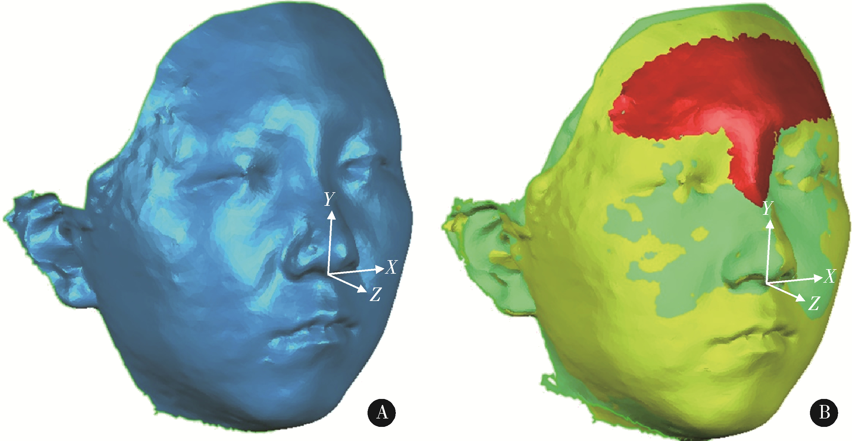
目的: 通过对骨性Ⅱ类高角患者拔牙矫治前后面部三维扫描模型与头颅侧位片进行测量分析, 探讨软硬组织变化趋势、影响因素及其作用。方法: 选取18例骨性Ⅱ类高角行单纯拔牙矫治的成人女性患者, 对矫治前后的头颅侧位片定点描记并进行硬组织测量, 同时获得纳入患者矫治前后三维颜面数据, 在完成模型重定位及模型重叠后, 对三维软组织标志点标定并测量相应三维软组织。硬组织测量包括17个测量指标(蝶鞍点-鼻根点-上齿槽座点的夹角、蝶鞍点-鼻根点-下齿槽座点的夹角、上齿槽座点-鼻根点-下齿槽座点间的夹角、面角、颌凸角、眶耳平面-下领平面角、Y轴角、前颅底平面-下颌平面角、颏前点到鼻根点-下齿槽座点连线的垂直距离、上中切牙到鼻根点-上齿槽座点连线的垂直距离、上中切牙与鼻根点-上齿槽座点的夹角、下中切牙到鼻根点-下齿槽座点连线的垂直距离、下中切牙与鼻根点-下齿槽座点连线交角、上下中切牙角、上切牙倾斜度、下中切牙-下颌平面角和Z角), 其中11个指标测量其治疗前后变化量。测量20个软组织标志点(左/右颧骨点、左/右口角点、左/右唇峰点、软组织颏顶点、左/右下颌角点、额点、下唇下缘点、上唇上缘点、软组织颏下点、左/右下颌边缘中点、软组织颏前点、口裂点、唇下点、鼻下点和上唇点)治疗前后三维位移量, 并取9项软组织测量指标(下唇高、面突角、下唇红高、下颌轮廓角、鼻唇角、人中长度、人中宽度、上唇高和上唇红高)。对矫治前后各指标变化量进行线性回归分析, 并运用Pearson系数分析各指标间相关性。结果: 矫治前后的硬组织及软组织20个测量指标中有18个测量值变化量差异有统计学意义(P<0.05), 主要变化表现为唇部的整体内收, 软组织颏前点存在垂直向的明显上移[(1.88±2.61) mm, P<0.05], 同时左/右口角点[(-2.95±1.9) mm、(-2.90±1.92) mm]、上下唇缘点[(-4.94±1.95) mm、(-3.25±1.44) mm]、唇下点[(-3.10±3.5) mm]、鼻下点[(-1.23±1.06) mm]在矢状向明显内收(P<0.05), 其他点差异无统计学意义; Z角平均增加4.10°±2.57°(P<0.05), 其他差异无统计学意义; 矫治后颏下点移动量与眶耳平面-下领平面角(比例为:-0.183 :1)、与前颅底平面-下领平面角(比例为:-0.157 :1)呈高度相关(r>0.7), 其余存在相关性的测量指标间呈中度相关(0.7≥r>0.4), 且差异均有统计学意义(P<0.05)。结论: 骨性Ⅱ类高角患者通过单纯拔牙矫治也可得到一定的面型改善, 主要表现为鼻、唇、颏三者矢状方向上关系的改善, 其软组织移动量与硬组织移动量间存在着一定的线性比例关系。

关键词: 骨性Ⅱ类, 高角, 正畸, 三维面型

Abstract:

Objective: To investigate the hard and soft tissue changing trend and contributing factors of skeletal class Ⅱ hyperdivergent patients before and after orthodontic camouflage treatment by analyzing the cephalogram and the three dimensional (3D) facial scan data. Methods: Eighteen skeletal class Ⅱ hyperdivergent adult female patients who finished camouflage orthodontic treatment were selected. Skeletal and dental measurements were carried out with the cephalometric analysis before and after the treatment. 3D facial data before and after orthodontic treatment were acquired and the anatomical landmarks were set after the repositioning and superimposition process. Hard tissue measurement included 17 mea-surement indicators (sella-nasion-subspinale angle, sella-nasion-supramental angle, subspinale-nasion-supramental angle, facial angle, angle of convexity, Frankfort horizontal plane-mandibular plane angle (FH-MP), Y axis angle, sella-nasion plane-mandibular plane angle (MP-SN), pogonion-nasion-supramental distance, upper incisor-nasion-subspinale distance, upper incisor to sella-nasion, lower incisor-nasion-supramental distance, lower incisor-nasion-supramental angle, upper incisor to lower incisor, upper incisor to sella-nasion, lower incisor-mandibular plane angle, and Z angle), and the changes before and after treatment were measured for 11 of them. Twenty soft tissue landmarks (left/right cheekbone, left/right chelion, left/right crista philtra, soft tissue gnathion, left/right gonion, glabella, labrale infe-rius, labrale superius, soft tissue menton, left/right mid-mandibular border, soft tissue pogonion, stomion superius, sublabial, subnasale, and supralabial) and 9 soft tissue indicators (lower lip height, facial convexity, lower vermilion height, mandibular contour, nasolabial angle, philtral length, philtral width, upper lip height, and upper vermilion height) were measured and recorded for treatment changes. Linear-regression analysis and correlation analysis were carried out for analyzing the relationship between hard and soft tissue changes before and after the treatment. Results: Significant differences were noticed for 18 out of the 20 cephalometric measurements and facial measurements before and after the treatment (P < 0.05), which mainly represented the sagittal retraction of lip area after the treatment. Significant vertical displacements were revealed for soft tissue menton after treatment [(1.88±2.61) mm, P < 0.05]. Significant sagittal displacements were revealed for left/right cheilion [(-2.95±1.9) mm, (-2.90±1.92) mm], labrale inferius[(-4.94±1.95) mm], labrale superius[(-3.25±1.44) mm], sublabial [(-3.10±3.5) mm], and subnasale [(-1.23±1.06) mm] after treatment (P < 0.05). An average of 4.10°±2.57° increasement was noticed for Z angle after treatment. High correlation (r>0.7) was noticed for the displacement of menton after treatment with FH-MP, with the rate of -0.183 :1, and MP-SN, with the rate of -0.157 :1. Moderate correlations (0.7≥r>0.4) were noticed for the other measurements with correlations (P < 0.05). Conclusion: A certain extent of facial improvements could be achieved with orthodontic camouflage treatment for skeletal class Ⅱ hyperdivergent patients, which were mostly represented by the improvement of sagittal relationship of nose, lips, and chin. Certain correlations were noticed for the hard and soft tissue changes.

Key words: Skeletal class Ⅱ, Hyperdivergent, Orthodontics treatment, Three-dimensional facial ana-lysis

中图分类号: 

  • R783.5

表1

头影测量项目及定义"

Measurement Definition
Hard tissue measurements before treatment
  SNA/(°) Sella-nasion-subspinale angle
  SNB/(°) Sella-nasion-supramental angle
  ANB/(°) Subspinale-nasion-supramental angle
  FH-NPo/(°) The angle formed by the Frankfort horizontal line and nasion-pogonion line
  NA-APo/(°) The angle formed by the nasion-subspinale line and subspinale-pogonion line
  FH-MP/(°) The angle formed by the Frankfort horizontal plane and mandibular plane
  SGn-FH/(°) The angle formed by the sella-gnathion line and Frankfort horizontal line
  MP-SN/(°) The angle formed by the sella-nasion plane and mandibular plane
  Po-NB/mm Vertical distance between pogonion and nasion-supramental line
Changes of measurements before and after treatment
  ΔU1-NA/mm The changes of vertical distance between the maxillary central incisor and nasion-subspinale line
  ΔU1-NA/(°) The changes of angle formed by the nasion-subspinale line and maxillary central incisor
  ΔL1-NB/mm The changes of vertical distance between the mandibular central incisor and nasion-supramental line
  ΔL1-NB/(°) The changes of angle formed by the nasion-supramental line and mandibular central incisor
  ΔU1-L1/(°) The changes of interincisal angle
  ΔU1-SN/(°) The changes of angle formed by the sella-nasion plane and maxillary central incisor
  ΔIMPA/(°) The changes of angle formed by the mandibular plane and mandibular central incisor
  ΔZ angle/(°) The changes of angle formed by Frankfort horizontal line and line marking the most front point of chin and lip

图1

三维面部模型处理"

表2

三维软组织面部特征点及测量项目"

Name Definition
Soft tissue landmark
  Left/right cheekbone (L/R Chb) Point of maximal convexity on the facial contour of the cheekbone region
  Left/right chelion (L/R Chl) Point marking the most lateral extent of the labial fissure
  Left/right crista philtri (L/R CPh) Point on each elevated margin of the philtrum at the vermilion border of the upper lip
  Soft tissue gnathion(Gn’) The halfway point on the medial curve between Pg’ and Me’ under the chin
  Left/right gonion (L/R Go) The inferior aspect of the mandible at its most acute (mandibular angle) and lateral point
  Glabella (Gl) The most convex sagittal midline point on the forehead
  Labrale inferius (Li) The midline point on the lower vermilion border of the lower lip
  Labrale superius (Ls) The midline point on the upper vermilion border of the upper lip
  Soft tissue menton (Me’) The most inferior midline point of the chin
  Left/right mid-mandibular border(L/R Mid-Mb) Point on the mandibular border, midway between Pg’ and Go, along the jaw line
  Soft tissue pogonion (Pg’) The point of maximal curvature on the midline chin curve
  Stomion superius (Sto) Midline point on the lower margin of bottom of the upper lip
  Sublabial (Sl) The deepest point of the mentolabial sulcus
  Subnasale (Sn) The apex of the nasolabial angle in the midline, where the inferior border of the nasal septum merges with the upper cutaneous lip
  Supralabial (Spl) Midline point of maximal concavity on the facial contour between Sn and Ls
Soft tissue measurement
  Lower lip height (LL H) Distance between Li and Sl
  Facial convexity (FC) Angle at Sn subtended by Gl and Pg’
  Lower vermilion height (L Verm H) Distance between Li and Sto
  Mandibular contour (Mand Cont) Angle at Me’ subtended by L/R Go projected to coronal plane
  Nasolabial angle(Nasolabial A) Angle at Sn subtended by Columella and Ls
  Philtral length (Philtral L) Distance between Sn and Ls
  Philtral width (Philtral W) Distance between L/R CPh
  Upper lip height (ULH) Distance between Sn and Sto
  Upper vermilion height (U Verm H) Distance between Ls and Sto

表3

头影测量分析中基准项目测量值"

Items Value, ˉx±s
SNA/ (°) 80.94±3.35
SNB/ (°) 73.12±4.42
ANB/ (°) 7.82±2.25
FH-NPo /(°) 81.13±3.18
NA-APo /(°) 17.15±6.26
FH-MP/ (°) 37.07±6.42
SGn-FH /(°) 69.00±3.59
MP-SN/ (°) 45.40±7.34
Po-NB/mm 1.53±1.11

表4

头影测量值及三维面部测量值"

Items Value, ˉx±s t P
Cephalometric measurement
  ΔANB/ (°) 0.43±0.44 3.68 0.003*
  ΔMP-SN/ (°) 0.51±0.83 1.78 0.099
  ΔFH-MP/ (°) 0.46±0.92 1.86 0.085
  ΔU1-NA/mm -6.22±1.03 22.69 <0.001*
  ΔU1-NA /(°) -7.87±5.17 5.70 <0.001*
  ΔL1-NB /mm -3.67±1.35 10.13 <0.001*
  ΔL1-NB/ (°) -7.33±3.38 8.11 <0.001*
  ΔU1-L1/ (°) 16.91±5.27 -12.01 <0.001*
  ΔU1-SN/(°) -10.51±4.45 8.83 <0.001*
  ΔIMPA/(°) -6.33±3.09 7.67 <0.001*
  ΔZ angle/ (°) 4.10±2.57 5.97 <0.001*
Soft tissue measurement
  ΔLL H/mm -0.82±0.63 4.87 <0.001*
  ΔFC/mm 0.76±0.79 -3.60 0.003*
  ΔL Verm H/mm -0.87±0.88 3.72 0.003*
  ΔMand Cont /(°) 3.54±2.82 -4.69 <0.001*
  ΔNasolabial A /(°) 6.40±4.81 -4.97 <0.001*
  ΔPhiltral L /mm 0.03±0.88 -0.13 0.896
  ΔPhiltral W/mm -0.60±0.33 6.82 <0.001*
  ΔULH/mm 0.33±1.04 -1.18 0.261
  ΔU Verm H/mm 0.29±1.00 -1.09 0.296

表5

15个三维面部特征点矫治前后的三维偏差"

Items Value, ˉx±s t P Items Value, ˉx±s t P
R Chb Li
  X -0.34±0.8 -1.60 0.134   X 0.15±0.74 0.73 0.477
  Y 0.14±1.53 0.35 0.734   Y -0.16±1.99 -0.31 0.764
  Z 0.04±0.91 0.18 0.859   Z -4.94±1.95 -9.50 <0.001*
  D 1.81±0.67 10.11 <0.001*   D 5.36±1.92 10.45 <0.001*
L Chb Ls
  X 0.43±0.77 2.08 0.058   X -0.07±0.57 -0.43 0.677
  Y 0.08±1.12 0.28 0.786   Y -0.43±1.99 -0.80 0.437
  Z -0.68±0.83 -3.09 0.009*   Z -3.25±1.44 -8.44 <0.001*
  D 1.58±0.74 8.04 <0.001*   D 3.81±1.51 9.47 <0.001*
R Chl Me’
  X -0.11±1.64 -0.26 0.802   X 0.60±1.35 1.68 0.118
  Y -0.18±1.38 -0.48 0.639   Y 1.02±2.4 1.60 0.134
  Z -2.90±1.92 -5.65 <0.001*   Z -0.86±2.44 -1.32 0.210
  D 3.66±1.75 7.83 <0.001*   D 3.46±1.63 7.97 <0.001*
L Chl R Mid-Mb
  X 0.37±1.25 1.09 0.294   X -0.82±1.32 -2.31 0.038*
  Y 0.66±0.98 2.49 0.027*   Y 0.11±1.74 0.24 0.816
  Z -2.95±1.9 -5.79 <0.001*   Z -1.05±1.88 -2.10 0.056
  D 3.48±1.76 7.42 <0.001*   D 2.99±1.76 6.36 <0.001*
Gn’ L Mid-Mb
  X 0.17±0.84 0.78 0.449   X 0.87±1.59 2.05 0.061
  Y 1.16±2.73 1.59 0.135   Y -0.34±1.63 -0.79 0.444
  Z -0.04±2.55 -0.06 0.957   Z -1.16±0.83 -5.21 <0.001*
  D 3.32±2.07 6.00 <0.001*   D 2.42±1.14 7.91 <0.001*
R Go Pg’
  X -0.48±1.41 -1.27 0.227   X 0.08±0.8 0.37 0.717
  Y 0.39±0.96 1.50 0.157   Y 1.88±2.61 2.69 0.019*
  Z -1.35±1.31 -3.86 0.002*   Z -0.15±2.02 -0.28 0.783
  D 2.46±0.86 10.73 <0.001*   D 3.45±1.6 8.05 <0.001*
L Go Sl
  X 0.41±1.29 1.18 0.258   X 0.24±1.01 0.90 0.383
  Y -0.44±1.08 -1.52 0.152   Y -0.50±2.03 -0.92 0.372
  Z -0.40±1.53 -0.98 0.347   Z -3.10±3.5 -3.32 0.006*
  D 2.03±1.13 6.70 <0.001*   D 4.72±2.02 8.76 <0.001*
Sn
  X 0.20±0.47 1.58 0.139
  Y -0.33±0.7 -1.78 0.099
  Z -1.23±1.06 -4.34 0.001*
  D 1.64±0.85 7.25 <0.001*

表6

三维面部测量值及特征点三维偏差量与头影测量值的线性回归分析"

Dependent variable Pearson correlation coefficient Prediction equation R2 Standard error of estimate P
Constant term Independent variable
ΔLL H 0.613 -0.227 0.287×ΔL1-NB (mm) 0.376 0.52 0.010*
ΔL Verm H 0.643 -0.656 0.417×ΔL1-NB (mm) 0.413 0.70 0.007**
ΔL Verm H 0.494 -0.068 0.127×ΔL1-NB 0.244 0.80 0.036*
ΔL Verm H -0.550 -0.678 -0.092×ΔU1-L1 0.303 0.76 0.021*
ΔPhiltral L -0.642 0.827 -0.109×ΔU1-NA 0.412 0.70 0.007**
ΔPhiltral L 0.473 1.304 0.079×ΔU1-L1 0.224 0.81 0.044*
ΔPhiltral L -0.488 0.981 -0.096×ΔU1-SN 0.238 0.80 0.038*
ΔULH -0.553 0.548 -0.111×ΔU1-NA 0.306 0.90 0.020*
ΔULH 0.482 1.281 0.095×ΔU1-L1 0.232 0.95 0.040*
ΔU Verm H -0.474 0.185 -0.187×ΔU1-NA (mm) 0.224 0.91 0.044*
R Chl D -0.566 5.021 -0.891×Po-NB (mm) 0.320 1.50 0.017*
L Chl D -0.692 5.157 -1.095×Po-NB (mm) 0.479 1.32 0.003**
Gn’ D -0.469 9.324 -0.132×MP-SN 0.220 1.90 0.045*
Gn’ D -0.463 5.394 -0.283×ΔL1-NB 0.214 1.91 0.048*
Me’ D -0.723 10.258 -0.183×FH-MP 0.523 1.17 0.002**
Me’ D -0.707 10.578 -0.157×MP-SN 0.501 1.20 0.002**
Pg’ D -0.483 7.925 -0.121×FH-MP 0.234 1.46 0.040*
Pg’ D -0.509 8.492 -0.111×MP-SN 0.259 1.44 0.032*
Sn D 0.560 0.614 0.140×ΔL1-NB 0.313 0.73 0.019*
Sn D 0.532 0.718 0.146×ΔIMPA 0.283 0.75 0.025*
R Chl Z 0.560 -4.390 0.970×Po-NB(mm) 0.313 1.66 0.019*
L Chl Z 0.695 -4.772 1.191×Po-NB(mm) 0.482 1.43 0.003**
Li Z 0.509 -6.304 0.892×Po-NB(mm) 0.259 1.74 0.032*
Li Z -0.509 -3.938 -0.394×ΔU1-NA(mm) 0.259 1.74 0.031*
Sn Z -0.571 0.083 -0.179×ΔL1-NB 0.326 0.90 0.016*
Sn Z -0.553 -0.028 -0.189×ΔIMPA 0.306 0.92 0.020*
1 Reis PMP , Lima P , Garcia FCP , et al. Effect of maxillary median diastema on the esthetics of a smile[J]. Am J Orthod Dentofacial Orthop, 2020, 158 (4): E37- E42.
doi: 10.1016/j.ajodo.2020.07.028
2 施丹妮, 杨鑫, 吴建勇. 锥形束CT三维头影测量参考坐标系的研究进展[J]. 国际口腔医学杂志, 2021, 48 (4): 398- 404.
3 Ferrario VF , Sforza C , Schmitz JH , et al. A three-dimensional computerized mesh diagram analysis and its application in soft tissue facial morphometry[J]. Am J Orthod Dentofacial Orthop, 1998, 114 (4): 404- 413.
doi: 10.1016/S0889-5406(98)70185-4
4 Xiao Z , Liu Z , Gu Y . Integration of digital maxillary dental casts with 3D facial images in orthodontic patients: A three-dimensional validation study[J]. Angle Orthod, 2020, 90 (3): 397- 404.
doi: 10.2319/071619-473.1
5 Janson G , Mendes LM , Junqueira CHZ , et al. Soft-tissue changes in class Ⅱ malocclusion patients treated with extractions: A systematic review[J]. Eur J Orthod, 2016, 38 (6): 631- 637.
doi: 10.1093/ejo/cjv083
6 Konstantonis D , Vasileiou D , Papageorgiou SN , et al. Soft tissue changes following extraction vs. nonextraction orthodontic fixed appliance treatment: A systematic review and meta-analysis[J]. Eur J Oral Sci, 2018, 126 (3): 167- 179.
doi: 10.1111/eos.12409
7 谢天伊, 贾绮林. 成年女性正畸患者颊颞部软组织变化的三维测量研究[J]. 中华口腔正畸学杂志, 2020, 27 (4): 181- 186.
8 Lu WX , Zhang X , Mei L , et al. Orthodontic incisor retraction caused changes in the soft tissue chin area: A retrospective study[J]. BMC Oral Health, 2020, 20 (1): 108.
doi: 10.1186/s12903-020-01099-2
9 Tadic N , Woods MG . Incisal and soft tissue effects of maxillary premolar extraction in class Ⅱ treatment[J]. Angle Orthod, 2007, 77 (5): 808- 816.
doi: 10.2319/081706-336
10 Veltkamp T , Buschang PH , English JD , et al. Predicting lower lip and chin response to mandibular advancement and genioplasty[J]. Am J Orthod Dentofacial Orthop, 2002, 122 (6): 627- 634.
doi: 10.1067/mod.2002.128864
11 Choi SH , Lee H , Hwang JJ , et al. Differences in soft-tissue thickness changes after bimaxillary surgery between patients with vertically high angle and normal angle[J]. Am J Orthod Dentofacial Orthop, 2021, 159 (1): 30- 40.
doi: 10.1016/j.ajodo.2019.10.028
12 Holdaway RA . A soft-tissue cephalometric analysis and its use in orthodontic treatment planning. Part Ⅰ[J]. Am J Orthod, 1983, 84 (1): 1- 28.
doi: 10.1016/0002-9416(83)90144-6
13 Hodges A , Rossouw PE , Campbell PM , et al. Prediction of lip response to four first premolar extractions in white female adolescents and adults[J]. Angle Orthod, 2009, 79 (3): 413- 421.
doi: 10.2319/050208-247.1
14 Upadhyay M , Yadav S , Nagaraj K , et al. Treatment effects of mini-implants for en-mass eretraction of anterior teeth in bialveolar dental protrusion patients: A randomized controlled trial[J]. Am J Orthod Dentofacial Orthop, 2008, 134 (1): 18- 29.
doi: 10.1016/j.ajodo.2007.03.025
15 Sivakumar A , Valiathan A . Cephalometric assessment of dentofacial vertical changes in class Ⅰ subjects treated with and without extraction[J]. Am J Orthod Dentofacial Orthop, 2008, 133 (6): 869- 875.
doi: 10.1016/j.ajodo.2006.05.041
[1] 雷玥,杨颖婷,雷海华. 异位萌出诱导矫治器在第一恒磨牙异位萌出治疗中的应用[J]. 北京大学学报(医学版), 2023, 55(4): 766-封三.
[2] 付玉,胡鑫浓,崔圣洁,施捷. 骨性Ⅱ类高角错患者术前正畸下前牙去代偿效果及牙槽骨改建分析[J]. 北京大学学报(医学版), 2023, 55(1): 62-69.
[3] 高娟,吕航苗,马慧敏,赵一姣,李小彤. 锥形束CT三维体积测量评估骨性Ⅲ类错正畸正颌治疗后的牙根吸收[J]. 北京大学学报(医学版), 2022, 54(4): 719-726.
[4] 杨雨卉,黄一平,李巍然. 骨皮质切开加速正畸牙齿移动对牙根吸收的影响[J]. 北京大学学报(医学版), 2021, 53(2): 434-437.
[5] 周境,刘怡. 不同垂直骨面型骨性Ⅱ类青少年女性颞下颌关节锥形束CT测量分析[J]. 北京大学学报(医学版), 2021, 53(1): 109-119.
[6] 王高南,焦剑,周彦恒,施捷. 正畸牙齿位置的移动对角化龈宽度的影响[J]. 北京大学学报(医学版), 2019, 51(5): 931-936.
[7] 戴帆帆,刘怡,许天民,陈贵. 探索成人正畸前后下颌三维数字化模型的重叠方法[J]. 北京大学学报(医学版), 2018, 50(2): 271-278.
[8] 张又文,辛天艺,焦剑,周彦恒,施捷. 慢性牙周炎的减数正畸治疗[J]. 北京大学学报(医学版), 2018, 50(2): 308-313.
[9] 柳玉树,李峥,赵一姣,叶红强,周彦秋,胡文杰,刘云松,寻春雷,周永胜. 数字化正畸修复联合治疗设计在前牙美学重建中的应用[J]. 北京大学学报(医学版), 2018, 50(1): 78-84.
[10] 柳大为,李晶,郭亮,荣起国,周彦恒. 舌侧矫治器关闭间隙上前牙牙周膜应力变化的三维有限元分析[J]. 北京大学学报(医学版), 2018, 50(1): 141-147.
[11] 张达,王林川,周彦恒,刘晓默,李晶. 3D打印间接粘接托槽精度[J]. 北京大学学报(医学版), 2017, 49(4): 704-708.
[12] 沈潇,施捷,徐莉,焦剑,路瑞芳,孟焕新. 伴错牙合畸形的侵袭性牙周炎患者牙周-正畸联合治疗的临床评价[J]. 北京大学学报(医学版), 2017, 49(1): 60-066.
[13] 郑旭,胡兴学,马宁,陈晓红. 正畸矫治牙性牙合平面倾斜的新方法——波浪形弓[J]. 北京大学学报(医学版), 2017, 49(1): 176-180.
[14] 温馥嘉,陈贵,刘怡. 基于锥形束CT的强支抗内收上前牙病例牙根及牙槽骨的形态学分析[J]. 北京大学学报(医学版), 2016, 48(4): 702-708.
[15] 黄俊强, 刘施瑶, 江久汇. Tweed-Merrifield技术矫治成人严重双颌前突的疗效评价[J]. 北京大学学报(医学版), 2016, 48(3): 555-561.
Viewed
Full text


Abstract

Cited

  Shared   
  Discussed   
No Suggested Reading articles found!