北京大学学报(医学版) ›› 2024, Vol. 56 ›› Issue (4): 735-740. doi: 10.19723/j.issn.1671-167X.2024.04.030
基于卷积神经网络实现锥形束CT牙齿分割及牙位标定
薄士仕1,2,高承志2,*( )
)
- 1. 北京大学口腔医学院·口腔医院综合二科, 国家口腔医学中心, 国家口腔疾病临床医学研究中心, 口腔生物材料和数字诊疗装备国家工程研究中心, 北京 100081
2. 北京大学人民医院口腔科, 北京 100044
Tooth segmentation and identification on cone-beam computed tomography with convolutional neural network based on spatial embedding information
Shishi BO1,2,Chengzhi GAO2,*()
- 1. Department of General Dentistry Ⅱ, Peking University School and Hospital of Stomatology & National Center for Stomatology & National Clinical Research Center for Oral Diseases & National Engineering Research Center of Oral Biomaterials and Digital Medical Devices, Beijing 100081, China
2. Department of Dentistry, Peking University People' s Hospital, Beijing 100044, China
摘要:
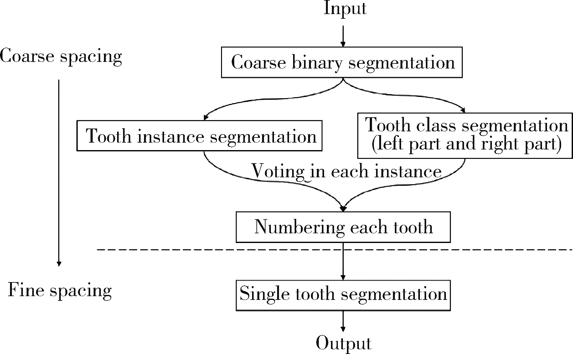
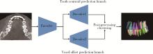
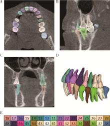
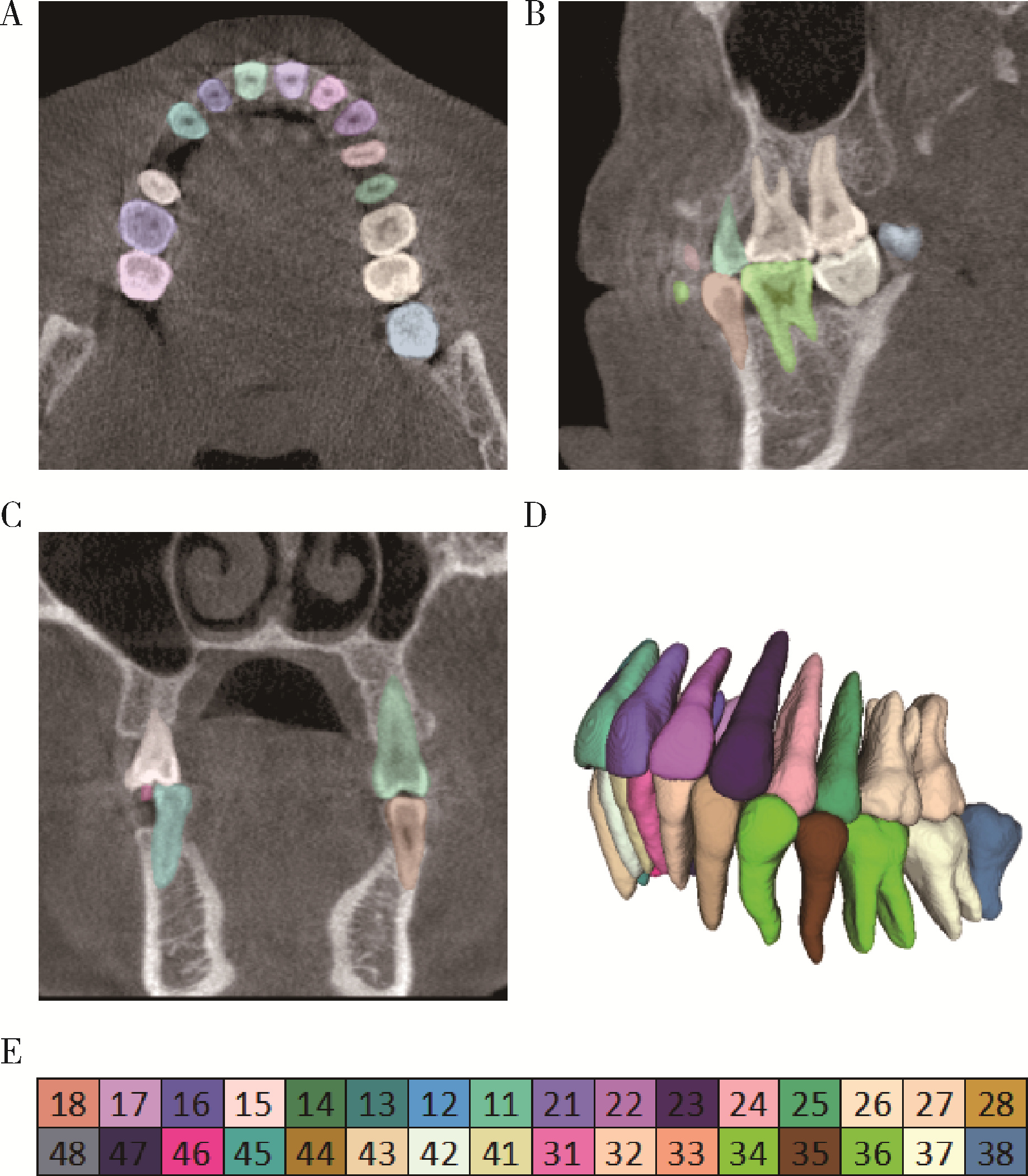
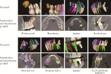
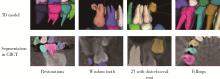
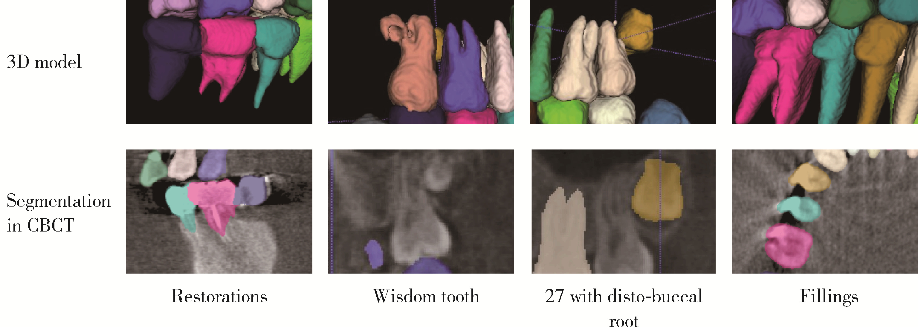
目的: 利用卷积神经网络实现基于锥形束CT(cone-beam computed tomography, CBCT)体素数据的牙齿实例分割和牙位标定。方法: 本文所提出的牙齿算法包含三个不同的卷积神经网络,网络架构以Resnet为基础模块,首先对CBCT图像进行降采样,然后确定一个包含CBCT图像中所有牙齿的感兴趣区域(region of interest, ROI)。通过训练模型,ROI利用一个双分支“编码器-解码器”结构网络,预测输入数据中每个体素所对应的相关空间位置信息,进行聚类后实现牙齿的实例分割。牙位标定则通过另一个多类别分割任务设计的U-Net模型实现。随后,在原始空间分辨率下,训练了一个用于精细分割的U-Net网络,得到牙齿的高分辨率分割结果。本实验收集了59例带有简单冠修复体及种植体的CBCT数据进行人工标注作为数据库,对牙齿算法的预测结果使用实例Dice相似系数(instance Dice similarity coefficient, IDSC)用来评估牙齿分割结果,使用平均Dice相似系数(the average Dice similarity coefficient,ADSC)评估牙齿分割及牙位标定的共同结果并进行评定。结果: 量化指标显示,IDSC为89. 35%,ADSC为84. 74%。剔除了带有修复体伪影的数据后生成了有43例样本的数据库,训练网络得到了更优良的性能,IDSC为90. 34%,ADSC为87.88%。将得到的结果进行可视化分析,牙齿算法不仅可以清晰地分割出CBCT中牙齿的形态,而且可以对牙齿的分类进行准确的编号。结论: 该牙齿算法不仅可以成功实现三维图像的牙齿及修复体分割,还可以准确标定所有恒牙的牙位,具有临床实用性。
中图分类号:
- R780.1


| 1 |
Liao FZ , Liang M , Li Z , et al. Evaluate the malignancy of pulmonary nodules using the 3-D deep leaky noisy-OR network[J]. IEEE Trans Neural Netw Learn Syst, 2019, 30 (11): 3484- 3495.
doi: 10.1109/TNNLS.2019.2892409 |
| 2 |
Long J , Shelhamer E , Darrell T . Fully convolutional networks for semantic segmentation[J]. IEEE Trans Pattern Anal Mach Intell, 2017, 39 (4): 640- 651.
doi: 10.1109/TPAMI.2016.2572683 |
| 3 | Ronneberger O, Fischer P, Brox T. U-Net: Convolutional networks for biomedical image segmentation: International conference on medical image computing and computer-assisted intervention[C]. Cham: Springer, 2015. |
| 4 | Neven D, Brabandere BD, Proesmans M, et al. Instance segmentation by jointly optimizing spatial embeddings and clustering bandwidth: 2019 IEEE/CVF conference on computer vision and pattern recognition (CVPR)[C]. Long Beach, CA: IEEE, 2019. |
| 5 |
Pei YR , Ai XS , Zha HB , et al. 3D exemplar-based random walks for tooth segmentation from cone-beam computed tomography images[J]. Med Phys, 2016, 43 (9): 5040- 5050.
doi: 10.1118/1.4960364 |
| 6 | Patil S , Kulkarni V , Bhise A . Algorithmic analysis for dental caries detection using an adaptive neural network architecture[J]. Heliyon, 2019, 5 (5): e1579. |
| 7 |
Zhang KL , Wu J , Chen H , et al. An effective teeth recognition method using label tree with cascade network structure[J]. Comput Med Imaging Graph, 2018, 68, 61- 70.
doi: 10.1016/j.compmedimag.2018.07.001 |
| 8 |
Hosntalab M , Aghaeizadeh ZR , Abbaspour TA , et al. Classification and numbering of teeth in multi-slice CT images using wavelet-Fourier descriptor[J]. Int J Comput Assist Radiol Surg, 2010, 5 (3): 237- 249.
doi: 10.1007/s11548-009-0389-8 |
| 9 |
Hwang JJ , Jung YH , Cho BH , et al. An overview of deep learning in the field of dentistry[J]. Imaging Sci Dent, 2019, 49 (1): 1- 7.
doi: 10.5624/isd.2019.49.1.1 |
| 10 |
Chen H , Zhang KL , Lyu PJ , et al. A deep learning approach to automatic teeth detection and numbering based on object detection in dental periapical films[J]. Sci Rep, 2019, 9 (1): 3840.
doi: 10.1038/s41598-019-40414-y |
| 11 |
Miki Y , Muramatsu C , Hayashi T , et al. Classification of teeth in cone-beam CT using deep convolutional neural network[J]. Comput Biol Med, 2017, 80, 24- 29.
doi: 10.1016/j.compbiomed.2016.11.003 |
| 12 | Cui ZM, Li CJ, Wang WP. ToothNet: Automatic tooth instance segmentation and identification from cone beam CT images: 2019 IEEE/CVF conference on computer vision and pattern recognition (CVPR)[C]. Long Beach, CA: IEEE, 2019. |
| 13 | He KM, Zhang XY, Ren SQ, et al. Deep residual learning for image recognition: 2016 IEEE conference on computer vision and pattern recognition (CVPR)[C]. Las Vegas: IEEE, 2016. |
| 14 | Dentistry-designation system for teeth and areas of the oral cavity: ISO 3950: 2009[S/OL]. [2016-03-01]. https://www.iso.org/standard/68292.html. |
| 15 |
Dice LR . Measures of the amount of ecologic association between species[J]. Ecology, 1945, 26 (3): 297- 302.
doi: 10.2307/1932409 |
| [1] | 王月, 梁宇红. 繁茂型牙骨质-骨结构不良1例[J]. 北京大学学报(医学版), 2026, 58(1): 220-224. |
| [2] | 刘杰, 马茗微, 王庆安, 石明, 尹金鹏, 王占平, 申静涛, 高献书. 基于锥形束CT的前列腺癌放射治疗两种体位固定方式摆位误差比较[J]. 北京大学学报(医学版), 2025, 57(4): 692-697. |
| [3] | 章锦花,潘洁,孙志鹏,王霄. 不同根管内容物对口腔颌面锥形束CT诊断牙根纵裂准确性的影响[J]. 北京大学学报(医学版), 2023, 55(2): 333-338. |
| [4] | 潘孟乔,刘建,徐莉,徐筱,侯建霞,李小彤,王晓霞. 牙周-正畸-正颌联合治疗骨性安氏Ⅲ类错 |
| [5] | 叶佳学,梁宇红. 牙髓专科医师应用锥形束CT的现况调查[J]. 北京大学学报(医学版), 2023, 55(1): 114-119. |
| [6] | 高娟,吕航苗,马慧敏,赵一姣,李小彤. 锥形束CT三维体积测量评估骨性Ⅲ类错 |
| [7] | 刘伟涛,王怡然,王雪东,周彦恒. 锥形束CT研究上颌反复扩缩前方牵引后上颌骨缝的三维变化[J]. 北京大学学报(医学版), 2022, 54(2): 346-355. |
| [8] | 孟圆,张丽琪,赵雅宁,柳登高,张祖燕,高岩. 67例上颌根尖周囊肿的三维影像特点分析[J]. 北京大学学报(医学版), 2021, 53(2): 396-401. |
| [9] | 曹畅,王菲,王恩博,刘宇. β-磷酸三钙用于下颌第三磨牙拔除术后骨缺损修复的自身对照研究[J]. 北京大学学报(医学版), 2020, 52(1): 97-102. |
| [10] | 颜野,夏海缀,李旭升,何为,朱学华,张智荧,肖春雷,刘余庆,黄华,何良华,卢剑. 基于U型卷积神经网络学习的前列腺癌影像重建模型在手术导航中的应用[J]. 北京大学学报(医学版), 2019, 51(3): 596-601. |
| [11] | 谢晓艳,贾淑梅,孙志辉,张祖燕. 分辨率设置与锥形束CT检测牙根外吸收的可靠性[J]. 北京大学学报(医学版), 2019, 51(1): 75-79. |
| [12] | 赵一姣,刘怡,孙玉春,王勇. 一种基于曲率连续算法的冠、根三维数据融合方法[J]. 北京大学学报(医学版), 2017, 49(4): 719-723. |
| [13] | 钟金晟, 欧阳翔英, 柳登高, 曹采方. 锥形束CT测量离体下颌磨牙Ⅱ°根分叉病变效果的评价[J]. 北京大学学报(医学版), 2010, 42(1): 41-45. |
| [14] | 刘怡, James MAH, 许天民. 锥形束计算机断层扫描中牙齿的分割精度[J]. 北京大学学报(医学版), 2010, 42(1): 98-102. |
| [15] | 王瑞永, 马绪臣, 张万林, 柳登高. 健康成年人颞下颌关节间隙锥形束计算机体层摄影术测量分析[J]. 北京大学学报(医学版), 2007, 39(5): 503-506. |
|
||
