北京大学学报(医学版) ›› 2026, Vol. 58 ›› Issue (2): 380-387. doi: 10.19723/j.issn.1671-167X.2026.02.024

• 技术方法 • 上一篇    下一篇

兔食管良性环周狭窄模型的建立

孟伶宇, 黄永辉*(), 闫秀娥, 王迎春   

  1. 北京大学第三医院消化科, 北京 100191
  • 收稿日期:2023-11-12 出版日期:2026-04-18 发布日期:2025-11-10
  • 通讯作者: 黄永辉
  • 基金资助:
    国家自然科学基金(82070653); 北京市自然科学基金(7222207); 首都卫生发展科研专项(2022-2Z-40914)

Establishment of rabbit model of benign circumferential esophageal stricture

Lingyu MENG, Yonghui HUANG*(), Xiu'e YAN, Yingchun WANG   

  1. Department of Gastroenterology, Peking University Third Hospital, Beijing 100191, China
  • Received:2023-11-12 Online:2026-04-18 Published:2025-11-10
  • Contact: Yonghui HUANG
  • Supported by:
    the National Natural Science Foundation of China(82070653); Beijing Natural Science Foundation(7222207); Capital's Funds for Health Improvement and Research(2022-2Z-40914)

RICH HTML


摘要:

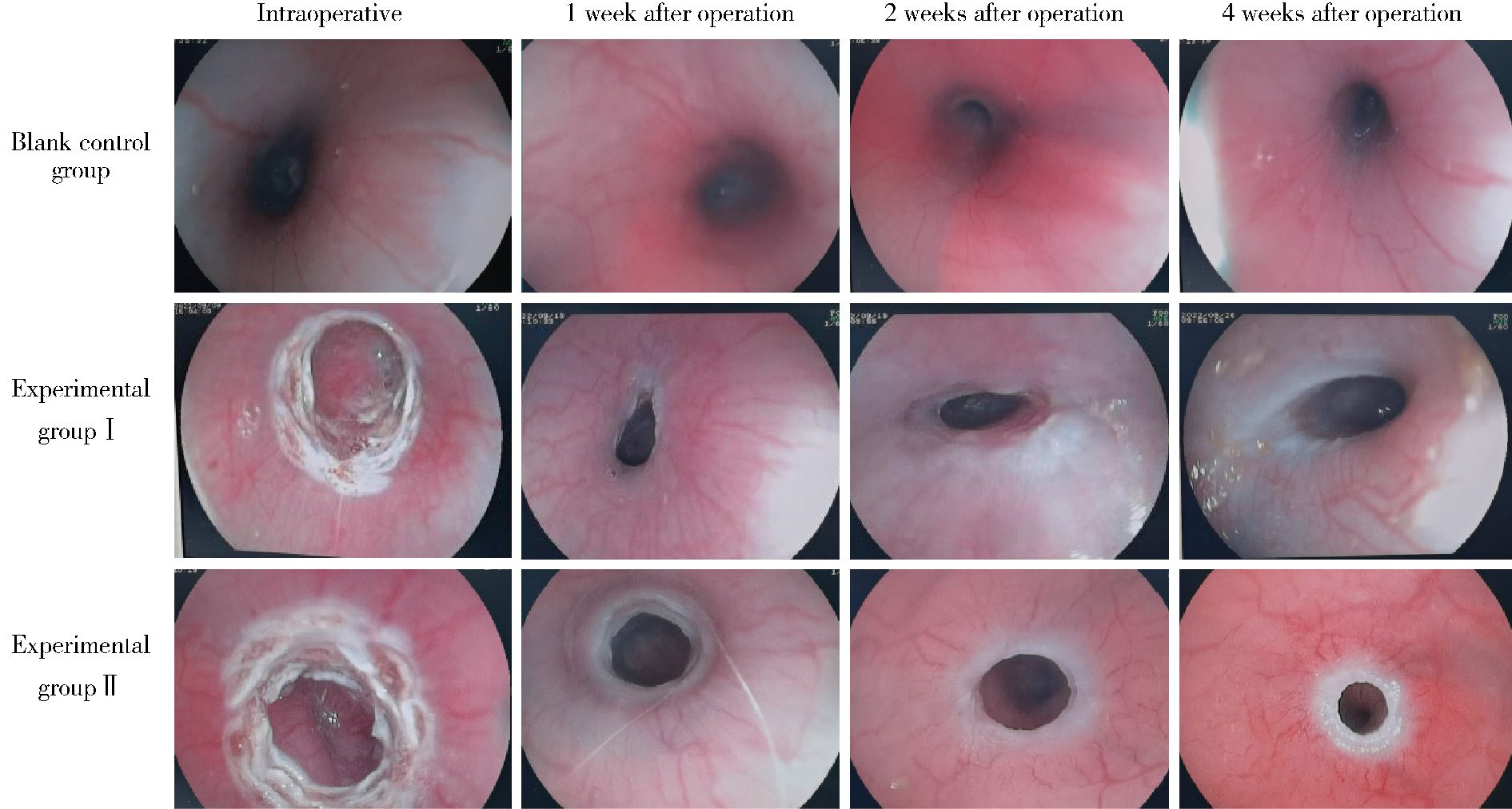
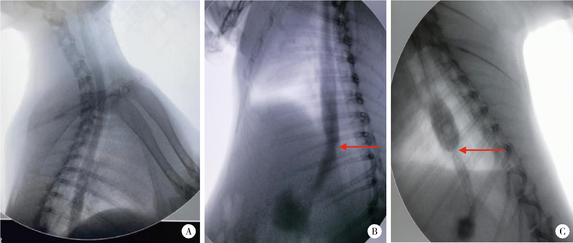
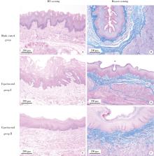
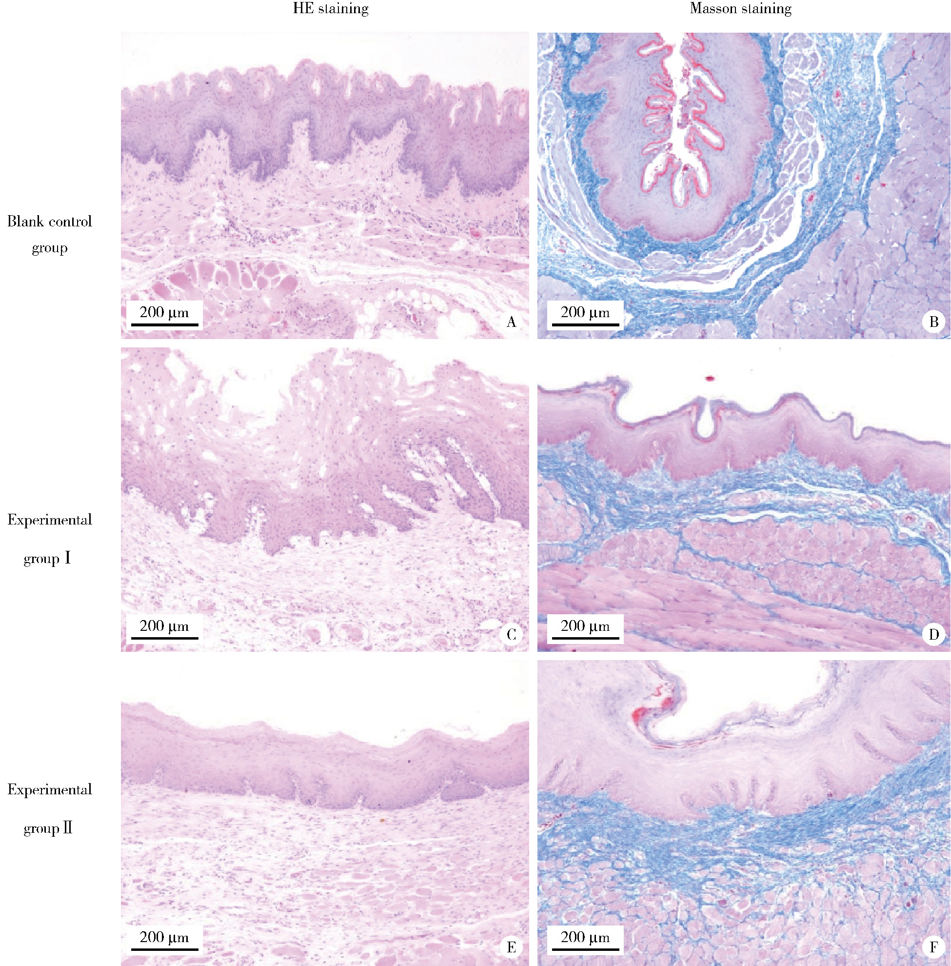
目的: 探索内镜下应用氩离子凝固术建立兔食管良性狭窄的方法,为后续研究食管狭窄的预防提供便捷、稳定的动物模型。方法: 雄性新西兰兔22只,完善X线下食管造影检查后随机分为3组,空白对照组(n=4)仅接受内镜下检查,实验Ⅰ组(n=9)和实验Ⅱ组(n=9)分别于内镜下应用30 W和50 W功率氩气刀对食管进行逐点、环周烧灼。术后第1、2、4周复查内镜,观察食管改变,并记录体重及精神状态。术后第4周完善X线下食管造影,测定食管狭窄处内径并计算狭窄指数;将所有实验动物处死后解剖获得食管标本,进行组织病理学检查并检测食管组织羟脯氨酸含量。结果: 空白对照组在实验后第4周时的体重较实验前明显增加[(4.13±0.25) kg vs. (3.10±0.39) kg,P < 0.05],食管内径与实验前相比无明显变化[(12.89±0.83) mm vs. (12.83±1.07) mm,P>0.05]。实验Ⅰ组在实验后第4周时与实验前相比,体重和食管内径均未见明显变化[(2.91±0.28) kg vs. (2.91±0.54) kg;(11.19±0.97) mm vs. (12.06±0.32) mm;P均>0.05];实验Ⅱ组在实验后第4周时与实验前相比,体重和食管内径均明显减小[(2.02±0.31) kg vs. (3.51±0.37) kg;(10.49±1.76) mm vs. (12.58±1.11) mm;P均<0.05]。术后第4周的食管狭窄指数,实验Ⅱ组明显高于实验Ⅰ组(1.242±0.148 vs. 1.083±0.104,P<0.05)。组织病理学评分和羟脯氨酸含量,实验Ⅰ组[2.55±0.52,(182.90±72.75) μg/g]和实验Ⅱ组[4.55±0.52,(210.81±54.28) μg/g]均较空白对照组[0,(91.37±29.74)μg/g]明显增加(P均<0.05)。结论: 内镜下应用50 W功率氩气刀进行环周烧灼诱导雄性新西兰兔食管狭窄方法可行,深度可控,且可大量重复,该方案可为研究食管狭窄提供便捷、稳定的动物模型。

关键词: 食管狭窄, 动物模型, 氩离子凝固术, 食管镜

Abstract:

Objective: To explore the method of establishing benign esophageal stricture in rabbits by using argon plasma coagulation under endoscopy, and to provide a convenient and stable animal model for subsequent research on the prevention of esophageal stricture. Methods: Twenty-two male New Zealand rabbits were randomly divided into three groups after completing esophageal radiography under X-ray. The blank control group (n=4) only received endoscopic examination. In the experimental group Ⅰ (n=9) and experimental group Ⅱ (n=9), argon knife was performed on the esophagus at 30 W and 50 W power, respectively, under endoscopy. Endoscopy was performed 1, 2, and 4 weeks after the operation to observe the changes in the esophagus, and the body weight and mental state were recorded. Four weeks after the operation, esophageal radiography under X-ray was performed to measure the inner diameter of the esophageal stricture and calculate the stricture index. All the experimental animals were sacrificed and esophageal specimens were obtained for histopathological examination and detection of hydroxyproline content in esophageal tissue. Results: In the blank control group, the body weight significantly increased 4 weeks after the operation compared with that before the operation [(4.13±0.25) kg vs. (3.10±0.39) kg, P < 0.05], and there was no significant change in the esophageal inner diameter [(12.89±0.83) mm vs. (12.83±1.07) mm, P>0.05]. In the experimental group Ⅰ, there was no significant change in the body weight and esophageal inner diameter 4 weeks after the operation compared with that before the operation [(2.91±0.28) kg vs. (2.91±0.54) kg; (11.19±0.97) mm vs. (12.06±0.32) mm; P>0.05]. In the experimental group Ⅱ, the body weight and esophageal inner diameter significantly decreased 4 weeks after the operation compared with that before the operation [(2.02±0.31) kg vs. (3.51±0.37) kg; (10.49±1.76) mm vs. (12.58±1.11) mm; P < 0.05]. The esophageal stricture index 4 weeks after the operation was significantly higher in the experimental group Ⅱ than in the experimental group Ⅰ (1.242±0.148 vs. 1.083±0.104, P < 0.05). The histopathological score and hydroxyproline content in the experimental group Ⅰ [2.55±0.52, (182.90±72.75) μg/g] and experimental group Ⅱ [4.55±0.52, (210.81±54.28) μg/g] were significantly higher than those in the blank control group [0, (91.37±29.74) μg/g] (P < 0.05). Conclusion: The induction of esophageal stricture in male New Zealand rabbits using 50 W argon knife under endoscopic guidance is a feasible, depth-controllable, and reproducible method. This animal model provides a reliable platform for the development and evaluation of novel therapeutic strategies for esophageal stricture.

Key words: Esophageal stenosis, Animal models, Argon plasma coagulation, Esophagoscopy

中图分类号: 

  • R571.1

表1

病变损伤评分标准"

Damage category Lesion degree Score
Submucosal collagen deposition No submucosal collagen deposition 0
Mild (collagen deposition in submucosa < twice the thickness of muscularis mucosae) 1
Severe (collagen deposition in submucosa>twice the thickness of muscularis mucosae) 2
Mucosal muscularis injury No 0
Yes 1
Muscularis propria injury and collagen deposition No 0
Mild (collagen deposition around smooth muscle fibers) 1
Severe (in addition to the above changes, some smooth muscle fibers are replaced by collagen) 2

表2

实验前后各组体重"

Group Before the operation 1 week after the operation 2 weeks after the operation 4 weeks after the operation
n Weight/kg n Weight/kg n Weight/kg n Weight/kg
Blank control group 4 3.10±0.39 4 3.25±0.34 4 3.48±0.33 4 4.13±0.25*
Experimental group Ⅰ 9 2.91±0.54 9 2.52±0.59 8 2.72±0.54 8 2.91±0.28
Experimental group Ⅱ 9 3.51±0.37 9 2.90±0.35 8 2.62±0.37 7 2.02±0.31*

图1

各组术中及术后食管内镜影像对比"

图2

各组术后4周食管造影"

表3

造影下测得的食管内径及狭窄指数"

Group Esophageal inner diameter/mm Stricture index
Preoperative 4 weeks after operation
Blank control group 12.83±1.07 12.89±0.83 0.995±0.028
Experimental group Ⅰ 12.06±0.32 11.19±0.97 1.083±0.104
Experimental group Ⅱ 12.58±1.11 10.49±1.76* 1.242±0.148#△

图3

各组病理HE染色和Masson染色结果"

表4

各组食管组织病理学评分和羟脯氨酸含量"

Group Pathology score Hydroxyproline/(μg/g)
Blank control group (n=4) 0 91.37±29.74
Experimental group Ⅰ (n=9) 2.55±0.52* 182.90±72.75*
Experimental group Ⅱ (n=9) 4.55±0.52*# 210.81±54.28*
1
Ferlay J , Soerjomataram I , Dikshit R , et al. Cancer incidence and mortality worldwide: Sources, methods and major patterns in GLOBOCAN 2012[J]. Int J Cancer, 2015, 136 (5): E359- E386.
2
Yamashina T , Ishihara R , Nagai K , et al. Long-term outcome and metastatic risk after endoscopic resection of superficial esophageal squamous cell carcinoma[J]. Am J Gastroenterol, 2013, 108 (4): 544- 551.

doi: 10.1038/ajg.2013.8
3
Motoori M , Yano M , Ishihara R , et al. Comparison between radical esophagectomy and definitive chemoradiotherapy in patients with clinical T1bN0M0 esophageal cancer[J]. Ann Surg Oncol, 2012, 19 (7): 2135- 2141.

doi: 10.1245/s10434-012-2231-8
4
Oyama T , Tomori A , Hotta K , et al. Endoscopic submucosal dissection of early esophageal cancer[J]. Clin Gastroenterol Hepatol, 2005, 3 (7 Suppl 1): S67- S70.
5
Katada C , Muto M , Manabe T , et al. Esophageal stenosis after endoscopic mucosal resection of superficial esophageal lesions[J]. Gastrointest Endosc, 2003, 57 (2): 165- 169.

doi: 10.1067/mge.2003.73
6
Hashimoto S , Kobayashi M , Takeuchi M , et al. The efficacy of endoscopic triamcinolone injection for the prevention of esophageal stricture after endoscopic submucosal dissection[J]. Gastrointest Endosc, 2011, 74 (6): 1389- 1393.

doi: 10.1016/j.gie.2011.07.070
7
Ezoe Y , Muto M , Horimatsu T , et al. Efficacy of preventive endoscopic balloon dilation for esophageal stricture after endoscopic resection[J]. J Clin Gastroenterol, 2011, 45 (3): 222- 227.

doi: 10.1097/MCG.0b013e3181f39f4e
8
Hanaoka N , Ishihara R , Takeuchi Y , et al. Intralesional steroid injection to prevent stricture after endoscopic submucosal dissection for esophageal cancer: A controlled prospective study[J]. Endoscopy, 2012, 44 (11): 1007- 1011.

doi: 10.1055/s-0032-1310107
9
Wu R , Fu M , Tao HM , et al. Benign esophageal stricture model construction and mechanism exploration[J]. Sci Rep, 2023, 13, 11769.

doi: 10.1038/s41598-023-38575-y
10
Grooteman KV , Wong Kee Song LM , Vleggaar FP , et al. Non-adherence to the rule of 3 does not increase the risk of adverse events in esophageal dilation[J]. Gastrointest Endosc, 2017, 85 (2): 332- 337.

doi: 10.1016/j.gie.2016.07.062
11
Yamaguchi N , Isomoto H , Nakayama T , et al. Usefulness of oral prednisolone in the treatment of esophageal stricture after endoscopic submucosal dissection for superficial esophageal squamous cell carcinoma[J]. Gastrointest Endosc, 2011, 73 (6): 1115- 1121.

doi: 10.1016/j.gie.2011.02.005
12
Iizuka T , Kikuchi D , Yamada A , et al. Polyglycolic acid sheet application to prevent esophageal stricture after endoscopic sub-mucosal dissection for esophageal squamous cell carcinoma[J]. Endoscopy, 2015, 47 (4): 341- 344.
13
Ohki T , Yamato M , Ota M , et al. Prevention of esophageal stricture after endoscopic submucosal dissection using tissue-engineered cell sheets[J]. Gastroenterology, 2012, 143 (3): 582- 588.

doi: 10.1053/j.gastro.2012.04.050
14
Saito Y , Tanaka T , Andoh A , et al. Novel biodegradable stents for benign esophageal strictures following endoscopic submucosal dissection[J]. Dig Dis Sci, 2008, 53 (2): 330- 333.

doi: 10.1007/s10620-007-9873-6
15
Nonaka K , Miyazawa M , Ban S , et al. Different healing process of esophageal large mucosal defects by endoscopic mucosal dissection between with and without steroid injection in an animal model[J]. BMC Gastroenterol, 2013, 13, 72.

doi: 10.1186/1471-230X-13-72
16
Guven A , Demirbag S , Uysal B , et al. Effect of 3-amino benzamide, a poly(adenosine diphosphate-ribose) polymerase inhibitor, in experimental caustic esophageal burn[J]. J Pediatr Surg, 2008, 43 (8): 1474- 1479.

doi: 10.1016/j.jpedsurg.2007.10.001
17
Berthet B , di Costanzo J , Arnaud C , et al. Influence of epidermal growth factor and interferon gamma on healing of oesophageal corrosive burns in the rat[J]. Br J Surg, 1994, 81 (3): 395- 398.

doi: 10.1002/bjs.1800810325
18
Goto A , Okamoto T , Ogawa R , et al. Intralesional steroid infusion using a spray tube to prevent stenosis after endoscopic submucosal dissection of esophageal cancer[J]. Clin Endosc, 2022, 55 (4): 520- 524.

doi: 10.5946/ce.2021.262
19
Nie D , Yan X , Huang Y . Efficacy of hydrocortisone sodium succinate and aluminum phosphate gel for stricture prevention after ≥3/4 circumferential endoscopic submucosal dissection[J]. J Int Med Res, 2020, 48 (4): 300060519894122.

doi: 10.1177/0300060519894122
20
Hong C , Zhuang H , Cai B , et al. β-elemene attenuates fibrosis after esophageal endoscopic submucosal dissection via modulating the HIF-1α/HK2/p38-MAPK signaling axis[J]. ACS Biomater Sci Eng, 2021, 7 (7): 3399- 3408.

doi: 10.1021/acsbiomaterials.1c00047
21
Hwang JC , Jin B , Kim JH , et al. Esophageal stricture induced by an ultraslim upper endoscope in a novel rabbit model of corrosive injury[J]. Scand J Gastroenterol, 2014, 49 (1): 30- 34.
22
Perry Y , Epperly MW , Fernando HC , et al. Photodynamic therapy induced esophageal stricture—an animal model: From mouse to pig[J]. J Surg Res, 2005, 123 (1): 67- 74.

doi: 10.1016/j.jss.2004.05.006
23
Türkyilmaz Z , Sönmez K , Karabulut R , et al. Mitomycin C decreases the rate of stricture formation in caustic esophageal burns in rats[J]. Surgery, 2009, 145 (2): 219- 225.

doi: 10.1016/j.surg.2008.10.007
24
Honda M , Hori Y , Nakada A , et al. Use of adipose tissue-derived stromal cells for prevention of esophageal stricture after circum-ferential EMR in a canine model[J]. Gastrointest Endosc, 2011, 73 (4): 777- 784.

doi: 10.1016/j.gie.2010.11.008
25
Takagi R , Yamato M , Kanai N , et al. Cell sheet technology for regeneration of esophageal mucosa[J]. World J Gastroenterol, 2012, 18 (37): 5145- 5150.
26
孟科. 内镜下注射A型肉毒毒素对兔食管良性狭窄影响的研究[D]. 北京: 军医进修学院, 2012.
27
Yang K , Li X , Zhou B , et al. Sodium hydroxide-induced esophageal stricture via an endoscopic injection needle: A novel rabbit model of corrosive injury[J]. J Interv Med, 2018, 1 (1): 5- 8.
28
Liu BR , Liu D , Yang W , et al. Mucosal loss as a critical factor in esophageal stricture formation after mucosal resection: A pilot experiment in a porcine model[J]. Surg Endosc, 2020, 34 (2): 551- 556.

doi: 10.1007/s00464-019-06793-z
29
国家消化系统疾病临床医学研究中心(上海), 中华医学会消化内镜学分会, 中国医师协会内镜医师分会消化内镜专业委员会, 等. 消化内镜高频电技术临床应用专家共识(2020, 上海)[J]. 中华消化内镜杂志, 2020, 37 (7): 457- 465.
[1] 秦秋实, 李蕊, 周妍希, 张玥, 韩铭, 朱鏐娈. 敲减Blimp1基因表达对CCl4诱导的小鼠肝纤维化模型早期肝损伤的保护作用[J]. 北京大学学报(医学版), 2025, 57(4): 727-734.
[2] 柯涵炜, 王起, 许克新. 优化环磷酰胺剂量在间质性膀胱炎/膀胱疼痛综合征啮齿动物模型中的应用[J]. 北京大学学报(医学版), 2024, 56(5): 908-912.
[3] 孟令玮,李雪,高胜寒,李悦,曹瑞涛,张毅,潘韶霞. 三种方法建立大鼠种植体周炎模型的比较[J]. 北京大学学报(医学版), 2023, 55(1): 22-29.
[4] 朱琳,张维宇,许克新. 环磷酰胺诱导SD大鼠膀胱疼痛综合征模型的有效性[J]. 北京大学学报(医学版), 2022, 54(4): 735-740.
[5] 王贵红,左婷,李然,左正才. 瑞巴派特在大鼠痛风性关节炎急性发作中的作用[J]. 北京大学学报(医学版), 2021, 53(4): 716-720.
[6] 胡卫国, 王晓峰, 徐涛, 李建兴, 陈亮, 于澄钒, 黄晓波. 纳米细菌大鼠肾结石模型初步建立及成石因素分析[J]. 北京大学学报(医学版), 2010, 42(4): 433-435.
[7] 熊小平. 镍钛合金覆膜支架治疗食管恶性狭窄[J]. 北京大学学报(医学版), 2008, 40(2): 205-207.
Viewed
Full text


Abstract

Cited

  Shared   
  Discussed   
No Suggested Reading articles found!