,正畸正颌治疗,锥形束CT,牙根吸收," /> ,正畸正颌治疗,锥形束CT,牙根吸收,"/> Evaluation of root resorption after surgical orthodontic treatment of skeletal Class Ⅲ malocclusion by three-dimensional volumetric measurement with cone-beam CT

Journal of Peking University (Health Sciences) ›› 2022, Vol. 54 ›› Issue (4): 719-726. doi: 10.19723/j.issn.1671-167X.2022.04.022

Previous Articles     Next Articles

Evaluation of root resorption after surgical orthodontic treatment of skeletal Class Ⅲ malocclusion by three-dimensional volumetric measurement with cone-beam CT

Juan GAO1,2,Hang-miao LV1,Hui-min MA1,Yi-jiao ZHAO3,Xiao-tong LI1,*()   

  1. 1. Department of Orthodontics, Peking University School and Hospital of Stomatology & National Center of Stomatology & National Clinical Research Center for Oral Diseases & National Engineering Research Center of Oral Biomaterials and Digital Medical Devices & Beijing Key Laboratory of Digital Stomatology & NHC Research Center of Engineering and Technology for Computerized Dentistry, Beijing 100081, China
    2. Department of Orthodontics, Guiyang Stomatological Hospital, Guiyang 550002, China
    3. Digital Technology of Stomatology, Peking University School and Hospital of Stomatology, Beijing 100081, China
  • Received:2021-05-12 Online:2022-08-18 Published:2022-08-11
  • Contact: Xiao-tong LI E-mail:xiaotonglee@hotmail.com
  • Supported by:
    the New Technology and New Therapy Fund of Peking University Stomatology Hospital(PKUSSNCT-19B04);the National Multi-disciplinary Collaborative Diagnosis and Treatment Capacity Building Project for Major Diseases(PKUSSNMP-201902);the Guiyang Science and Technology Project([2019]9-7-9)

RICH HTML


Abstract:

Objective: To explore the method of measuring root volume with cone-beam computed tomography (CBCT) three-dimensional reconstruction technology, and to study root length and root volume of upper and lower central incisors in patients with skeletal Class Ⅲ malocclusion treated by surgical orthodontic treatment. Methods: Twenty patients with skeletal Class Ⅲ malocclusion undergoing surgical orthodontic treatment were selected. CBCT data at three time points, before decompensation treatment (T0), after decompensation treatment (before orthognathic surgery, T1), and the end of post-operative orthodontic treatment (T2) were collected. Three-dimensional reconstruction technology was used to measure the root length and root volume of the upper and lower central incisors (including total root volume, cervical root and apical root), calculate the percentage of reduction volume, and measure the distance of tooth movement after orthodontic treatment. Data were statistically analyzed by SPSS 20.0 software. Least significant difference (LSD) method was used for pair comparison between the groups subject to normal distribution, and non-parametric test was used for comparison between the groups not subject to normal distribution. The differences of root length and root volume of upper and lower incisors were compared, and the characteristics of root absorption were analyzed. Results: Root length and root volume of the upper and lower central incisors were reduced during the surgical orthodontic treatment (P < 0.05) in cases. Both the root volume of cervical root and apical root were significantly reduced (P < 0.05), the reduction of apical root was more significant. The percentage of root volume reduction of the upper central incisor was (30.51±23.23)%, and lower central incisor (23.24±11.96)%. Compared with the upper central incisor, the root volume reduction amount and percentage of the lower central incisor were smaller, and apical root volume reduction of the upper central incisor was greater than that of the lower central incisor, which was statistically significant (P < 0.05). During pre-surgical orthodontic treatment, maxillary central incisor palatal moving was in a controlled tipping manner, and the mandibular central incisor tipped labially. Conclusion: In patients with skeletal Class Ⅲ malocclusion, root length and total root volume of upper and lower central incisors decreased during surgical orthodontic treatment. Root volume measurement indicated that the cervical root also had root resorption. The difference in root resorption of the upper and lower central incisors might be related to the distance and direction of teeth movement. CBCT three-dimensional reconstruction will compensate for the limitation of root length measurement in evaluating root resorption.

Key words: Skeletal Class Ⅲ malocclusion, Surgical orthodontic treatment, Cone-beam computed tomography, Tooth root resorption

CLC Number: 

  • R783.5

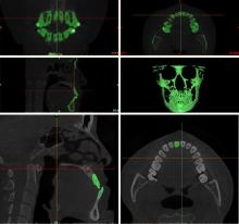
Figure 1

Schematic diagram of 3D reconstruction of upper and under anterior teeth"

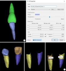
Figure 2

Mimics software modeling, virtual segmentation and measurement A, modeling of upper and lower anterior teeth; B, volume measurement; C, schematic diagram of virtual root segmentation."

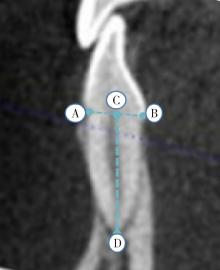
Figure 3

Measurement of root length in two dimensions A, the lowest point of labial enamel cementum boundary; B, the lowest point of lingual enamel cementum boundary; C, the midpoint of A and B lines; D, the apical point. C and D lines are the root length."

Table 1

Changes of root volume and length of upper and under central incisors in patients with skeletal Class Ⅲ malocclusion during orthodontic combined orthodontic treatment"

Change parts T0, $\bar x \pm s$ T1, $\bar x \pm s$ T2, $\bar x \pm s$ P Comparison among groups
Upper central incisor
    Root length/mm 10.47±2.13 9.41±2.36 8.95±2.37 < 0.001 T0>T1>T2
    Total root volume/mm3 156.90±31.77 145.29±31.49 135.16±31.46 < 0.001 T0>T1>T2
    Root apical volume/mm3 62.55±21.74 53.13±22.47 46.88±21.96 < 0.001 T0>T1>T2
    Root cervical volume/mm3 94.34±15.16 92.15±15.83 88.27±15.41 0.001 (T0, T1)>T2
Under central incisor
    Root length/mm 10.97±0.91 10.16±0.93 9.72±0.86 < 0.001 T0>T1>T2
    Total root volume/mm3 98.07±15.18 89.04±17.10 82.69±16.37 < 0.001 T0>T1>T2
    Root apical volume/mm3 43.71±8.95 37.72±9.28 33.72±9.25 < 0.001 T0>T1>T2
    Root cervical volume/mm3 54.35±8.28 51.32±10.27 48.96±9.05 < 0.001 (T0, T1)>(T1, T2)

Table 2

Difference of root volume and root length reduction and percentage of upper and under central incisors after orthodontic combined orthodontic treatment (T2-T0) in patients with skeletal Class Ⅲ malocclusion"

Items Upper central incisor, $\bar x \pm s$ Under central incisor, $\bar x \pm s$ P
Root length/mm 1.51±1.34 1.24±0.70 0.41
Total root volume/mm3 21.73±13.81 15.38±7.78 0.08
Root apical volume/mm3 15.66±9.63 9.99±5.54 0.04
Root cervical volume/mm3 6.07±7.06 5.39±4.54 0.69
Percentage of root length/% 14.86±12.39 11.21±5.94 0.24
Volume percentage of total root/% 13.96±8.44 15.93±8.15 0.39
Volume percentage of root apical/% 30.51±23.23 23.24±11.96 0.15
Volume percentage of root neck/% 6.37±7.55 10.03±8.52 0.11

Table 3

Comparison of volume reduction and percentage of root neck and root body of upper and under central incisors after combined orthodontic treatment (T2-T0) in patients with skeletal Class Ⅲ malocclusion"

Items Root neck, $\bar x \pm s$ Root body, $\bar x \pm s$ P
Upper central incisor
    Volume change/mm3 6.07±7.06 15.66±9.63 < 0.001
    Volume reduction percentage/% 6.37±7.54 30.51±23.23 < 0.001
Under central incisor
    Volume change/mm3 5.39±4.53 9.99±5.54 0.005
    Volume reduction percentage/% 10.02±8.51 23.23±11.96 < 0.001

Table 4

Comparison of tooth movement of upper and under central incisors before and after orthodontic combined orthodontic treatment in patients with skeletal Class Ⅲ malocclusion"

Items T0, $\bar x \pm s$ T1, $\bar x \pm s$ T1′, $\bar x \pm s$ T2, $\bar x \pm s$ ΔT1-T0, $\bar x \pm s$ ΔT2-T1′, $\bar x \pm s$ P1 (T1-T0) P2 (T2-T1)
Upper central incisor
    S-edge/mm 70.43±5.72 66.51±5.63 64.24±6.09 65.66±5.58 -3.92±1.78 1.42±2.73 < 0.001 0.048
    V-edge/mm 76.14±3.72 77.26±3.54 75.24±3.67 74.11±3.21 1.12±1.63 -1.13±2.25 0.012 0.055
    S-root/mm 57.75±5.54 56.31±6.00 57.55±4.79 56.82±4.77 -1.44±1.63 -0.73±1.42 0.002 0.050
    V-root/mm 57.17±4.04 57.84±3.79 56.12±4.13 56.36±3.78 0.67±1.14 0.24±1.57 0.027 0.537
Under central incisor
    S-edge/mm 71.44±7.72 74.61±8.45 61.81±6.17 62.49±5.67 3.16±1.97 1.06±2.79 < 0.001 0.151
    V-edge/mm 77.16±5.07 78.06±4.49 74.36±3.84 72.25±3.54 0.89±1.82 -2.30±2.63 0.070 0.003
    S-root/mm 64.79±8.09 64.42±9.03 53.18±6.33 54.57±5.81 -0.37±2.05 1.91±3.68 0.485 0.056
    V-root/mm 96.76±4.02 95.55±3.95 91.32±3.70 89.55±3.82 -1.22±1.60 -1.84±2.31 0.011 0.006
1 Weltman B , Vig KW , Fields HW , et al. Root resorption associated with orthodontic tooth movement: A systematic review[J]. Am J Orthod Dentofacial Orthop, 2010, 137 (4): 462- 476.
doi: 10.1016/j.ajodo.2009.06.021
2 赵志河. 口腔正畸学[M]. 7版 北京: 人民卫生出版社, 2020.
3 Sawicka M , Bedini R , Pecci R , et al. The application of X-ray microtomography for the assessement of root resorption caused by the orthodontic treatment of premolars[J]. Ann Ist Super Sanita, 2012, 48 (1): 71- 74.
4 Zahed Zahedani S , Oshagh M , Momeni Danaei S , et al. Roeinpeikar SMM: A comparison of apical root resorption in incisors after fixed orthodontic treatment with standard edgewise and straight wire (MBT) method[J]. J Dent Shiraz Univ Med Sci, 2013, 14 (3): 103- 110.
5 Hodges RJ , Atchison KA , White SC . Impact of cone-beam computed tomography on orthodontic diagnosis and treatment planning[J]. Am J Orthod Dentofacial Orthop, 2013, 143 (5): 665- 674.
doi: 10.1016/j.ajodo.2012.12.011
6 张婕, 李小彤. 骨性安氏Ⅲ类手术患者前牙区的牙槽骨厚度[J]. 北京大学学报(医学版), 2016, 48 (1): 111- 115.
doi: 10.3969/j.issn.1671-167X.2016.01.020
7 Giudice AL , Quinzi V , Ronsivalle V , et al. Evaluation of imaging software accuracy for 3-dimensional analysis of the mandibular condyle. A comparative study using a surface-to-surface matching technique[J]. Int J Environ Res Public Health, 2020, 17 (13): 4789.
doi: 10.3390/ijerph17134789
8 Deng Y , Sun Y , Xu T . Evaluation of root resorption after comprehensive orthodontic treatment using cone beam computed tomography (CBCT): A meta-analysis[J]. BMC Oral Health, 2018, 18 (1): 116.
doi: 10.1186/s12903-018-0579-2
9 熊再道, 赵桂芝, 柯杰, 等. 上颌快速扩弓后牙根吸收变化的三维测量研究[J]. 实用口腔医学杂志, 2018, 34 (1): 65- 68.
doi: 10.3969/j.issn.1001-3733.2018.01.014
10 Yildirim M , Akin M . Comparison of root resorption after bone-borne and tooth-borne rapid maxillary expansion evaluated with the use of microtomography[J]. Am J Orthod Dentofacial Orthop, 2019, 155 (2): 182- 190.
doi: 10.1016/j.ajodo.2018.03.021
11 Lo Giudice A , Galletti C , Gay-Escoda C , et al. CBCT assessment of radicular volume loss after rapid maxillary expansion: A syste-matic review[J]. J Clin Exp Dent, 2018, 10 (5): e484- e494.
12 Puttaravuttiporn P , Wongsuwanlert M , Charoemratrote C , et al. Volumetric evaluation of root resorption on the upper incisors using cone beam computed tomography after 1 year of orthodontic treatment in adult patients with marginal bone loss[J]. Angle Orthod, 2018, 88 (6): 710- 718.
doi: 10.2319/121717-868.1
13 Aras I , Unal I , Huniler G , et al. Root resorption due to orthodontic treatment using self-ligating and conventional brackets: A cone-beam computed tomography study[J]. J Orofac Orthop, 2018, 79 (3): 181- 190.
doi: 10.1007/s00056-018-0133-5
14 Ma H , Li W , Xu L , et al. Morphometric evaluation of the alveolar bone around central incisors during surgical orthodontic treatment of high-angle skeletal Class Ⅲ malocclusions[J]. Orthod Craniofac Res, 2021, 24 (1): 87- 95.
doi: 10.1111/ocr.12408
15 Kook YA , Kim G , Kim Y . Comparison of alveolar bone loss around incisors in normal occlusion samples and surgical skeletal Class Ⅲ patients[J]. Angle Orthod, 2012, 82 (4): 645- 652.
doi: 10.2319/070111-424.1
16 吕航苗, 高娟, 马慧敏, 等. 锥束CT三维重建技术应用于骨性Ⅲ类错患者上前牙牙根吸收的研究[J]. 中华口腔正畸学杂志, 2020, 27 (3): 129- 133.
doi: 10.3760/cma.j.cn115797-20200407-20303
17 王芳, 王建国, 张锡忠. 成人骨性Ⅲ类错术前正畸后切牙牙根吸收的CBCT研究[J]. 天津医药, 2015, 43 (4): 390- 392.
18 Suteerapongpun P , Sirabanchongkran S , Wattanachai T , et al. Root surface areas of maxillary permanent teeth in anterior normal overbite and anterior open bite assessed using cone-beam computed tomography[J]. Imaging Sci Dent, 2017, 47 (4): 241- 246.
doi: 10.5624/isd.2017.47.4.241
19 Bartley N , Türk T , Colak C , et al. Physical properties of root cementum: Part 17. Root resorption after the application of 2. 5° and 15° of buccal root torque for 4 weeks: A microcomputed tomography study[J]. Am J Orthod Dentofacial Orthop, 2011, 139 (4): e353- e360.
doi: 10.1016/j.ajodo.2010.01.033
20 Hohmann A , Wolfram U , Geiger M , et al. Periodontal ligament hydrostatic pressure with areas of root resorption after application of a continuous torque moment[J]. Angle Orthod, 2007, 77 (4): 653- 659.
doi: 10.2319/060806-234
21 Segal GR , Schiffman PH , Tuncay OC . Meta analysis of the treatment-related factors of external apical root resorption[J]. Orthod Craniofac Res, 2004, 7 (2): 71- 78.
doi: 10.1111/j.1601-6343.2004.00286.x
22 Martins DR , Tibola D , Janson G , et al. Effects of intrusion combined with anterior retraction on apical root resorption[J]. Eur J Orthod, 2012, 34 (2): 170- 175.
doi: 10.1093/ejo/cjq178
23 Li X , Xu J , Yin Y , et al. Association between root resorption and tooth development: A quantitative clinical study[J]. Am J Orthod Dentofacial Orthop, 2020, 157 (5): 602- 610.
doi: 10.1016/j.ajodo.2019.11.011
24 马宁, 李巍然, 陈晓红, 等. 上切牙内收前后的牙根吸收研究[J]. 现代口腔医学杂志, 2015, 29 (6): 330- 334.
[1] Shishi BO,Chengzhi GAO. Tooth segmentation and identification on cone-beam computed tomography with convolutional neural network based on spatial embedding information [J]. Journal of Peking University (Health Sciences), 2024, 56(4): 735-740.
[2] Xiaotong LING,Liuyang QU,Danni ZHENG,Jing YANG,Xuebing YAN,Denggao LIU,Yan GAO. Three-dimensional radiographic features of calcifying odontogenic cyst and calcifying epithelial odontogenic tumor [J]. Journal of Peking University (Health Sciences), 2024, 56(1): 131-137.
[3] Deng-hui DUAN,Hom-Lay WANG,En-bo WANG. Role of collagen membrane in modified guided bone regeneration surgery using buccal punch flap approach: A retrospective and radiographical cohort study [J]. Journal of Peking University (Health Sciences), 2023, 55(6): 1097-1104.
[4] Jin-hua ZHANG,Jie PAN,Zhi-peng SUN,Xiao WANG. Effect of various intracanal materials on the diagnostic accuracy of cone-beam computed tomography in vertical root fractures [J]. Journal of Peking University (Health Sciences), 2023, 55(2): 333-338.
[5] Jia-xue YE,Yu-hong LIANG. A prevalence survey of cone-beam computed tomography use among endodontic practitioners [J]. Journal of Peking University (Health Sciences), 2023, 55(1): 114-119.
[6] Meng-qiao PAN,Jian LIU,Li XU,Xiao XU,Jian-xia HOU,Xiao-tong LI,Xiao-xia WANG. A long-term evaluation of periodontal phenotypes before and after the periodontal-orthodontic-orthognathic combined treatment of lower anterior teeth in patients with skeletal Angle class Ⅲ malocclusion [J]. Journal of Peking University (Health Sciences), 2023, 55(1): 52-61.
[7] Yu FU,Xin-nong HU,Sheng-jie CUI,Jie SHI. Decompensation effectiveness and alveolar bone remodeling analysis of mandibular anterior teeth after preoperative orthodontic treatment in high-angle patients with skeletal class Ⅱ malocclusion [J]. Journal of Peking University (Health Sciences), 2023, 55(1): 62-69.
[8] LIU Wei-tao,WANG Yi-ran,WANG Xue-dong,ZHOU Yan-heng. A cone-beam computed tomography evaluation of three-dimensional changes of circummaxillary sutures following maxillary protraction with alternate rapid palatal expansions and constrictions [J]. Journal of Peking University (Health Sciences), 2022, 54(2): 346-355.
[9] Gang YANG,Wen-jie HU,Jie CAO,Deng-gao LIU. Three-dimensional morphology analysis of the supraosseous gingival profile of periodontally healthy maxillary anterior teeth [J]. Journal of Peking University (Health Sciences), 2021, 53(5): 990-994.
[10] MENG Yuan,ZHANG Li-qi,ZHAO Ya-ning,LIU Deng-gao,ZHANG Zu-yan,GAO Yan. Three-dimentional radiographic features of 67 maxillary radicular cysts [J]. Journal of Peking University (Health Sciences), 2021, 53(2): 396-401.
[11] ZHOU Jing,LIU Yi. Cone-beam CT evaluation of temporomandibular joint in skeletal class Ⅱ female adolescents with different vertical patterns [J]. Journal of Peking University (Health Sciences), 2021, 53(1): 109-119.
[12] GAO Lu,GU Yan. Chinese morphological stages of midpalatal suture and its correlation with Demirjian dental age [J]. Journal of Peking University (Health Sciences), 2021, 53(1): 133-138.
[13] WANG Yi-ran, ZHOU Yan-heng, WANG Xue-dong, WEI Song, LIU Wei-tao. Evaluation of maxillary three-dimensional changes in maxillary protraction with alternating rapid palatal expansion and constriction based on the cone-beam computed tomography [J]. Journal of Peking University(Health Sciences), 2018, 50(4): 685-693.
[14] JIA Peng-chen, YANG Gang, HU Wen-jie, ZHAO Yi-jiao, LIU Mu-qing. Preliminary study on the accuracy of infrabony root surface area of single-root teeth by periapical films [J]. Journal of Peking University(Health Sciences), 2018, 50(1): 91-97.
[15] MA Jing, JIANG Jiu-hui. Morphological analysis of alveolar bone of anterior mandible in high-angle skeletal class Ⅱ and class Ⅲ malocclusions assessed with cone-beam computed tomography [J]. Journal of Peking University(Health Sciences), 2018, 50(1): 98-103.
Viewed
Full text


Abstract

Cited

  Shared   
  Discussed   
No Suggested Reading articles found!