Journal of Peking University (Health Sciences) ›› 2023, Vol. 55 ›› Issue (1): 114-119. doi: 10.19723/j.issn.1671-167X.2023.01.017

Previous Articles     Next Articles

A prevalence survey of cone-beam computed tomography use among endodontic practitioners

Jia-xue YE,Yu-hong LIANG*()   

  1. Department of Cariology and Endodontology, Peking University School and Hospital of Stomatology & National Center of Stomatology & National Clinical Research Center for Oral Diseases & National Engineering Research Center of Oral Biomaterials and Digital Medical Devices & Beijing Key Laboratory of Digital Stomatology & NHC Research Center of Engineering and Technology for Computerized Dentistry & NMPA Key Laboratory for Dental Materials, Beijing 100081, China
  • Received:2022-10-11 Online:2023-02-18 Published:2023-01-31
  • Contact: Yu-hong LIANG E-mail:leungyuhong@sina.com

RICH HTML


Abstract:

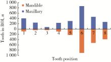
Objective: To investigate the clinical application of cone-beam computed tomography (CBCT) among endodontic practitioners, and to analyze the indications and reasonability of CBCT in the diagnosis and treatment of pulpal and periapical diseases. Methods: The clinical data were collected from patients who visited the Department of Cariology and Endodontology, Peking University School and Hospital of Stomatology and underwent CBCT examination from January to December, 2021. The data with their complete clinical information (including clinical records, radiology request forms/reports, two-dimensional and three-dimensional imaging data) were included. Those who underwent CBCT examination for orthodontic or prosthodontics were excluded. The experience and training background of the endodontic specialists, the number of patients treated in the whole year, the objective and region of interest (ROI) of CBCT examination, technical parameters, such as machine type, field of view (FoV) and radiographic reports were collected and analyzed to evaluate the impact on diagnosis. Wilcoxon and Mann-Whitney tests were used to compare the distribution of CBCT ROI. Chi-squared test and pairwise comparison were used to compare the application of CBCT by endodontic specialists with different clinical experience (senior, middle and junior). Results: In 2021, a total of 3 308 CBCT scans were prescribed by 61 endodontic specialists who treated 34 952 patients throughout the year. 3 218 patients (male ∶female about 1 ∶2) amounting for 10% of the patients treated in the whole year who received CBCT scans with an median age of 35 years (28, 49). Around 98% CBCT examinations were performed after clinical examination and two-dimensional periapical radiographs were taken. The FoV of CBCT scanning less than 10 cm×10 cm accounted for 96% of the total number of the images. Among the 3 308 CBCT scans, 83% of the ROI were in posterior teeth, with a higher number of anterior teeth (Z=-2.278, P < 0.05). Maxillary and mandibular first molars accounted for 35% of the examined teeth. The objectives of CBCT scanning included three aspects: clarifying clinical diagnosis, guiding surgical and non-surgical endodontic treatment (including management of endodontic complications), and outcome assessment, accounting for 1 111 (34%), 1 745 (54%), 311 (10%), respectively. and the others 2%. In the diagnosis process, CBCT was mainly used for the diagnosis of chronic periapical periodontitis, root fracture, root resorption and dental trauma. In the study, 353 CBCT were used in the diagnosis of root fracture, with a positive diagnosis rate of 35% (125/353). 846 CBCT used to reveal the anatomy of the root canal system, of which 297 cases were used to find missed/extra canals after treatment failure, and 58% (171/297) were used to confirm the missed/extra canals. In the management of complications or errors, CBCT was mainly used to assist the diagnosis of perforation and to locate the separated instruments. In the study, 311 CBCT scans were used for outcome assessment, including 240 cases related to non-surgical treatment and 71 cases related to surgical endodontic treatment for follow-up or presence of clinical symptoms, and persistent lesions on 2D films. Among the 61 endodontic specialists who used CBCT, 23 (45%) were with senior experience, 15 (30%) with middle experience, and 23 (25%) with junior experience. The proportion of senior or junior experience prescribing CBCT examination was 10%, higher than that of middle experience (8%, χ12=39.4, χ22=29.1, P < 0.001). The application rate of chief endodontists was 18%, which was higher than that of associate chief endodontists (9%, χ12=139.4, P < 0.001). 31% (1 109/3 308) cases of diagnosis or treatment plans were changed after CBCT was taken. Conclusion: Use of CBCT in endodontic practice could provide more clinical information, which is helpful for diagnosis, accurate treatment and prognosis evaluation.

Key words: Cone-beam computed tomography, Endodontics, Endodontic practitioners

CLC Number: 

  • R781.3

Figure 1

The distribution of tooth examined by CBCT scan ROI, region of interest; CBCT, cone-beam computed tomography."

Table 1

Summary of use of CBCT for endodontists"

Items Examination content
Diagnosis and differential diagnosis
  Periapical lesions Determine the presence, scope and destruction of periapical lesions and alveolar bone
  Abnormal development Dens invaginatus, taurodontism, etc
  Root fracture Except or diagnose (vertical) root fracture
  Root resorption Except or diagnose root (internal or external) resorption
  Traumatic injuries of the tooth A clear history of trauma in the medical history and the target tooth was scanned
Guiding surgical and non-surgical endodontic treatment
  Retreatment decisions(prognosis) Guiding clinical treatment decisions (surgical/non-surgical/extraction)
  Anatomy of root canal Including retreatment, obstructed root canal, calcified canals, missed canal, C-shaped canal and other complex root canal system
  Preoperative evaluation of surgery Routine examination before surgery
  Management of complications Perforation, instrument separation, etc
Outcome evaluation
  Post-root canal treatment Follow-up after root canal treatment
  ost-endodontic surgery Follow-up after endodontic surgery
Others Examination of hyperdontia, tumour, temporal-mandibular joints, extent of caries lesions, root development, maxillary sinus, etc

Table 2

Information of CBCT use for radiographic endodontic outcome evaluation"

Treatments Amount, n Follow-up time/months Absence Reduction
Non-surgical 240 9.8(2-36) 54% 92%
Surgical 71 15.5(1-46) 44% 94%
1 Uraba S , Ebihara A , Komatsu K , et al. Ability of cone-beam computed tomography to detect periapical lesions that were not detected by periapical radiography: A retrospective assessment according to tooth group[J]. J Endod, 2016, 42 (8): 1186- 1190.
doi: 10.1016/j.joen.2016.04.026
2 姜岚, 陈晨, 高学军, 等. 锥形束CT与根尖X线片诊断根尖病变的准确性对比[J]. 中华口腔医学杂志, 2013, 48 (z1): 1- 5.
3 Patel S , Dawood A , Whaites E , et al. New dimensions in endodontic imaging: Part 1. Conventional and alternative radiographic systems[J]. Int Endod J, 2009, 42 (6): 447- 462.
doi: 10.1111/j.1365-2591.2008.01530.x
4 Patel S , Dawood A , Wilson R , et al. The detection and management of root resorption lesions using intraoral radiography and cone beam computed tomography: An in vivo investigation[J]. Int Endod J, 2009, 42 (9): 831- 838.
doi: 10.1111/j.1365-2591.2009.01592.x
5 Ahlowalia M , Patel S , Anwar H , et al. Accuracy of CBCT for volumetric measurement of simulated periapical lesions[J]. Int Endod J, 2013, 46 (6): 538- 546.
doi: 10.1111/iej.12023
6 Metska ME , Aartman IH , Wesselink PR , et al. Detection of vertical root fractures in vivo in endodontically treated teeth by cone-beam computed tomography scans[J]. J Endod, 2012, 38 (10): 1344- 1347.
doi: 10.1016/j.joen.2012.05.003
7 Özer SY . Detection of vertical root fractures by using cone beam computed tomography with variable voxel sizes in an in vitro model[J]. J Endod, 2011, 37 (1): 75- 79.
doi: 10.1016/j.joen.2010.04.021
8 Patel S , Brady E , Wilson R , et al. The detection of vertical root fractures in root filled teeth with periapical radiographs and CBCT scans[J]. Int Endod J, 2013, 46 (12): 1140- 1152.
doi: 10.1111/iej.12109
9 Zou X , Liu D , Yue L , et al. The ability of cone-beam compute-rized tomography to detect vertical root fractures in endodontically treated and nonendodontically treated teeth: A report of 3 cases[J]. Oral Surg Oral Med Oral Pathol Oral Radiol Endod, 2011, 111 (6): 797- 801.
doi: 10.1016/j.tripleo.2010.12.015
10 Chavda R , Mannocci F , Andiappan M , et al. Comparing the in vivo diagnostic accuracy of digital periapical radiography with cone-beam computed tomography for the detection of vertical root fracture[J]. J Endod, 2014, 40 (10): 1524- 1529.
doi: 10.1016/j.joen.2014.05.011
11 Bernardes RA , de Paulo RS , Pereira LO , et al. Comparative study of cone beam computed tomography and intraoral periapical radiographs in diagnosis of lingual-simulated external root resorptions[J]. Dent Traumatol, 2012, 28 (4): 268- 272.
doi: 10.1111/j.1600-9657.2011.01113.x
12 Estrela C , Bueno MR , Porto OC , et al. Influence of intracanal post on apical periodontitis identified by cone-beam computed tomography[J]. Braz Dent J, 2009, 20 (5): 370- 375.
doi: 10.1590/S0103-64402009000500003
13 American Association of Endodontists , American Academy of Oral and Maxillofacial Radiology . Use of cone-beam computed tomography in endodontics joint position statement of the American Association of Endodontists and the American Academy of Oral and Maxillofacial Radiology[J]. Oral Surg Oral Med Oral Pathol Oral Radiol Endod, 2011, 111 (2): 234- 237.
doi: 10.1016/j.tripleo.2010.11.012
14 Setzer FC , Hinckley N , Kohli MR , et al. A survey of cone-beam computed tomographic use among endodontic practitioners in the United States[J]. J Endod, 2017, 43 (5): 699- 704.
doi: 10.1016/j.joen.2016.12.021
15 Alzamazmi ZT , Abulhamael AM , Talim DJ , et al. Cone-beam computed tomographic usage: Survey of american endodontists[J]. J Contemp Dent Pract, 2019, 20 (10): 1132- 1137.
doi: 10.5005/jp-journals-10024-2661
16 梁宇红, 岳林. 锥形束CT在牙髓根尖周病诊治中的合理应用与思考[J]. 中华口腔医学杂志, 2019, 54 (9): 591- 597.
doi: 10.3760/cma.j.issn.1002-0098.2019.09.003
17 Ludlow JB , Timothy R , Walker C , et al. Correction to effective dose of dental CBCT: A meta analysis of published data and additional data for nine CBCT units[J]. Dentomaxillofac Radiol, 2015, 44 (7): 20159003.
doi: 10.1259/dmfr.20159003
18 Ludlow JB , Ivanovic M . Comparative dosimetry of dental CBCT devices and 64-slice CT for oral and maxillofacial radiology[J]. Oral Surg Oral Med Oral Pathol Oral Radiol Endod, 2008, 106 (1): 106- 114.
doi: 10.1016/j.tripleo.2008.03.018
19 Patel S , Brown J , Semper M , et al. European Society of Endodontology position statement: Use of cone beam computed tomography in endodontics European Society of Endodontology (ESE) deve-loped by[J]. Int Endod J, 2019, 52 (12): 1675- 1378.
doi: 10.1111/iej.13187
20 中华口腔医学会牙体牙髓病学专业委员会. 牙体牙髓病诊疗中口腔放射学的应用指南[J]. 中华口腔医学杂志, 2021, 56 (4): 311- 317.
doi: 10.3760/cma.j.cn112144-20210125-00039
21 Mathew AI, Lee SC, Ha WN, et al. Cone-beam computed tomography-predictors and characteristics of usage in Australia and New Zealand: A multifactorial analysis[J/OL]. Aust Endod J, 2022, 7 (2022-07-13)[2022-09-13]. https://pubmed.ncbi.nlm.nih.gov/35830370.
22 Rajeevan M , Chandler NP , Makdissi J , et al. A survey of cone beam computed tomography (CBCT) use among endodontic practitioners in the UK[J]. Endo-Endod Pract Tod, 2018, 12 (1): 29- 33.
23 Bhatt M , Coil J , Chehroudi B , et al. Clinical decision-making and importance of the AAE/AAOMR position statement for CBCT examination in endodontic cases[J]. Int Endod J, 2021, 54 (1): 26- 37.
doi: 10.1111/iej.13397
24 Mota de Almeida FJ , Knutsson K , Flygare L . The effect of cone beam CT (CBCT) on therapeutic decision-making in endodontics[J]. Dentomaxillofac Radiol, 2014, 43 (4): 20130137.
doi: 10.1259/dmfr.20130137
25 Rodriguez G , Patel S , Duran-Sindreu F , et al. Influence of cone-beam computed tomography on endodontic retreatment strategies among general dental practitioners and endodontists[J]. J Endod, 2017, 43 (9): 1433- 1437.
doi: 10.1016/j.joen.2017.04.004
26 Ee J , Fayad MI , Johnson BR . Comparison of endodontic diagnosis and treatment planning decisions using cone-beam volumetric tomography versus periapical radiography[J]. J Endodont, 2014, 40 (7): 910- 916.
doi: 10.1016/j.joen.2014.03.002
27 Aljuhani A , Dutta S , Mandorah A . Evaluation of knowledge and perspective of endodontic residents and general dentist towards the endodontic application of CBCT in Saudi Arabia[J]. J Res Med Dent Sci, 2020, 8 (7): 459- 464.
[1] Shishi BO,Chengzhi GAO. Tooth segmentation and identification on cone-beam computed tomography with convolutional neural network based on spatial embedding information [J]. Journal of Peking University (Health Sciences), 2024, 56(4): 735-740.
[2] Xiaotong LING,Liuyang QU,Danni ZHENG,Jing YANG,Xuebing YAN,Denggao LIU,Yan GAO. Three-dimensional radiographic features of calcifying odontogenic cyst and calcifying epithelial odontogenic tumor [J]. Journal of Peking University (Health Sciences), 2024, 56(1): 131-137.
[3] Deng-hui DUAN,Hom-Lay WANG,En-bo WANG. Role of collagen membrane in modified guided bone regeneration surgery using buccal punch flap approach: A retrospective and radiographical cohort study [J]. Journal of Peking University (Health Sciences), 2023, 55(6): 1097-1104.
[4] Yun-fei DAI,He LIU,Chu-fang PENG,Xi-jun JIANG. Retrospective evaluation of treatment outcomes in immature teeth treated with regenerative endodontic procedures with an over-36-month review [J]. Journal of Peking University (Health Sciences), 2023, 55(4): 729-735.
[5] Jin-hua ZHANG,Jie PAN,Zhi-peng SUN,Xiao WANG. Effect of various intracanal materials on the diagnostic accuracy of cone-beam computed tomography in vertical root fractures [J]. Journal of Peking University (Health Sciences), 2023, 55(2): 333-338.
[6] Meng-qiao PAN,Jian LIU,Li XU,Xiao XU,Jian-xia HOU,Xiao-tong LI,Xiao-xia WANG. A long-term evaluation of periodontal phenotypes before and after the periodontal-orthodontic-orthognathic combined treatment of lower anterior teeth in patients with skeletal Angle class Ⅲ malocclusion [J]. Journal of Peking University (Health Sciences), 2023, 55(1): 52-61.
[7] Yu FU,Xin-nong HU,Sheng-jie CUI,Jie SHI. Decompensation effectiveness and alveolar bone remodeling analysis of mandibular anterior teeth after preoperative orthodontic treatment in high-angle patients with skeletal class Ⅱ malocclusion [J]. Journal of Peking University (Health Sciences), 2023, 55(1): 62-69.
[8] Juan GAO,Hang-miao LV,Hui-min MA,Yi-jiao ZHAO,Xiao-tong LI. Evaluation of root resorption after surgical orthodontic treatment of skeletal Class Ⅲ malocclusion by three-dimensional volumetric measurement with cone-beam CT [J]. Journal of Peking University (Health Sciences), 2022, 54(4): 719-726.
[9] LIU Wei-tao,WANG Yi-ran,WANG Xue-dong,ZHOU Yan-heng. A cone-beam computed tomography evaluation of three-dimensional changes of circummaxillary sutures following maxillary protraction with alternate rapid palatal expansions and constrictions [J]. Journal of Peking University (Health Sciences), 2022, 54(2): 346-355.
[10] Gang YANG,Wen-jie HU,Jie CAO,Deng-gao LIU. Three-dimensional morphology analysis of the supraosseous gingival profile of periodontally healthy maxillary anterior teeth [J]. Journal of Peking University (Health Sciences), 2021, 53(5): 990-994.
[11] MENG Yuan,ZHANG Li-qi,ZHAO Ya-ning,LIU Deng-gao,ZHANG Zu-yan,GAO Yan. Three-dimentional radiographic features of 67 maxillary radicular cysts [J]. Journal of Peking University (Health Sciences), 2021, 53(2): 396-401.
[12] ZHOU Jing,LIU Yi. Cone-beam CT evaluation of temporomandibular joint in skeletal class Ⅱ female adolescents with different vertical patterns [J]. Journal of Peking University (Health Sciences), 2021, 53(1): 109-119.
[13] GAO Lu,GU Yan. Chinese morphological stages of midpalatal suture and its correlation with Demirjian dental age [J]. Journal of Peking University (Health Sciences), 2021, 53(1): 133-138.
[14] WANG Yi-ran, ZHOU Yan-heng, WANG Xue-dong, WEI Song, LIU Wei-tao. Evaluation of maxillary three-dimensional changes in maxillary protraction with alternating rapid palatal expansion and constriction based on the cone-beam computed tomography [J]. Journal of Peking University(Health Sciences), 2018, 50(4): 685-693.
[15] JIA Peng-chen, YANG Gang, HU Wen-jie, ZHAO Yi-jiao, LIU Mu-qing. Preliminary study on the accuracy of infrabony root surface area of single-root teeth by periapical films [J]. Journal of Peking University(Health Sciences), 2018, 50(1): 91-97.
Viewed
Full text


Abstract

Cited

  Shared   
  Discussed   
No Suggested Reading articles found!