Journal of Peking University (Health Sciences) ›› 2025, Vol. 57 ›› Issue (4): 692-697. doi: 10.19723/j.issn.1671-167X.2025.04.010

Previous Articles     Next Articles

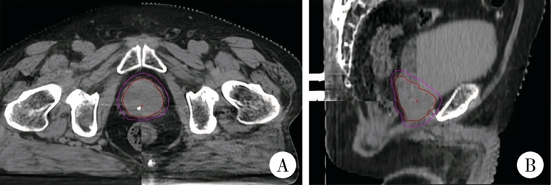
Comparison of setup errors between two immobilization methods in prostate cancer radiotherapy based on cone-beam computed tomography

Jie LIU, Mingwei MA*(), Qing'an WANG, Ming SHI, Jinpeng YIN, Zhanping WANG, Jingtao SHEN, Xianshu GAO   

  1. Department of Radiation Oncology, Peking University First Hospital, Beijing 100034, China
  • Received:2025-01-03 Online:2025-08-18 Published:2025-08-02
  • Contact: Mingwei MA
  • Supported by:
    the National Key Research and Development Program of China(2022YFC2404606); the Beijing Natural Science Foundation(L232126)

RICH HTML


Abstract:

Objective: To analyze and compare the interfractional setup errors between two body positioning fixation methods (lithotomy position with carbon fiber full-body fixation frame vs. conventional carbon fiber body fixation frame combined with thermoplastic membrane) in radical radiotherapy for prostate cancer, and to calculate the clinical target volume (CTV) to planning target volume (PTV) margin (MPTV) for both methods to optimize immobilization techniques and radiotherapy workflows. Methods: A retrospective analysis was conducted on 37 consecutive patients who underwent radical prostate radiotherapy at Peking University First Hospital between August 2021 and March 2023. The patients were divided into two groups based on the immobilization method: Group A (18 patients, 450 CBCT image sets) used a carbon fiber whole-body fixator in the lithotomy position, while Group B (19 patients, 461 CBCT image sets) used a conventional carbon fiber fixator combined with a thermoplastic mask. All the patients underwent daily cone-beam computed tomography (CBCT) image guidance. Bone registration combined with manual registration was used to obtain the setup error data in the left-right (X), cranio-caudal (Y) and anterior-posterior (Z) directions. The positioning errors of the two groups were compared by using the independent sample t-test, the Mann-Whitney U test and the chi-square test. The average positioning error, systematic positioning error (Σ) and random positioning error (δ) were calculated, and the CTV-PTV extension distance was calculated by using the (MPTV=2.5Σ+0.7δ). Results: The analysis of the setup errors in the three-dimensional direction showed significant differences between the two groups (all P < 0.01). Specifically, the median (quartile) absolute values of the errors in the X, Y, and Z directions of group A were [0.40 (0.20, 0.70) cm, 0.50 (0.30, 0.80) cm, and 0.35 (0.20, 0.60) cm], respectively. In group B, the corresponding values were significantly reduced to [0.20 (0.10, 0.40) cm, 0.40 (0.20, 0.70) cm and 0.20 (0.10, 0.40) cm]. The results of Mann-Whitney U test showed that the differences in each direction were highly statistically significant (X: z=-6.86; Y: z=-2.76; Z: z=-5.71). The cumulative distribution ratio of the setup error displacement within 0.5 cm in the X, Y, and Z directions in group A and group B were 297 (66.0%) and 408 (88.5%) (P < 0.01), 250 (55.6%) and 285 (61.8%) (P=0.055), 308 (68.4%) and 391 (84.8%) (P < 0.01), respectively. The CTV-PTV margins in three directions were X 0.66 cm in group A and 0.35 cm in group B; Y 0.67 cm and 0.45 cm; Z 0.54 cm and 0.42 cm. Conclusion: Conventional carbon fiber human body fixator combined with thermoplastic film can significantly reduce the setup error. However, the carbon fiber whole-body fixator in the lithotomy position demonstrated larger setup errors in the X, Y, and Z directions compared with the conventional fixator combined with a thermoplastic mask, indicating the need for further workflow optimization. Given that the lithotomy position is essential for transperineal ultrasound-guided techniques, further research and improvements are required to achieve higher positioning accuracy.

Key words: Prostate cancer, Cone-beam computed tomography, Setup error, Immobilization, Thermoplastic mask

CLC Number: 

  • R737.25

Figure 1

Positioning and fixation devices for prostate cancer radiotherapy A, a carbon fiber whole-body fixator in lithotomy position; B, a conventional carbon fiber human body fixator and thermoplastic film."

Figure 2

Localization computed tomography (CT) and magnetic resonance imaging (MRI) images and target volumes of a patient treated with definitive radiotherapy for prostate cancer A, axial CT images; B, sagittal CT images; C, axial MRI T2WI sequence images; D, sagittal MRI T2WI sequence images."

Figure 3

Cone-beam computed tomography(CBCT)-matched images of patients receiving radiotherapy treatment for prostate cancer A, axial matching images of the CBCT for the patient; B, sagittal matched images of the CBCT for the patients."

Table 1

Comparison of setup errors between group A and group B"

Parameters Group A Group B z P
X (left-right)/cm 0.40 (0.20, 0.70) 0.20 (0.10, 0.40) -6.86 < 0.001
Y (cranio-caudal)/cm 0.50 (0.30, 0.80) 0.40 (0.20, 0.70) -2.76 0.006
Z (anterior-posterior)/cm 0.35 (0.20, 0.60) 0.20 (0.10, 0.40) -5.71 < 0.001

Table 2

Cumulative distribution proportion of setup error displacement in different directions within 0.5 cm in group A and group B"

Parameters Group A (n=450) Group B (n=461) P
X (left-right) 297 (66.0) 408 (88.5) < 0.001
Y (cranio-caudal) 250 (55.6) 285 (61.8) 0.055
Z (anterior-posterior) 308 (68.4) 391 (84.8) < 0.001

Figure 4

Cumulative distribution of setup errors in groups A and B A, cumulative distribution probability of setup errors in the X-axis direction; B, cumulative distribution probability of setup errors in the Y-axis direction; C, cumulative distribution probability of setup errors in the Z-axis direction."

Table 3

CTV-PTV margin and inter-fractional setup Errors (Σ- and δ-clusters) in group A and group B"

Parameters MPTV/cm Average setup error/cm Σ/cm δ/cm
X (left-right)
  Group A 0.66 0.46 0.175 0.321
  Group B 0.35 0.285 0.084 0.206
Y (cranio-caudal)
  Group A 0.67 0.552 0.178 0.314
  Group B 0.45 0.48 0.094 0.311
Z (anterior-posterior)
  Group A 0.54 0.438 0.126 0.321
  Group B 0.42 0.302 0.107 0.218
1
Wolff RF, Ryder S, Bossi A, et al. A systematic review of randomised controlled trials of radiotherapy for localised prostate cancer[J]. Eur J Cancer, 2015, 51(16): 2345- 2367.
2
Butler WM, Merrick GS, Reed JL, et al. Intrafraction displacement of prone versus supine prostate positioning monitored by real-time electromagnetic tracking[J]. J Appl Clin Med Phys, 2013, 14(2): 4141.
3
Daşu A. Is the alpha/beta value for prostate tumours low enough to be safely used in clinical trials[J]. Clin Oncol (R Coll Radiol), 2007, 19(5): 289- 301.
4
van Herk M. Errors and margins in radiotherapy[J]. Semin Radiat Oncol, 2004, 14(1): 52- 64.
5
Pommer T, Oh JH, Rosenschöld PMA, et al. Simulating intrafraction prostate motion with a random walk model[J]. Adv Radiat Oncol, 2017, 2(3): 429- 436.
6
Aluwini S, Pos F, Schimmel E, et al. Hypofractionated versus conventionally fractionated radiotherapy for patients with prostate cancer (HYPRO): Late toxicity results from a randomised, non-inferiority, phase 3 trial[J]. Lancet Oncol, 2016, 17(4): 464- 474.
7
Aluwini S, Pos F, Schimmel E, et al. Hypofractionated versus conventionally fractionated radiotherapy for patients with prostate cancer (HYPRO): Acute toxicity results from a randomised non-inferiority phase 3 trial[J]. Lancet Oncol, 2015, 16(3): 274- 283.
8
Dearnaley D, Syndikus I, Mossop H, et al. Conventional versus hypofractionated high-dose intensity-modulated radiotherapy for prostate cancer: 5-year outcomes of the randomised, non-inferiority, phase 3 CHHiP trial[J]. Lancet Oncol, 2016, 17(8): 1047- 1060.
9
King CR, Freeman D, Kaplan I, et al. Stereotactic body radiotherapy for localized prostate cancer: Pooled analysis from a multi-institutional consortium of prospective phase Ⅱ trials[J]. Radio-ther Oncol, 2013, 109(2): 217- 221.
10
马广栋, 洪莉. 宫颈癌的图像引导放疗研究综述[J]. 中国医疗设备, 2018, 33(5): 117-120, 129.
11
于松茂, 孟繁里, 康加阜, 等. 真空垫制作方式对肺癌立体定向放疗摆位精度影响研究[J]. 医疗卫生装备, 2019, 40(12): 33-35, 40.
12
马茗微, 王淑莲, 覃仕瑞, 等. 面罩及乳腺托架固定下乳腺癌保乳术后放疗锁骨上下区摆位误差分析[J]. 中华放射肿瘤学杂志, 2019, 28(3): 217- 221.
13
Murray J, Griffin C, Gulliford S, et al. A randomised assessment of image guided radiotherapy within a phase 3 trial of conventional or hypofractionated high dose intensity modulated radiotherapy for prostate cancer[J]. Radiother Oncol, 2020, 142, 62- 71.
14
高研, 高献书, 马茗微, 等. 前列腺癌放疗时CBCT使用频率和匹配策略的剂量学分析[J]. 中华放射肿瘤学杂志, 2024, 33(8): 733- 739.
15
Ghilezan M, Yan D, Martinez A. Adaptive radiation therapy for prostate cancer[J]. Semin Radiat Oncol, 2010, 20(2): 130- 137.
[1] Ye YAN,Xiaolong LI,Haizhui XIA,Xuehua ZHU,Yuting ZHANG,Fan ZHANG,Ke LIU,Cheng LIU,Lulin MA. Analysis of risk factors for long-term overactive bladder after radical prostatectomy [J]. Journal of Peking University (Health Sciences), 2024, 56(4): 589-593.
[2] Shishi BO,Chengzhi GAO. Tooth segmentation and identification on cone-beam computed tomography with convolutional neural network based on spatial embedding information [J]. Journal of Peking University (Health Sciences), 2024, 56(4): 735-740.
[3] Xiaotong LING,Liuyang QU,Danni ZHENG,Jing YANG,Xuebing YAN,Denggao LIU,Yan GAO. Three-dimensional radiographic features of calcifying odontogenic cyst and calcifying epithelial odontogenic tumor [J]. Journal of Peking University (Health Sciences), 2024, 56(1): 131-137.
[4] Deng-hui DUAN,Hom-Lay WANG,En-bo WANG. Role of collagen membrane in modified guided bone regeneration surgery using buccal punch flap approach: A retrospective and radiographical cohort study [J]. Journal of Peking University (Health Sciences), 2023, 55(6): 1097-1104.
[5] Jin-hua ZHANG,Jie PAN,Zhi-peng SUN,Xiao WANG. Effect of various intracanal materials on the diagnostic accuracy of cone-beam computed tomography in vertical root fractures [J]. Journal of Peking University (Health Sciences), 2023, 55(2): 333-338.
[6] Jia-xue YE,Yu-hong LIANG. A prevalence survey of cone-beam computed tomography use among endodontic practitioners [J]. Journal of Peking University (Health Sciences), 2023, 55(1): 114-119.
[7] Meng-qiao PAN,Jian LIU,Li XU,Xiao XU,Jian-xia HOU,Xiao-tong LI,Xiao-xia WANG. A long-term evaluation of periodontal phenotypes before and after the periodontal-orthodontic-orthognathic combined treatment of lower anterior teeth in patients with skeletal Angle class Ⅲ malocclusion [J]. Journal of Peking University (Health Sciences), 2023, 55(1): 52-61.
[8] Yu FU,Xin-nong HU,Sheng-jie CUI,Jie SHI. Decompensation effectiveness and alveolar bone remodeling analysis of mandibular anterior teeth after preoperative orthodontic treatment in high-angle patients with skeletal class Ⅱ malocclusion [J]. Journal of Peking University (Health Sciences), 2023, 55(1): 62-69.
[9] Juan GAO,Hang-miao LV,Hui-min MA,Yi-jiao ZHAO,Xiao-tong LI. Evaluation of root resorption after surgical orthodontic treatment of skeletal Class Ⅲ malocclusion by three-dimensional volumetric measurement with cone-beam CT [J]. Journal of Peking University (Health Sciences), 2022, 54(4): 719-726.
[10] Sheng-jie LIU,Hui-min HOU,Zheng-tong LV,Xin DING,Lu WANG,Lei ZHANG,Ming LIU. Bipolar androgen therapy followed by immune checkpoint inhibitors in metastatic castration resistant prostate cancer: A report of 4 cases [J]. Journal of Peking University (Health Sciences), 2022, 54(4): 766-769.
[11] LIU Wei-tao,WANG Yi-ran,WANG Xue-dong,ZHOU Yan-heng. A cone-beam computed tomography evaluation of three-dimensional changes of circummaxillary sutures following maxillary protraction with alternate rapid palatal expansions and constrictions [J]. Journal of Peking University (Health Sciences), 2022, 54(2): 346-355.
[12] Gang YANG,Wen-jie HU,Jie CAO,Deng-gao LIU. Three-dimensional morphology analysis of the supraosseous gingival profile of periodontally healthy maxillary anterior teeth [J]. Journal of Peking University (Health Sciences), 2021, 53(5): 990-994.
[13] BAI Gao-chen,SONG Yi,JIN Jie,YU Wei,HE Zhi-song. Clinical efficacy of docetaxel combined with carboplatin in patients with metastatic castration-resistant prostate cancer [J]. Journal of Peking University (Health Sciences), 2021, 53(4): 686-691.
[14] MENG Yuan,ZHANG Li-qi,ZHAO Ya-ning,LIU Deng-gao,ZHANG Zu-yan,GAO Yan. Three-dimentional radiographic features of 67 maxillary radicular cysts [J]. Journal of Peking University (Health Sciences), 2021, 53(2): 396-401.
[15] ZHOU Jing,LIU Yi. Cone-beam CT evaluation of temporomandibular joint in skeletal class Ⅱ female adolescents with different vertical patterns [J]. Journal of Peking University (Health Sciences), 2021, 53(1): 109-119.
Viewed
Full text


Abstract

Cited

  Shared   
  Discussed   
No Suggested Reading articles found!