北京大学学报(医学版) ›› 2025, Vol. 57 ›› Issue (4): 650-661. doi: 10.19723/j.issn.1671-167X.2025.04.004

• 论著 • 上一篇    下一篇

TFE3重排肾细胞癌合并静脉癌栓患者的临床病理特征及生存分析

张展奕1,*, 陆敏2,3,*, 孙悦皓1, 董靖晗1, 侯小飞1, 肖春雷1, 王国良1, 田晓军1, 马潞林1, 张洪宪1, 张树栋1,4,*()   

  1. 1. 北京大学第三医院泌尿外科, 北京 100191
    2. 北京大学第三医院病理科, 北京 100191
    3. 北京大学基础医学院病理学系, 北京 100191
    4. 北京大学第三医院肿瘤中心, 北京 100191
  • 收稿日期:2025-02-28 出版日期:2025-08-18 发布日期:2025-08-02
  • 通讯作者: 张树栋
  • 作者简介:

    *These authors contributed equally to this work

  • 基金资助:
    国家自然科学基金(82273389); 北京市自然科学基金(7232212)

Clinicopathological features and survival analysis of TFE3-rearranged renal cell carcinoma with venous tumor thrombus

Zhanyi ZHANG1, Min LU2,3, Yuehao SUN1, Jinghan DONG1, Xiaofei HOU1, Chunlei XIAO1, Guoliang WANG1, Xiaojun TIAN1, Lulin MA1, Hongxian ZHANG1, Shudong ZHANG1,4,*()   

  1. 1. Department of Urology, Peking University Third Hospital, Beijing 100191, China
    2. Department of Pathology, Peking University Third Hospital, Beijing 100191, China
    3. Department of Pathology, Peking University School of Basic Medical Sciences, Beijing 100191, China
    4. Cancer Center, Peking University Third Hospital, Beijing 100191, China
  • Received:2025-02-28 Online:2025-08-18 Published:2025-08-02
  • Contact: Shudong ZHANG
  • Supported by:
    the National Natural Science Foundation of China(82273389); the Beijing Natural Science Foundation(7232212)

RICH HTML


摘要:

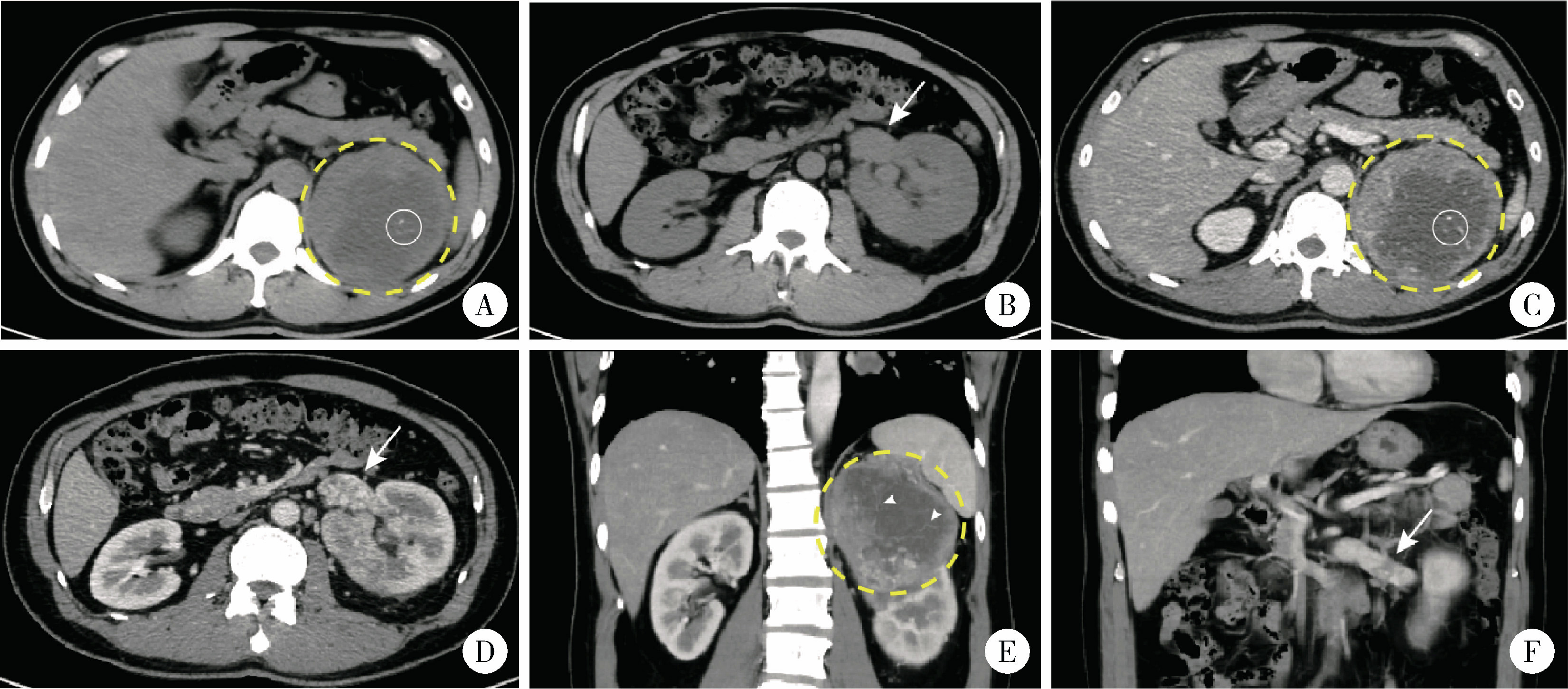
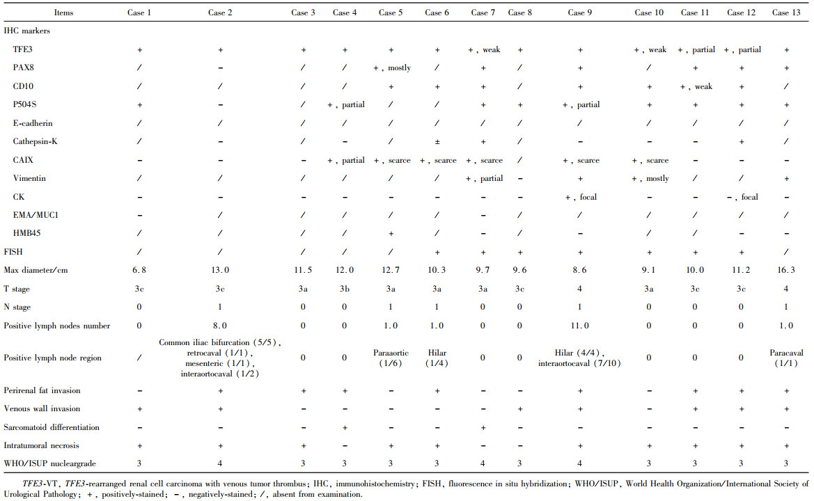
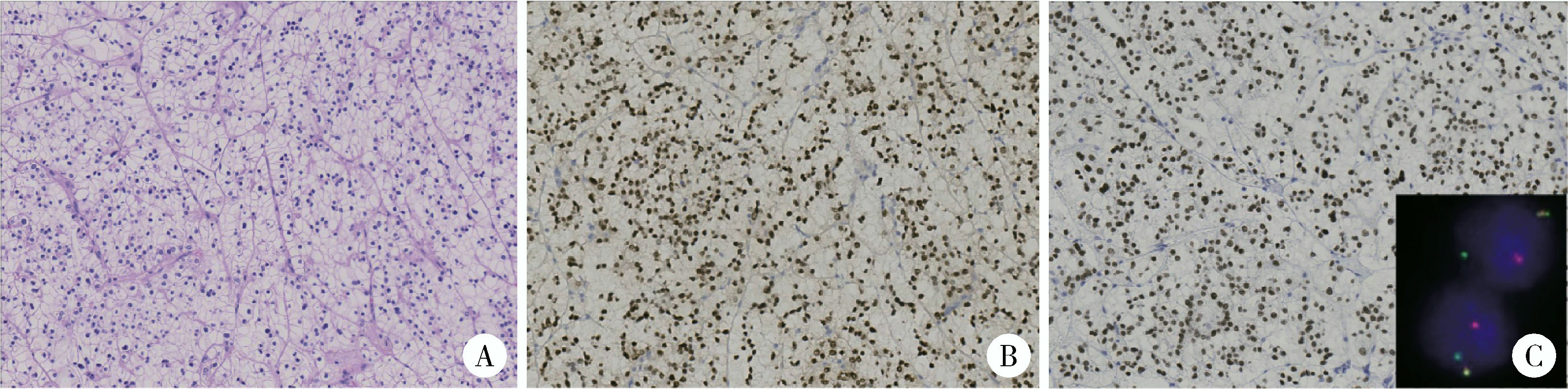
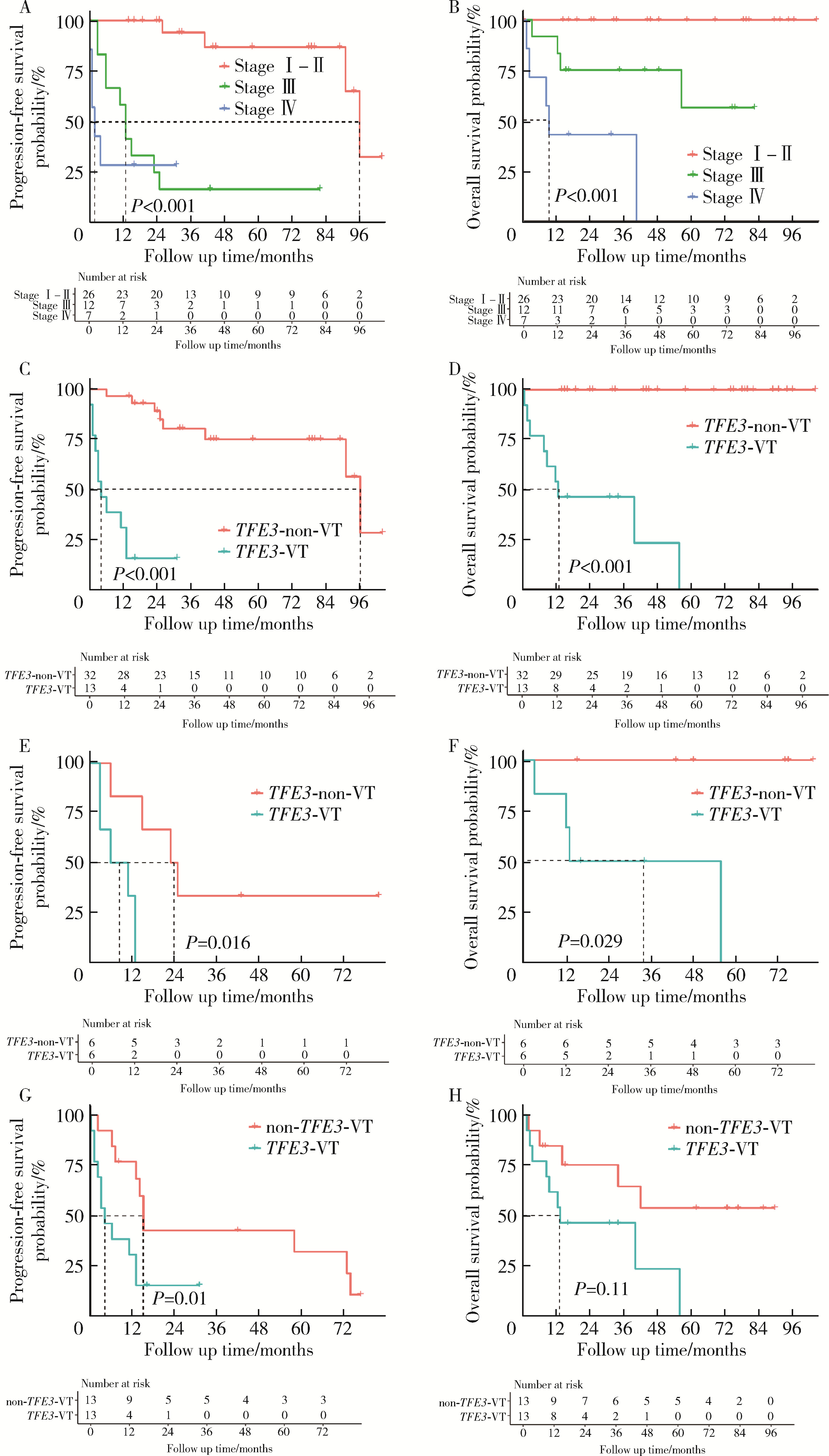
目的: 回顾TFE3重排肾细胞癌(TFE3-rearranged renal cell carcinoma, TFE3-RCC)合并静脉癌栓(venous tumor thrombus, VT)(TFE3-VT)患者的临床病理特征, 探索其治疗策略及预后特点, 为TFE3-VT患者的诊断和治疗提供参考。方法: 纳入2013年1月至2024年1月在北京大学第三医院泌尿外科接受手术且术后病理诊断为TFE3-VT的患者、诊断为TFE3-RCC但不合并VT(TFE3-non-VT)的患者, 以及诊断为非TFE3突变的肾细胞癌且合并VT(non-TFE3-VT)的患者。收集患者的临床病史资料、影像学资料、病理资料及随访资料。研究的主要结局为无进展生存期(progression free survival, PFS), 次要结局为总生存期(overall survival, OS)。(1)对TFE3-VT与TFE3-non-VT患者的基线资料进行比较, 服从正态分布的连续变量以均数±标准差表示, 组间比较采用Student’s t检验; 不服从正态分布的连续变量以中位数(P25, P75)表示, 组间比较采用Mann-Whitney U检验; 分类变量以样本数及百分比[n(%)]表示, 组间比较采用χ2检验或Fisher’s精确检验。(2)对13例TFE3-VT患者的临床病史、影像学表现、手术情况及组织病理学特征进行描述。(3)对TFE3-VT患者进行生存分析, 描述13例TFE3-VT患者的随访情况, 并与TFE3-non-VT及non-TFE3-VT进行生存情况比较。在与TFE3-non-VT患者进行比较时, 利用Kaplan-Meier法绘制临床分期Ⅰ~Ⅳ期TFE3-RCC患者、TFE3-VT与TFE3-non-VT患者, 以及临床分期Ⅲ期TFE3-VT与TFE3-non-VT亚组患者的PFS及OS曲线, 通过Log-rank检验比较各组患者生存曲线之间的差异是否有统计学意义; 在与non-TFE3-VT患者进行比较时, 采用1 ∶1倾向性评分匹配(propensity score matching, PSM)对两组患者进行配对, 利用Kaplan-Meier法绘制配对后两组患者的PFS及OS曲线, 通过Log-rank检验比较两组患者生存曲线之间的差异是否有统计学意义。所有统计分析均使用R语言(v 4.2.3)进行, 双侧检验P < 0.05为差异有统计学意义。结果: 共纳入45例TFE3-RCC患者, 其中TFE3-VT患者13例, TFE3-non-VT患者32例, 同时纳入non-TFE3-VT患者523例。13例TFE3-VT患者中女性9例(69.2%), 男性4例(30.8%), 平均年龄(37.9±14.4)岁, 平均体重指数(body mass index, BMI)为(22.2±3.5) kg/m2, 中位年龄校正Charlson合并症指数(age-adjusted Charlson comorbidity index, aCCI)为1.0(0.0, 1.0)分, 术前平均肌酐为(75.3±15.9) μmol/L; 7例(53.8%)患者肿瘤位于左肾, 6例(46.2%)位于右肾; 6例(46.2%)患者在术前存在远处转移(M1期), 7例(53.8%)术前未发现远处转移; 合并Mayo 0级VT的患者共7例(53.8%), 合并Mayo Ⅰ级及Mayo Ⅳ级VT的患者各1例(7.7%), 合并Mayo Ⅱ级及Mayo Ⅲ级VT的患者各2例(15.4%); 2例(15.4%)行开放手术, 6例(46.1%)行腹腔镜手术, 5例(38.5%)行机器人辅助腹腔镜手术, 平均手术时长(273±79) min, 平均出血量(722±570) mL; 13例患者肿瘤大体标本的平均最大直径为(10.8±2.4) cm。13例患者均进行了TFE3蛋白免疫组织化学(immunohistochemistry, IHC)染色, 其中7例患者进行了进一步的荧光原位杂交(fluorescence in situ hybridization, FISH)检测并证实为TFE3-RCC。13例患者在随访中有11例(84.6%)出现肿瘤的复发转移, 9例患者(69.2%)死亡, 中位PFS为4个月, 1年PFS率为31%;中位OS为13个月, 1年OS率为54%。在45例TFE3-RCC患者中, 不同临床分期患者的PFS及OS曲线之间差异有统计学意义(P < 0.001);TFE3-VT患者与TFE3-non-VT患者的PFS及OS曲线之间差异有统计学意义(P < 0.001);在临床Ⅲ期患者的亚组分析中, TFE3-VT患者与TFE3-non-VT患者的PFS及OS曲线之间差异仍有统计学意义(P < 0.05)。PSM后, TFE3-VT患者与non-TFE3-VT患者的PFS曲线之间差异有统计学意义(P=0.01), OS曲线之间的差异无统计学意义(P=0.11)。结论: TFE3-VT患者以中青年女性为主, 术前远处转移发生率高; IHC染色TFE3蛋白呈强阳性、FISH检测见红-绿分离信号可明确诊断; TFE3-VT患者与TFE3-non-VT患者相比生存预后较差; TFE3-VT患者较non-TFE3-VT患者更易发生早期进展。

关键词: 肾细胞癌, TFE3基因, 静脉癌栓, 免疫组织化学染色, 荧光原位杂交

Abstract:

Objective: To review the clinicopathological features of TFE3-rearranged renal cell carcinoma (TFE3-RCC) with venous tumor thrombus (VT) (TFE3-VT), to explore treatment strategies and to prognostic characteristics, and to provide diagnostic and therapeutic references for TFE3-VT patients. Methods: Patients who underwent surgery at Department of Urology, Peking University Third Hospital from January 2013 to January 2024 were enrolled, including three cohorts: Pathologically confirmed TFE3-VT patients, TFE3-RCC patients without VT (TFE3-non-VT), and non-TFE3-rearranged renal cell carcinoma patients with VT (non-TFE3-VT). Clinical history, imaging data, pathological data, and follow-up records were collected. Primary and secondary endpoints were progression-free survival (PFS) and overall survival (OS), respectively. (1) Baseline characteristics were compared between the TFE3-VT and TFE3-non-VT patients. Normally distributed continuous variables were expressed as mean±SD and compared using Student's t-test; non-normally distributed variables were expressed as M (P25, P75) and analyzed with Mann-Whitney U test; categorical variables were described as frequency and percentage [n (%)] and compared by χ2 test or Fisher's exact test. (2) Clinical history, radiological presentations, surgical data, and histopathological features of the TFE3-VT patients were comprehensively characterized. (3) Survival analysis was performed for the TFE3-VT patients. Follow-up data of the TFE3-VT patients were described in detail, and their survival outcomes were compared with the TFE3-non-VT and non-TFE3-VT patients. When compared with the TFE3-non-VT counterparts, Kaplan-Meier method was used to generate PFS and OS curves among: (1) the TFE3-RCC patients across clinical stages Ⅰ-Ⅳ; (2) TFE3-VT versus TFE3-non-VT cohorts; (3) stage Ⅲ subgroups of the TFE3-VT and TFE3-non-VT patients. Intergroup survival differences were statistically evaluated using Log-rank tests. For comparisons with the non-TFE3-VT patients, a 1 : 1 propensity score matching (PSM) was implemented to balance baseline characteristics between the two cohorts. Post-matching Kaplan-Meier curves were generated to compare PFS and OS between the matched groups, with Log-rank tests employed to determine statistical significance of survival disparities. All statistical analyses were conducted with R software (v 4.2.3), and two-tailed P < 0.05 was considered statistically significant. Results: The study included 45 TFE3-RCC patients: 13 TFE3-VT and 32 TFE3-non-VT cases. Additionally, 523 non-TFE3-VT patients were enrolled. Among the 13 TFE3-VT patients, 9 were female (69.2%) and 4 male (30.8%), with a mean age of (37.9±14.4) years, mean BMI of (22.2 ± 3.5) kg/m2, median age-adjusted Charlson comorbidity index (aCCI) of 1.0 (0.0, 1.0), and preoperative creatinine level of (75.3±15.9) μmol/L; tumors were located in the left kidney in 7 patients (53.8%) and right kidney in 6 (46.2%); preoperative distant metastasis (M1 stage) was present in 6 patients (46.2%), while 7 (53.8%) showed no metastasis; VT distribution by Mayo level comprised 7 cases (53.8%) at level 0, 1 case each at levels Ⅰ and Ⅳ (7.7% respectively), and 2 cases each at levels Ⅱ and Ⅲ (15.4% respectively); surgical approaches comprised open surgery (n=2, 15.4%), laparoscopic surgery (n=6, 46.1%), and robot-assisted laparoscopic surgery (n=5, 38.5%); mean operative time was (273±79) min, and intraoperative blood loss was (722±570) mL; mean maximum tumor diameter was (10.8±2.4) cm. All the 13 patients underwent TFE3 protein immunohistochemistry (IHC) staining, with 7 confirmed by fluorescence in situ hybridization (FISH). Tumor recurrence or metastasis occurred in 11 patients (84.6%), and 9 (69.2%) patients died during follow-up. Median PFS was 4 months (1 year PFS rate: 31%), and median OS was 13 months (1 year OS rate: 54%). Survival analysis of 45 TFE3-RCC patients revealed statistically significant differences in PFS and OS across all the clinical stages (P < 0.001). The TFE3-VT patients exhibited significantly worse PFS and OS than the TFE3-non-VT patients (P < 0.001), with persistent significance in stage Ⅲ subgroup analysis (P < 0.05). After PSM, TFE3-VT patients showed significantly inferior PFS compared with non-TFE3-VT (P=0.01), though no significant difference was shown between the OS curves (P=0.11). Conclusion: TFE3-VT predominantly occurs in young females with frequent preoperative metastases. Strongly-positive staining of TFE3 protein in IHC staining and red-green split signals in FISH tests are reliable diagnostic markers. TFE3-VT patients exhibit inferior survival compared with TFE3-non-VT patients and earlier progression than non-TFE3-VT patients.

Key words: Renal cell carcinoma, TFE3 gene, Venous tumor thrombus, Immunohistochemical staining, Fluorescence in situ hybridization

中图分类号: 

  • R737.11

表1

45例TFE3重排肾细胞癌患者(合并或不合并静脉癌栓)的基线特征"

Items TFE3-VT (n=13) TFE3-non-VT (n=32) P value
Age/years, ${\bar x}$±s 37.9 ±14.4 36.0 ±13.1 0.662
Gender, n (%) 0.638
  Male 4 (30.8) 14 (43.8)
  Female 9 (69.2) 18 (56.2)
BMI/(kg/m2), ${\bar x}$±s 22.2 ±3.5 24.8 ±3.4 0.023*
aCCI, M (P25, P75) 1.0 (0.0, 1.0) 0.0 (0.0, 1.0) 0.376
ASA level, n (%) 0.840
  1 7 (53.8) 20 (62.5)
  2 6 (46.2) 12 (37.5)
Cr/(μmoL/L), ${\bar x}$±s 75.3 ±15.9 75.6±16.6 0.953
Laterality, n (%) 0.924
  Left 7 (53.8) 15 (46.9)
  Right 6 (46.2) 17 (53.1)
Surgical approach, n (%) < 0.001*
  Laparoscopic 2 (15.4) 23 (71.9)
  Open 6 (46.1) 0 (0.0)
  Robotic 5 (38.5) 9 (28.1)
Operative time/min, M (P25, P75) 292.0 (194.0, 320.0) 138.0 (109.8, 167.8) < 0.001*
Blood loss/mL, M (P25, P75) 600.0 (300.0, 1 100.0) 40.0 (17.5, 100.0) < 0.001*
RBC transfusion/mL, M (P25, P75) 0.0 (0.0, 600.0) 0.0 (0.0, 0.0) < 0.001*
Clavien-Dindo level, n (%) 0.031*
  0-Ⅰ 9 (69.2) 31 (96.9)
  Ⅱ 4 (30.8) 1 (3.1)
Postoperative hospital stays/d, M (P25, P75) 9.0 (6.0, 14.0) 6.0 (5.0, 6.0) 0.009*
Tumor diameter/cm, M (P25, P75) 10.3 (9.6, 12.0) 4.4 (3.2, 6.5) < 0.001*
pT stage, n (%) < 0.001*
  pT1 0 (0.0) 23 (71.9)
  pT2 0 (0.0) 4 (12.5)
  pT3 11 (84.6) 5 (15.6)
  pT4 2 (15.4) 0 (0.0)
N1 stage, n (%) 5 (38.5) 3 (9.4) 0.060
M1 stage, n (%) 6 (46.2) 0 (0.0) < 0.001*
Clinical tumor stage, n (%) < 0.001*
  Stage Ⅰ 0 (0.0) 22 (68.8)
  Stage Ⅱ 0 (0.0) 4 (12.5)
  Stage Ⅲ 6 (46.2) 6 (18.8)
Stage Ⅳ 7 (53.8) 0 (0.0)
Nuclear grade, n (%) 0.003*
  Ⅰ-Ⅱ 0 (0.0) 17 (53.1)
  Ⅲ-Ⅳ 13 (100.0) 15 (46.9)
Sarcomatoid differentiation, n (%) 2 (15.4) 1 (3.1) 0.404

表2

13例TFE3-VT患者的临床病史、肿瘤影像学特征及手术资料"

图1

1例合并Mayo 0级肾静脉癌栓的TFE3重排肾细胞癌患者(病例8)的增强CT影像"

表3

13例TFE3-VT患者的组织病理学特征"

图2

1例合并Mayo Ⅱ级下腔静脉癌栓的TFE3重排肾细胞癌患者(病例9)的病理图像"

表4

13例TFE3 -VT患者的术后治疗及生存结局"

Items Case 1 Case 2 Case 3 Case 4 Case 5 Case 6 Case 7
Postoperative therapy TKI TKI TKI None TKI TKI TKI, ICI
Type of TKI Sunitinib, axitinib Sunitinib Sunitinib None Pazopanib NA Sunitinib, axitinib, anlotinib
Type of ICI None None None None None None Triprolizumab, bevacizumab (peritoneal perfusion)
Death + + + + + - +
Death reason Cancer NA Cancer Cancer Cancer None Cancer
OS/months 56 3 9 12 8 16 13
Progression + + + + + + +
Sites of tumor relapse or metastasis Lung, liver NA Abdominal wall, surgical region Lung, liver Liver Lung, liver Liver, peritoneum, omentum, retroperitoneal region
PFS/months 13 3 4 3 1 11 6
Items Case 8 Case 9 Case 10 Case 11 Case 12 Case 13
Postoperative therapy TKI TKI TKI TKI, radiotherapy, ICI None TKI, ICI
Type of TKI Sunitinib, axitinib Pazopanib Pazopanib Sunitinib, axitinib None Axitinib
Type of ICI None None None Pembrolizumab None Pembrolizumab
Death + + - - + -
Death reason Cancer Cancer None None Cancer None
OS/months 40 1 34 31 2 16
Progression + + + - + -
Sites of tumor relapse or metastasis Lung Liver, adrenal gland, abdominal wall Surgical region, cervical and mediastinal lymph nodes, lung None NA None
PFS/months 1 0 13 31 2 16

图3

TFE3重排肾细胞癌合并静脉癌栓患者、不合并静脉癌栓的TFE3重排肾细胞癌患者以及非TFE3突变的肾细胞癌合并静脉癌栓患者的生存曲线"

表5

倾向性评分匹配前后匹配因素的分布情况"

Items Before PSM SMD After PSM SMD
TFE3-VT (n=13) non-TFE3-VT (n=523) TFE3-VT (n=13) non-TFE3-VT (n=13)
Age/years, n (%) 1.432 < 0.001
  ≤45 8 (61.5) 60 (11.5) 8 (61.5) 8 (61.5)
  >45-65 5 (38.5) 312 (59.7) 5 (38.5) 5 (38.5)
  >65 0 (0.0) 151 (28.9) 0 (0.0) 0 (0.0)
Gender, n (%) 0.857 < 0.001
  Male 4 (30.8) 367 (70.2) 4 (30.8) 4 (30.8)
  Female 9 (69.2) 156 (29.8) 9 (69.2) 9 (69.2)
M1 stage, n (%) 7 (53.8) 144 (27.5) 0.556 7 (53.8) 7 (53.8) < 0.001
Neoadjuvant therapy, n (%) 4 (30.8) 66 (12.6) 0.451 4 (30.8) 4 (30.8) < 0.001
1
Cheng J , Han Z , Mehra R , et al. Computational analysis of pathological images enables a better diagnosis of TFE3 Xp11.2 translocation renal cell carcinoma[J]. Nat Commun, 2020, 11 (1): 1778.
2
He J , Chen X , Gan W , et al. Renal cell carcinoma associated with Xp11.2 translocation/TFE3 gene fusions: Clinical experience and literature review[J]. Future Oncol, 2015, 11 (24): 3243- 3252.
3
Komai Y , Fujiwara M , Fujii Y , et al. Adult Xp11 translocation renal cell carcinoma diagnosed by cytogenetics and immunohistochemistry[J]. Clin Cancer Res, 2009, 15 (4): 1170- 1176.
4
Zhong M , de Angelo P , Osborne L , et al. Translocation renal cell carcinomas in adults: A single-institution experience[J]. Am J Surg Pathol, 2012, 36 (5): 654- 662.
5
Cheng X , Gan W , Zhang G , et al. Clinical characteristics of XP11.2 translocation/TFE3 gene fusion renal cell carcinoma: A systematic review and meta-analysis of observational studies[J]. BMC Urol, 2016, 16 (1): 40.
6
王军卫, 夏丹, 汪朔, 等. 成人Xp11.2/TFE3基因融合相关性肾细胞癌的临床病理特征及预后分析[J]. 中华泌尿外科杂志, 2022, 43 (3): 165- 170.
7
简远熙, 吴俊霖, 杨素萍. MiT家族易位性肾细胞癌的影像学表现[J]. 临床放射学杂志, 2022, 41 (7): 1379- 1383.
8
He J , Zhou K , Zhu B , et al. Dynamic contrast-enhanced CT characterization of Xp11.2 translocation/TFE3 gene fusions versus papillary renal cell carcinomas[J]. Biomed Res Int, 2015, 2015, 298679.
9
Nyhan SM , Kostibas MP . Assessing the incidence of perioperative pulmonary embolism and optimal timing of surgery based on pre-operative renal cell cancer tumor thrombus level and growth kine-tics: Avenues to explore[J]. J Cardiothorac Vasc Anesth, 2019, 33 (10): 2735- 2736.
10
Argani P . MiT family translocation renal cell carcinoma[J]. Semin Diagn Pathol, 2015, 32 (2): 103- 113.
11
黄艳, 王玉环, 丁丽红, 等. TFE3重排肾细胞癌16例临床病理特征及预后分析[J]. 诊断病理学杂志, 2024, 31 (8): 732- 736.
12
Zhao J , Tang Y , Hu X , et al. Patients with ASPSCR1-TFE3 fusion achieve better response to ICI based combination therapy among TFE3-rearranged renal cell carcinoma[J]. Mol Cancer, 2024, 23 (1): 132.
13
Cheng AV, Wu DJ, Friedman LA, et al. MAPK1IP1L: TFE3-rearranged renal cell carcinoma: A novel fusion adding to the differential diagnosis of oncocytic renal neoplasms [J]. Virchows Arch, 2025, 1(2025-01-25)[2025-01-30]. https://pubmed.ncbi.nlm.nih.gov/39862330.
14
Moch H , Amin MB , Berney DM , et al. The 2022 World Health Organization classification of tumours of the urinary system and male genital organs. Part A: Renal, penile, and testicular tumours[J]. Eur Urol, 2022, 82 (5): 458- 468.
15
Chen W , Wu M , Du L , et al. Upregulation of GSTP1 mediated by chimeric TFE3 promotes TFE3-tRCC progression by targeting JNK signaling pathway[J]. World J Surg Oncol, 2024, 22 (1): 352.
16
Méjean A , Ravaud A , Thezenas S , et al. Sunitinib alone or after nephrectomy for patients with metastatic renal cell carcinoma: Is there still a role for cytoreductive nephrectomy?[J]. Eur Urol, 2021, 80 (4): 417- 424.
17
Bex A , Mulders P , Jewett M , et al. Comparison of immediate vs deferred cytoreductive nephrectomy in patients with synchronous metastatic renal cell carcinoma receiving sunitinib: The SURTIME randomized clinical trial[J]. JAMA Oncol, 2019, 5 (2): 164- 170.
18
Sun G , Chen J , Liang J , et al. Integrated exome and RNA sequencing of TFE3-translocation renal cell carcinoma[J]. Nat Commun, 2021, 12 (1): 5262.
19
Bakouny Z , Sadagopan A , Ravi P , et al. Integrative clinical and molecular characterization of translocation renal cell carcinoma[J]. Cell Rep, 2022, 38 (1): 110190.
[1] 郭博达, 陆敏, 王国良, 张洪宪, 刘磊, 侯小飞, 赵磊, 田晓军, 张树栋. 肾透明细胞癌与非透明细胞癌伴静脉癌栓患者的临床病理特征及预后比较[J]. 北京大学学报(医学版), 2025, 57(4): 644-649.
[2] 周泽臻, 葛力源, 张帆, 邓绍晖, 颜野, 张洪宪, 王国良, 刘磊, 黄毅, 张树栋. 病理T3a期肾细胞癌肾部分切除与根治性肾切除的回顾性匹配研究[J]. 北京大学学报(医学版), 2025, 57(4): 704-710.
[3] 周泽臻,邓绍晖,颜野,张帆,郝一昌,葛力源,张洪宪,王国良,张树栋. 非转移性T3a肾细胞癌患者3年肿瘤特异性生存期预测[J]. 北京大学学报(医学版), 2024, 56(4): 673-679.
[4] 兰东,刘茁,李宇轩,王国良,田晓军,马潞林,张树栋,张洪宪. 根治性肾切除和静脉癌栓取出术大出血的危险因素[J]. 北京大学学报(医学版), 2023, 55(5): 825-832.
[5] 沈棋,刘亿骁,何群. 肾黏液样小管状和梭形细胞癌的临床病理特点及预后[J]. 北京大学学报(医学版), 2023, 55(2): 276-282.
[6] 许云屹,苏征征,郑林茂,张孟尼,谭珺娅,杨亚蓝,张梦鑫,徐苗,陈铌,陈雪芹,周桥. 转录通读环状RNA rt-circ-HS促进低氧诱导因子1α表达和肾癌细胞增殖与侵袭[J]. 北京大学学报(医学版), 2023, 55(2): 217-227.
[7] 李东,邸吉廷,熊焰. 程序性细胞死亡1-配体1在不同免疫组织化学染色方法的一致性比较[J]. 北京大学学报(医学版), 2023, 55(2): 339-342.
[8] 张铨,宋海峰,马冰磊,张喆楠,周朝晖,李傲林,刘军,梁磊,朱时雨,张骞. 术前预后营养指数可作为预测非转移性肾细胞癌预后的指标[J]. 北京大学学报(医学版), 2023, 55(1): 149-155.
[9] 博尔术,洪鹏,张宇,邓绍晖,葛力源,陆敏,李楠,马潞林,张树栋. 乳头状肾细胞癌的临床病理特征和预后分析[J]. 北京大学学报(医学版), 2022, 54(4): 615-620.
[10] 周鑫,李文智. 肾细胞癌极致保肾时代的冷思考[J]. 北京大学学报(医学版), 2022, 54(4): 595-598.
[11] 田雨,程晓悦,贺慧颖,王国良,马潞林. 肾细胞癌合并尿路瘤栓的临床病理特征: 6例报道及文献回顾[J]. 北京大学学报(医学版), 2021, 53(5): 928-932.
[12] 韩松辰,黄子雄,刘慧鑫,徐涛. 单侧肾细胞癌根治性切除术后的肾功能代偿[J]. 北京大学学报(医学版), 2021, 53(4): 680-685.
[13] 赵勋,颜野,黄晓娟,董靖晗,刘茁,张洪宪,刘承,马潞林. 癌栓粘连血管壁对非转移性肾细胞癌合并下腔静脉癌栓患者手术及预后的影响[J]. 北京大学学报(医学版), 2021, 53(4): 665-670.
[14] 孙争辉,黄晓娟,董靖晗,刘茁,颜野,刘承,马潞林. 临床T1期肾细胞癌肾窦侵犯的危险因素[J]. 北京大学学报(医学版), 2021, 53(4): 659-664.
[15] 于妍斐,何世明,吴宇财,熊盛炜,沈棋,李妍妍,杨风,何群,李学松. 延胡索酸水合酶缺陷型肾细胞癌的临床病理特征及预后[J]. 北京大学学报(医学版), 2021, 53(4): 640-646.
Viewed
Full text


Abstract

Cited

  Shared   
  Discussed   
No Suggested Reading articles found!