北京大学学报(医学版) ›› 2026, Vol. 58 ›› Issue (2): 372-379. doi: 10.19723/j.issn.1671-167X.2026.02.023

• 论著 • 上一篇    下一篇

浓缩生长因子联合引导性组织再生术治疗下颌磨牙根分叉病变的疗效

王昕莹1,*, 程雪原2,*, 张孟钧2, 李菲2, 段晋瑜2, 乔静2,*()   

  1. 1. 北京大学口腔医学院·口腔医院医学影像科, 国家口腔医学中心, 国家口腔疾病临床医学研究中心, 口腔生物材料和数字诊疗装备国家工程研究中心, 北京 100081
    2. 北京大学口腔医学院·口腔医院门诊部, 北京 100034
  • 收稿日期:2025-07-09 出版日期:2026-04-18 发布日期:2026-03-12
  • 通讯作者: 乔静
  • 作者简介:

    *These authors contributed equally to this work

  • 基金资助:
    北京大学口腔医院新技术新疗法重点项目(PKUSSNCT-17G01); 北京大学临床医学+X青年专项(PKU2025PKULCXQ007); 北京市自然科学基金-海淀原始创新联合基金(L252167)

Effect of concentrated growth factors in guided tissue regeneration for the treatment of mandibular molar furcation lesions

Xinying WANG1, Xueyuan CHENG2, Mengjun ZHANG2, Fei LI2, Jinyu DUAN2, Jing QIAO2,*()   

  1. 1. Department of Oral and Maxillofacial Radiology, Peking University School and Hospital of Stomatology & National Cli-nical Research Center for Oral Diseases & National Engineering Laboratory for Digital and Material Technology of Stomatology & Beijing Key Laboratory of Digital Stomatology, Beijing 100081, China
    2. First Clinical Division, Peking University School and Hospital of Stomatology, Beijing 100034, China
  • Received:2025-07-09 Online:2026-04-18 Published:2026-03-12
  • Contact: Jing QIAO
  • Supported by:
    the Program of New Clinical Techniques and Therapies of Peking University School and Hospital of Stomatology(PKUSSNCT-17G01); Peking University Clinical Medicine Plus X-Youth Scholars Project(PKU2025PKULCXQ007); Beijing Natural Science Foundation-Haidian Original Innovation Joint Fund(L252167)

RICH HTML


摘要:

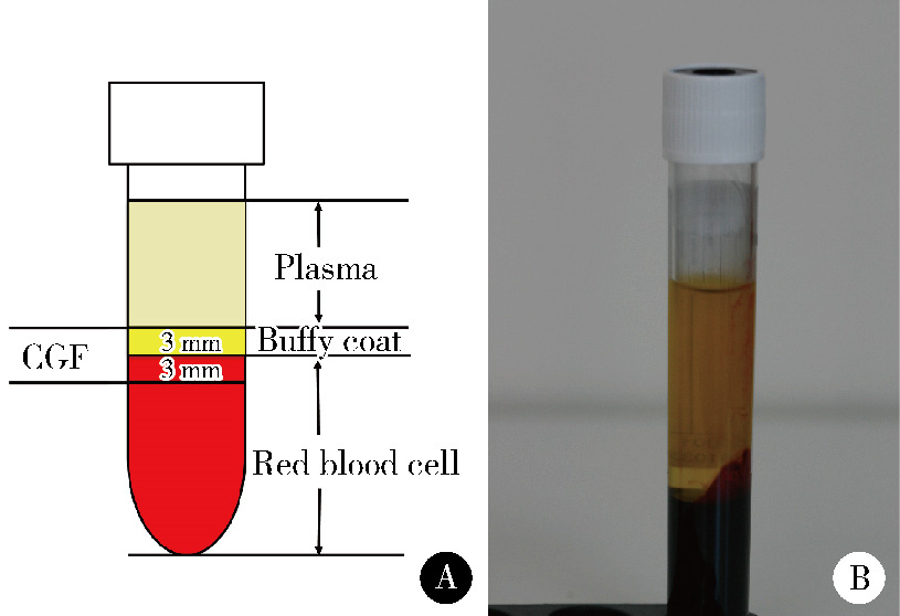
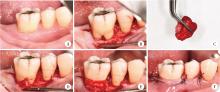
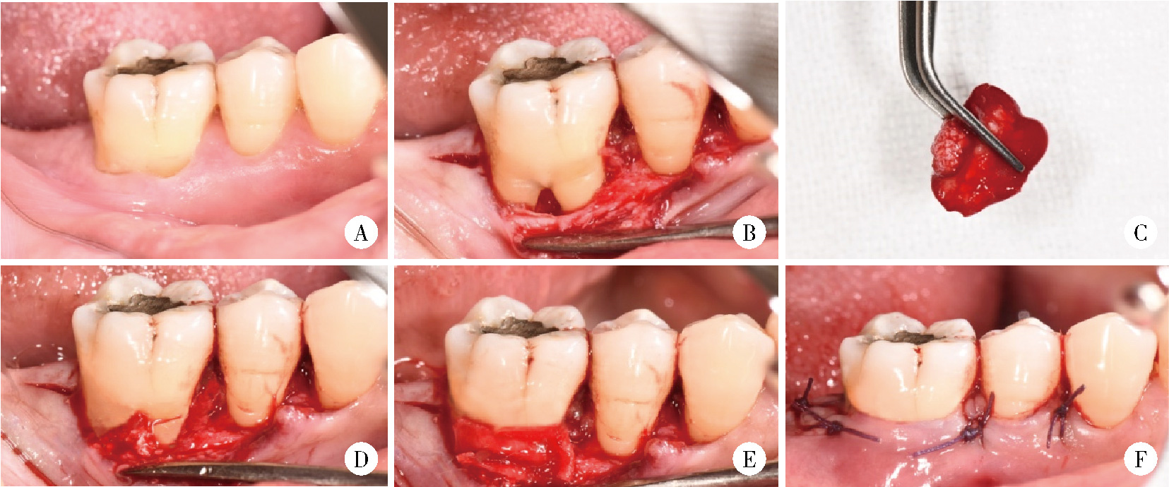
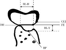
目的: 评价浓缩生长因子(concentrated growth factors,CGF)促进引导性组织再生术(guided tissue regeneration,GTR)联合植骨术治疗下颌磨牙Ⅱ度根分叉病变的临床效果,为根分叉病变的再生寻求更好的治疗方法。方法: 纳入16例需进行牙周手术的慢性牙周炎患者(共20颗下颌磨牙,含36例颊舌侧Ⅱ度根分叉病变),随机分为两组(每组18例病变),试验组采用GTR+植骨术+CGF进行治疗,对照组采用GTR+植骨术进行治疗。分别于术前、术后6个月和1年对患牙进行临床检查,并拍摄锥形束CT(cone beam CT,CBCT)。比较试验组和对照组手术前后临床和CBCT数据的变化。结果: 基线时两组的探诊深度、垂直和水平附着丧失,以及CBCT所示的垂直向和水平向骨丧失差异均无统计学意义(P>0.05)。术后6个月和1年时两组的临床指标及术后1年时两组的CBCT指标较基线时均有显著改善(P < 0.01),且试验组改善程度显著优于对照组(P < 0.05)。其中,术后1年时试验组探诊深度减少(4.75±1.87) mm,垂直和水平附着丧失分别减少(5.55±1.04) mm和(3.89±1.22) mm;对照组探诊深度减少(3.43±1.76) mm,垂直和水平附着丧失分别减少(4.41±1.08) mm和(3.07±1.02) mm。CBCT显示,试验组垂直向和水平向骨丧失分别减少(4.05±1.37) mm和(4.02±1.32) mm,对照组分别减少(3.17±1.09) mm和(3.27±1.08) mm。结论: 为期1年的观察结果显示,CGF可以促进GTR+植骨术在下颌磨牙Ⅱ度根分叉病变中的再生治疗效果。

关键词: 浓缩生长因子, 根分叉部缺损, 引导组织再生术, 磨牙, 下颌骨

Abstract:

Objective: To evaluate the potential of concentrated growth factors (CGF) to enhance the regenerative efficacy of guided tissue regeneration (GTR) when combined with bone graft in the treatment of grade Ⅱ furcation defects in mandibular molars. Methods: This study was approved by the Ethics Committee of Peking University School and Hospital of Stomatology. A total of 16 patients (aged 20-60 years) with chronic periodontitis requiring periodontal surgical intervention were enrolled. All the participants had completed initial periodontal therapy. This involved a total of 20 mandibular molars, which comprised 36 instances of grade Ⅱ furcation lesions located on the buccal or lingual aspects. The 36 furcation lesions were randomly assigned to two groups, with each group containing 18 lesions. The experimental group received treatment with CGF combined with GTR and bone grafting, while the control group was treated with GTR and bone grafting alone. Clinical examinations and cone beam CT (CBCT) assessments were conducted on the affected teeth prior to the surgery, 6 months and 1 year post surgery. Clinical parameters recorded included probing depth (PD), vertical clinical attachment level (CAL-V), horizontal clinical attachment level (CAL-H). CBCT scans were acquired. The radiographic outcomes assessed included bone loss in the vertical direction (BL-V) and horizontal direction (BL-H). Changes in both clinical parameters and CBCT data at baseline and 1 year post surgery were compared between the experimental group and control group. Results: At baseline, no statistically significant differences were observed between the two groups in terms of PD, CAL-V, CAL-H, and BL-V, BL-H as assessed by CBCT (P>0.05), indicating good baseline balance. Six months and 1 year post surgery, both groups demonstrated significant improvements in clinical indicators compared with baseline (P < 0.01). Notably, one year post surgery, the enhancement observed in the experimental group was significantly greater than that of the control group (P < 0.05): the reduction in PD was (4.75±1.87) mm in the experimental group versus (3.43±1.76) mm in the control group; the decrease in CAL-V was (5.55±1.04) mm in the experimental group versus (4.41±1.08) mm in the control group; the decrease in CAL-H was (3.89±1.22) mm in the experimental group versus (3.07±1.02) mm in the control group. One year post surgery, CBCT results demonstrated that the reduction in BL-V was (4.05±1.37) mm in the experimental group compared with (3.17±1.09) mm in the control group, and the reduction in BL-H was (4.02±1.32) mm versus (3.27±1.08) mm. Conclusion: The one-year observational findings demonstrate that CGF enhances the regenerative efficacy of GTR combined with bone graft in the treatment of grade Ⅱ furcation defects in mandibular molars.

Key words: Concentrated growth factors, Furcation defect, Guided tissue regeneration, Molar, Mandible

中图分类号: 

  • R781.4

图1

CGF制备图"

图2

骨粉与可吸收生物膜浸泡于液态CGF内"

图3

试验组手术过程"

图4

CBCT测量示意图"

表1

试验组和对照组基线和术后的出血指数和菌斑指数"

Items GTR+Bone graft+CGF (n=18) GTR+Bone graft (n=18)
Plaque index
  Baseline 0.7±0.2 0.9±0.5
  6 months after surgery 0.7±0.3 1.0±0.6
  1 year after surgery 0.9±0.4 1.1±0.4
Bleeding index
  Baseline 1.0±0.5 1.2±0.6
  6 months after surgery 1.1±0.6 0.9±0.5
  1 year after surgery 0.8±0.6 1.1±0.7

表2

试验组与对照组基线和术后的临床指标"

Items GTR+Bone graft+CGF (n=18) GT+Bone graft (n=18) P value
PD/mm
  Baseline 7.95±2.69 7.69±2.74 0.965
  6 months after surgery 3.14±0.95* 4.09±1.37* 0.032
  1 year after surgery 3.20±1.06* 4.26±1.55* 0.004
  Decrease at 1 year after surgery 4.75±1.87 3.43±1.76 0.013
REC/mm
  Baseline 1.87±0.99 1.95±0.96 0.245
  6 months after surgery 0.98±0.58* 1.10±0.42* 0.878
  1 year after surgery 1.07±0.62* 0.97±0.44* 0.327
  Decrease at 1 year after surgery 0.80±0.54 0.98±0.57 0.984
CAL-V/mm
  Baseline 9.82±2.78 9.64±2.53 0.052
  6 months after surgery 4.12±1.83* 5.19±1.94* 0.039
  1 year after surgery 4.27±1.79* 5.23±1.88* 0.034
  Decrease at 1 year after surgery 5.55±1.04 4.41±1.08 0.027
CAL-H/mm
  Baseline 6.32±2.74 6.45±2.87 0.977
  6 months after surgery 2.49±1.03* 3.44±1.35* 0.028
  1 year after surgery 2.43±1.06* 3.38±1.19* 0.009
  Decrase at 1 year after surgery 3.89±1.22 3.07±1.02 0.035

表3

试验组与对照组基线和术后1年时的CBCT指标"

Items GTR+Bone graft+CGF (n=18) GTR+Bone graft (n=18) P value
BL-V/mm
  Baseline 7.03±2.86 7.35±2.77 0.684
  1 year after surgery 2.98±1.01* 4.18±1.34* 0.004
  Change of BL-V 4.05±1.37 3.17±1.09 0.009
BL-H/mm
  Baseline 6.94±2.77 6.89±2.87 0.509
  1 year after surgery 2.92±1.15* 3.62±1.09* 0.012
  Change of BL-H 4.02±1.32 3.27±1.08 0.027

图5

基线和术后1年时试验组CBCT图像"

1
Eickholz P , Cordis T , Dannewitz B , et al. Long-term prognosis of teeth with class Ⅱ furcation involvement: A retrospective cohort study[J]. J Clin Periodontol, 2025, 52 (9): 1298- 1305.

doi: 10.1111/jcpe.14186
2
Trombelli L , Farina R . Clinical outcomes with bioactive agents alone or in combination with grafting or guided tissue regeneration[J]. J Clin Periodontol, 2008, 35 (Suppl 8): 117- 135.
3
Stoecklin-Wasmer C , Rutjes AWS , da Costa BR , et al. Absorb-able collagen membranes for periodontal regeneration: A systematic review[J]. J Dent Res, 2013, 92 (9): 773- 781.

doi: 10.1177/0022034513496428
4
Irma J , Kartasasmita AS , Kartiwa A , et al. From growth factors to structure: PDGF and TGF-β in granulation tissue formation. A literature review[J]. J Cell Mol Med, 2025, 29 (11): e70374.

doi: 10.1111/jcmm.70374
5
Chandra P , Faizan M , Porwal M , et al. An overview and review of growth factors in wound healing: Emerging trends and innovations[J]. Curr Diabetes Rev, 2026, 22 (1): e15733998332692.

doi: 10.2174/0115733998332692241202072249
6
Zhou D , Liu ZW , Jie X , et al. Advances in research on concentrated growth factor applications for androgenetic alopecia treatment: A review[J]. Med Sci Monit, 2025, 31, e948054.
7
Kinaia BM , Steiger J , Neely AL , et al. Treatment of class Ⅱ molar furcation involvement: Meta-analyses of reentry results[J]. J Periodontol, 2011, 82 (3): 413- 428.

doi: 10.1902/jop.2010.100306
8
Pajnigara N , Kolte A , Kolte R , et al. Volumetric assessment of regenerative efficacy of demineralized freeze-dried bone allograft with or without amnion membrane in grade Ⅱ furcation defects: A cone beam computed tomography study[J]. Int J Periodontics Restorative Dent, 2017, 37 (2): 255- 262.

doi: 10.11607/prd.2901
9
Jepsen S , Gennai S , Hirschfeld J , et al. Regenerative surgical treatment of furcation defects: A systematic review and Bayesian network meta-analysis of randomized clinical trials[J]. J Clin Periodontol, 2020, 47 (S22): 352- 374.

doi: 10.1111/jcpe.13238
10
Huidrom E , Srivastava V , Meenawat A , et al. Evaluation of the efficacy of concentrated growth factor along with bovine-derived xenograft and collagen membrane in the treatment of Degree Ⅱ mandibular molar furcation defect: A clinicoradiographic study[J]. J Indian Soc Periodontol, 2022, 26 (2): 130- 136.

doi: 10.4103/jisp.jisp_44_21
11
Pradeep AR , Pai S , Garg G , et al. A randomized clinical trial of autologous platelet-rich plasma in the treatment of mandibular degree Ⅱ furcation defects[J]. J Clin Periodontol, 2009, 36 (7): 581- 588.

doi: 10.1111/j.1600-051X.2009.01428.x
12
Sharma A , Pradeep AR . Autologous platelet-rich fibrin in the treatment of mandibular degree Ⅱ furcation defects: A randomized clinical trial[J]. J Periodontol, 2011, 82 (10): 1396- 1403.

doi: 10.1902/jop.2011.100731
13
Gawlak-Socka S , Jeżewska K , Bielecka-Kowalska N , et al. Adjunctive use of platelet-derived concentrates (platelet-rich plasma, platelet-rich fibrin, concentrated growth factor, platelet-poor plasma) in non-surgical periodontal therapy: Current evidence and comparative analysis[J]. J Clin Med, 2026, 15 (2): 554.

doi: 10.3390/jcm15020554
14
Iao S , Ouyang X , Qiao J , et al. Growth factors release in concentrated growth factor and advanced platelet-rich fibrin sticky bone[J]. Oral Dis, 2025, 31 (8): 2531- 2539.

doi: 10.1111/odi.15325
15
Qiao J , An N . Effect of concentrated growth factors on function and Wnt3a expression of human periodontal ligament cells in vitro[J]. Platelets, 2017, 28 (3): 281- 286.

doi: 10.1080/09537104.2016.1213381
16
Kuybu İ , Alan H , Şimşek MS . Comparison of growth factor concentrations in platelet-rich fibrin biomaterial in different systemic diseases[J]. Clin Oral Investig, 2025, 29 (12): 588.

doi: 10.1007/s00784-025-06673-y
17
Panda S , Karanxha L , Goker F , et al. Autologous platelet concentrates in treatment of furcation defects: A systematic review and meta-analysis[J]. Int J Mol Sci, 2019, 20 (6): 1347.

doi: 10.3390/ijms20061347
18
Yu M , Luo D , Qiao J , et al. A hierarchical bilayer architecture for complex tissue regeneration[J]. Bioact Mater, 2022, 10, 93- 106.
19
Tabatabaei F , Aghamohammadi Z , Tayebi L . In vitro and in vivo effects of concentrated growth factor on cells and tissues[J]. J Biomed Mater Res Part A, 2020, 108 (6): 1338- 1350.

doi: 10.1002/jbm.a.36906
20
Rodella LF , Favero G , Boninsegna R , et al. Growth factors, CD34 positive cells, and fibrin network analysis in concentrated growth factors fraction[J]. Microsc Res Tech, 2011, 74 (8): 772- 777.

doi: 10.1002/jemt.20968
21
Yu B , Wang Z . Effect of concentrated growth factors on beagle periodontal ligament stem cells in vitro[J]. Mol Med Rep, 2014, 9 (1): 235- 242.

doi: 10.3892/mmr.2013.1756
22
Li X , Yang H , Zhang Z , et al. Concentrated growth factor exudate enhances the proliferation of human periodontal ligament cells in the presence of TNF-α[J]. Mol Med Rep, 2019, 19 (2): 943- 950.
23
Chen X , Wang J , Yu L , et al. Effect of concentrated growth factor (CGF) on the promotion of osteogenesis in bone marrow stromal cells (BMSC) in vivo[J]. Sci Rep, 2018, 8, 5876.

doi: 10.1038/s41598-018-24364-5
24
Yu M , Wang X , Liu Y , et al. Cytokine release kinetics of concentrated growth factors in different scaffolds[J]. Clin Oral Investig, 2019, 23 (4): 1663- 1671.

doi: 10.1007/s00784-018-2582-z
[1] 王月, 梁宇红. 繁茂型牙骨质-骨结构不良1例[J]. 北京大学学报(医学版), 2026, 58(1): 220-224.
[2] 仇师禹, 练洋, 康一帆, 张雷, 蔡义望, 单小峰, 蔡志刚. 基于下颌骨数据库和全连接神经网络的三维检索模型辅助下的下颌骨个性化重建[J]. 北京大学学报(医学版), 2025, 57(2): 360-368.
[3] 王菲, 张馨月, 刘木清, 王恩博, 段登辉. 顺牙长轴拔牙法在下颌近中与水平智齿拔除术中的应用及三维有限元分析[J]. 北京大学学报(医学版), 2025, 57(1): 106-112.
[4] 石宇彤, 危伊萍, 胡文杰, 徐涛, 张浩筠. 罹患重度牙周炎下颌磨牙拔牙微翻瓣牙槽嵴保存效果评价[J]. 北京大学学报(医学版), 2025, 57(1): 33-41.
[5] 王昕莹, 程雪原, 张勇, 李菲, 段晋瑜, 乔静. 浓缩生长因子与自固化磷酸钙人工骨联合治疗牙周骨下袋缺损的疗效:临床和影像学评价[J]. 北京大学学报(医学版), 2025, 57(1): 42-50.
[6] 康一帆, 葛严军, 吕晓鸣, 谢尚, 单小峰, 蔡志刚. 即刻种植体支持式义齿修复的血管化髂骨瓣重建下颌骨缺损[J]. 北京大学学报(医学版), 2025, 57(1): 78-84.
[7] 李红光,韩玮华,吴训,冯继玲,李刚,孟娟红. 关节腔冲洗联合液态浓缩生长因子注射治疗单侧颞下颌关节骨关节炎的初步研究[J]. 北京大学学报(医学版), 2024, 56(2): 338-344.
[8] 陈晨,梁宇红. 复杂根管上颌磨牙的根管治疗3例[J]. 北京大学学报(医学版), 2024, 56(1): 190-195.
[9] 徐心雨,吴灵,宋凤岐,李自力,张益,刘筱菁. 基于下颌运动轨迹的正颌外科术中下颌骨髁突定位方法及初步精度验证[J]. 北京大学学报(医学版), 2024, 56(1): 57-65.
[10] 王聪伟,高敏,于尧,章文博,彭歆. 游离腓骨瓣修复下颌骨缺损术后义齿修复的临床分析[J]. 北京大学学报(医学版), 2024, 56(1): 66-73.
[11] 卢汉,张建运,杨榕,徐乐,李庆祥,郭玉兴,郭传瑸. 下颌牙龈鳞状细胞癌患者预后的影响因素[J]. 北京大学学报(医学版), 2023, 55(4): 702-707.
[12] 雷玥,杨颖婷,雷海华. 异位萌出诱导矫治器在第一恒磨牙异位萌出治疗中的应用[J]. 北京大学学报(医学版), 2023, 55(4): 766-封三.
[13] 高梓翔,王勇,温奥楠,朱玉佳,秦庆钊,张昀,王晶,赵一姣. 基于三维下颌骨平均模型的颌骨标志点自动确定方法[J]. 北京大学学报(医学版), 2023, 55(1): 174-180.
[14] 田靖,秦满,陈洁,夏斌. 失活剂烧伤致乳磨牙早失及恒牙胚丧失2例[J]. 北京大学学报(医学版), 2022, 54(2): 381-385.
[15] 陈震,谷宝鑫,汤玉芳,闫子玉,倪方端,崔念晖. 应用Delphi法构建下颌阻生第三磨牙拔除难度评分量表[J]. 北京大学学报(医学版), 2022, 54(1): 100-104.
Viewed
Full text


Abstract

Cited

  Shared   
  Discussed   
No Suggested Reading articles found!