北京大学学报(医学版) ›› 2026, Vol. 58 ›› Issue (1): 220-224. doi: 10.19723/j.issn.1671-167X.2026.01.030

• 病例报告 • 上一篇    下一篇

繁茂型牙骨质-骨结构不良1例

王月1, 梁宇红2,*()   

  1. 1. 北京大学国际医院口腔科, 北京 102206
    2. 北京大学口腔医学院·口腔医院急诊科, 国家口腔医学中心, 国家口腔疾病临床医学研究中心, 口腔生物材料和数字诊疗装备国家工程研究中心, 口腔数字医学北京市重点实验室, 国家卫生健康委员会口腔数字医学重点实验室, 国家药品监督管理局口腔材料重点实验室, 北京 100081
  • 收稿日期:2025-09-25 出版日期:2026-02-18 发布日期:2025-11-28
  • 通讯作者: 梁宇红

Florid cemento-osseous dysplasia: A case report

Yue WANG1, Yuhong LIANG2,*()   

  1. 1. Department of Stomatology, Peking University International Hospital, Beijing 102206, China
    2. Department of Oral Emergency, Peking University School and Hospital of Stomatology & National Center for Stomatology & National Clinical Research Center for Oral Diseases & National Engineering Research Center of Oral Biomaterials and Digital Medical Devices & Beijing Key Laboratory of Digital Stomatology & NHC Key Laboratory of Digital Stomatology & NMPA Key Laboratory for Dental Materials, Beijing 100081, China
  • Received:2025-09-25 Online:2026-02-18 Published:2025-11-28
  • Contact: Yuhong LIANG

RICH HTML


摘要:

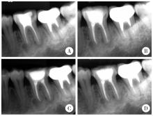
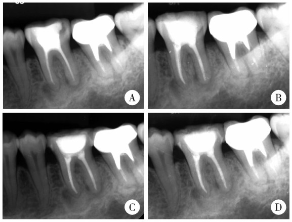
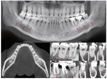
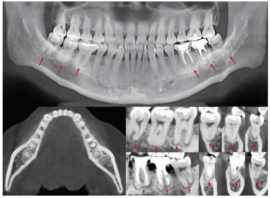
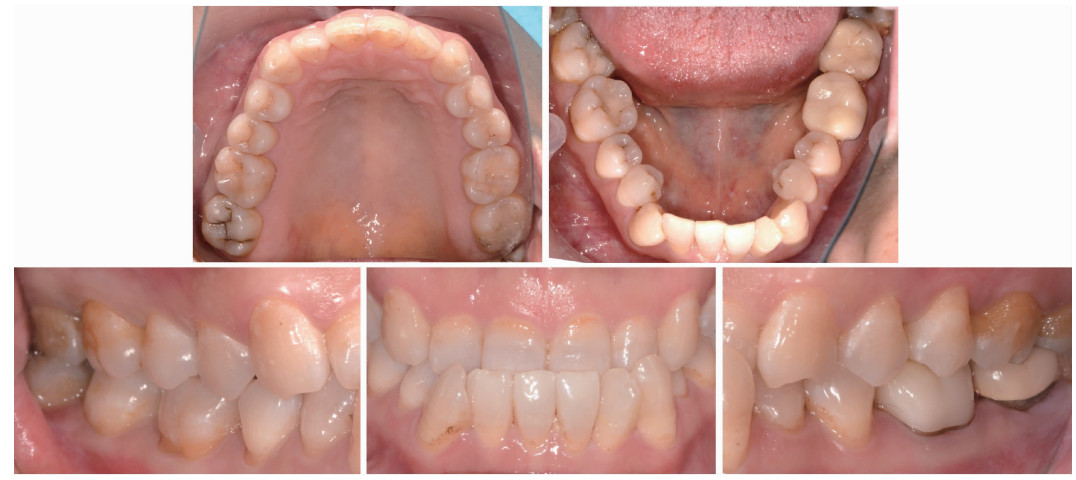
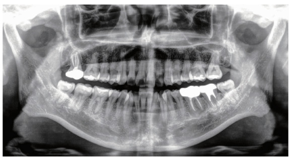
牙骨质-骨结构不良(cemento-osseous dysplasia, COD)属于临床少见的一种纤维-骨肿瘤及结构不良, 病变通常无症状, 多通过X线检查偶然发现。本文报道1例就诊于北京大学国际医院口腔科的45岁女性繁茂型牙骨质-骨结构不良(florid cemento-osseous dysplasia, FLCOD)病例, 患者因左下后牙充填体脱落3周就诊, 临床检查发现左下颌第一磨牙(36)存在继发龋坏达髓腔, 但无牙髓炎或根尖周病症状。邻近牙齿(左下颌第三磨牙)及对侧磨牙(右下颌第一、二、三磨牙)牙髓活力测试反应正常, 无肿胀、窦道或牙齿松动。锥形束CT(cone beam computed tomography, CBCT)检查显示, 双侧下颌磨牙根尖周区域存在多灶性混合密度病变, 符合世界卫生组织制定的FLCOD诊断标准。病变累及的6颗牙齿(36、37、38、46、47和48)根尖周影像学表现多样, 呈现骨质溶解破坏期、类牙骨质小体形成期与钙化成熟期三期并存的特征。本病例为临床少见的FLCOD病例, 诊断主要依据其无临床症状的病程、牙髓状态及典型的影像学表现。CBCT在清晰显示病变、提供诊断依据方面发挥了关键作用。治疗策略上应着重处理患者的继发龋等伴发病变, 控制局部与全身风险因素, 而非针对COD病变本身进行干预, 预后还需长期随访。

关键词: 繁茂型牙骨质-骨结构不良, 锥形束CT, 鉴别诊断, 下颌磨牙

Abstract:

Cemento-osseous dysplasia (COD) is an uncommon, non-neoplastic benign fibro-osseous lesion of the jaws, characterized by cementum-like tissue deposition. It primarily affects middle-aged women of African and East Asian descent. Generally asymptomatic, this condition is frequently identified incidentally through radiographs showing radiopacities with radiolucent rims. This report presented a case of florid cemento-osseous dysplasia (FLCOD) in a 45-year-old Asian female. The patient exhibited secondary caries extending to the pulp chamber on the left mandibular first molar, showed no signs of pulpitis or periapical pathosis. Adjacent tooth (the left mandibular third molar) and contralateral molars (the right mandibular molars) responded normally to pulp vitality testing, with no swelling, sinus tracts, or mobility observed. Cone beam computed tomography (CBCT) revealed multifocal lesions with amorphous radiopacities and thin radiolucent rims without root resorption or cortical perforation in the periapical regions of bilateral mandibular molars. These findings reflected characteristic radiographic features of FLCOD, aligning with the World Health Organization (WHO) 2022 classification criteria. Significantly, three radiographic stages of COD were concurrently demonstrated through multifocal lesions involving six mandibular molars, providing a representative model of its natural progression. The initial osteolytic stage in teeth 38, 46, and 48 manifested as well-defined periapical radiolucencies with sclerotic borders; the intermediate stage in 36 and 37 featured punctate or nodular radiopacities within periapical radiolucency; while the mature terminal stage in 47 presented a homogeneous radiopaque mass bordered by a thin radiolucent rim. The diagnostic process prioritized exclusion of chronic apical periodontitis through confirmed pulp vitality and absence of infection. Cemento-ossifying fibroma (COsF) was dismissed based on solitary presentation and cortical expansion. Familial gigantiform cementoma (FGC) was differentiated by early diffuse jaw expansion and extensive involvement beyond COD. Cementoblastoma exclusion relied on pathognomonic features, specifically root resorption or compromised pulp vitality. Given the asymptomatic, non-progressive behavior of the FLCOD lesions, annual surveillance was implemented, with intervention limited to the restorative failure in tooth 36. At 12-month follow-up, the FLCOD lesions showed clinical-radiographic stability without secondary infection or progression. This case reflected the classic presentation of FLCOD as incidental, multifocal mandibular posterior radiopacities bordered by radiolucent rims. CBCT proved instrumental in delineating the lesions, providing diagnostic evidence. Long-term surveillance remains the cornerstone of management for COD. Invasive procedures are not recommended in cases with asymptomatic non-progressive behavior to prevent surgical complications.

Key words: Florid cemento-osseous dysplasia, Cone-beam computed tomography, Differential diagnosis, Mandibular molars

中图分类号: 

  • R782.1

图1

左下颌第一磨牙(36)的术前和术后根尖片"

图2

患者初诊时的CBCT图像"

图3

1年随访复查口内照"

图4

1年随访复查曲面断层图像"

表1

牙骨质-骨结构不良三个分期的病理学和影像学表现"

Disease stage Pathological manifestations Radiographic manifestations
Osteolytic stage Replacement of normal bone by cellular fibrous tissue without calcification Well-demarcated periapical radiolucency with sclerotic borders
Cementoblastic stage Progressive deposition of cementum-like droplets and woven bone within the fibrous stroma Punctate or flocculent radiopacities within a radiolucent halo
Mature stage Densely mineralized masses with peripheral fibrous encapsulation Homogeneous radiopacities bordered by thin radiolucent rims
1
Pereira DL , Pires FR , Lopes MA , et al. Clinical, demographic, and radiographic analysis of 82 patients affected by florid osseous dysplasia: An international collaborative study[J]. Oral Surg Oral Med Oral Pathol Oral Radiol, 2016, 122 (2): 250- 257.

doi: 10.1016/j.oooo.2016.04.013
2
WHO Classification of Tumours Editorial Board . Head and neck tumours[M]. 5th ed Lyon (France): IARC Press, 2022: 389- 390.
3
史册, 李志民, 孙宏晨. 纤维-骨肿瘤及结构不良的临床病理特征与鉴别诊断[J]. 中华口腔医学杂志, 2023, 58 (2): 124- 130.
4
Pick E , Schäfer T , Al-Haj Husain A , et al. Clinical, radiological, and pathological diagnosis of fibro-osseous lesions of the oral and maxillofacial region: A retrospective study[J]. Diagnostics, 2022, 12 (2): 238.

doi: 10.3390/diagnostics12020238
5
Cotti E . Advanced techniques for detecting lesions in bone[J]. Dent Clin North Am, 2010, 54 (2): 215- 235.

doi: 10.1016/j.cden.2009.12.007
6
Gumru B , Akkitap MP , Deveci S , et al. A retrospective cone beam computed tomography analysis of cemento-osseous dysplasia[J]. J Dent Sci, 2021, 16 (4): 1154- 1161.

doi: 10.1016/j.jds.2021.03.009
7
Titinchi F , Alturki N , Morkel J , et al. Cemento-osseous dysplasia: A multi-centre analysis of surgical management[J]. Oral Maxillofac Surg, 2025, 29 (1): 96.

doi: 10.1007/s10006-025-01394-8
8
Haefliger S , Turek D , Andrei V , et al. Cemento-osseous dysplasia is caused by RAS-MAPK activation[J]. Pathology, 2023, 55 (3): 324- 328.

doi: 10.1016/j.pathol.2022.10.006
9
Aiuto R , Gucciardino F , Rapetti R , et al. Management of symptomatic florid cemento-osseous dysplasia: Literature review and a case report[J]. J Clin Exp Dent, 2018, 10 (3): e291- e295.
10
Olgac V , Sinanoglu A , Selvi F , et al. A clinicopathologic analysis of 135 cases of cemento-osseous dysplasia: To operate or not to operate?[J]. J Stomatol Oral Maxillofac Surg, 2021, 122 (3): 278- 282.

doi: 10.1016/j.jormas.2020.06.002
11
Decolibus K , Shahrabi-Farahani S , Brar A , et al. Cemento-osseous dysplasia of the jaw: Demographic and clinical analysis of 191 new cases[J]. Dent J, 2023, 11 (5): 138.

doi: 10.3390/dj11050138
12
Gabay M , DiPede L , Fornatora M , et al. Treatment indications for symptomatic versus asymptomatic florid cemento-osseous dysplasia in adult patients: A systematic review[J]. Oral Surg Oral Med Oral Pathol Oral Radiol, 2024, 138 (6): 686- 701.

doi: 10.1016/j.oooo.2024.06.011
13
Ali D , Figeac F , Caci A , et al. High-fat diet-induced obesity augments the deleterious effects of estrogen deficiency on bone: Evidence from ovariectomized mice[J]. Aging Cell, 2022, 21 (12): e13726.

doi: 10.1111/acel.13726
14
Wang Y , LaMonte MJ , Hovey KM , et al. Association of serum 17β-estradiol concentration, hormone therapy, and alveolar crest height in postmenopausal women[J]. J Periodontol, 2015, 86 (4): 595- 605.

doi: 10.1902/jop.2015.140533
15
Sarmento DJ , Monteiro BV , de Medeiros AM , et al. Severe florid cemento-osseous dysplasia: A case report treated conservatively and literature review[J]. Oral Maxillofac Surg, 2013, 17 (1): 43- 46.

doi: 10.1007/s10006-012-0314-0
16
Robinson HB . Osseous dysplasia: Reaction of bone to injury[J]. J Oral Surg, 1956, 14 (1): 3- 14.
17
El-Naggar AK , Chan JKC , Grandis JR , et al. WHO classification of head and neck tumours[M]. 4th ed Lyon (France): IARC Press, 2017: 254.
[1] 肖晓笛, 夏有辰, 柳剑英, 付鹏. 左侧胸锁乳突肌间血管内乳头状内皮增生1例[J]. 北京大学学报(医学版), 2025, 57(5): 1002-1004.
[2] 刘杰, 马茗微, 王庆安, 石明, 尹金鹏, 王占平, 申静涛, 高献书. 基于锥形束CT的前列腺癌放射治疗两种体位固定方式摆位误差比较[J]. 北京大学学报(医学版), 2025, 57(4): 692-697.
[3] 石宇彤, 危伊萍, 胡文杰, 徐涛, 张浩筠. 罹患重度牙周炎下颌磨牙拔牙微翻瓣牙槽嵴保存效果评价[J]. 北京大学学报(医学版), 2025, 57(1): 33-41.
[4] 钟华, 李原, 徐丽玲, 白明欣, 苏茵. 18F-FDG PET/CT在风湿免疫病中的应用[J]. 北京大学学报(医学版), 2024, 56(5): 853-859.
[5] 薄士仕,高承志. 基于卷积神经网络实现锥形束CT牙齿分割及牙位标定[J]. 北京大学学报(医学版), 2024, 56(4): 735-740.
[6] 哈雪梅,姚永正,孙莉华,辛春杨,熊焰. 实性肺胎盘样变形1例及文献复习[J]. 北京大学学报(医学版), 2023, 55(2): 357-361.
[7] 章锦花,潘洁,孙志鹏,王霄. 不同根管内容物对口腔颌面锥形束CT诊断牙根纵裂准确性的影响[J]. 北京大学学报(医学版), 2023, 55(2): 333-338.
[8] 潘孟乔,刘建,徐莉,徐筱,侯建霞,李小彤,王晓霞. 牙周-正畸-正颌联合治疗骨性安氏Ⅲ类错畸形患者下前牙牙周表型的长期观察[J]. 北京大学学报(医学版), 2023, 55(1): 52-61.
[9] 叶佳学,梁宇红. 牙髓专科医师应用锥形束CT的现况调查[J]. 北京大学学报(医学版), 2023, 55(1): 114-119.
[10] 高娟,吕航苗,马慧敏,赵一姣,李小彤. 锥形束CT三维体积测量评估骨性Ⅲ类错正畸正颌治疗后的牙根吸收[J]. 北京大学学报(医学版), 2022, 54(4): 719-726.
[11] 刘伟涛,王怡然,王雪东,周彦恒. 锥形束CT研究上颌反复扩缩前方牵引后上颌骨缝的三维变化[J]. 北京大学学报(医学版), 2022, 54(2): 346-355.
[12] 孟圆,张丽琪,赵雅宁,柳登高,张祖燕,高岩. 67例上颌根尖周囊肿的三维影像特点分析[J]. 北京大学学报(医学版), 2021, 53(2): 396-401.
[13] 曹畅,王菲,王恩博,刘宇. β-磷酸三钙用于下颌第三磨牙拔除术后骨缺损修复的自身对照研究[J]. 北京大学学报(医学版), 2020, 52(1): 97-102.
[14] 谢晓艳,贾淑梅,孙志辉,张祖燕. 分辨率设置与锥形束CT检测牙根外吸收的可靠性[J]. 北京大学学报(医学版), 2019, 51(1): 75-79.
[15] 卢昕,张立宁. 肌活检在特发性炎性肌病诊断和临床分型中的价值[J]. 北京大学学报(医学版), 2018, 50(6): 949-951.
Viewed
Full text


Abstract

Cited

  Shared   
  Discussed   
No Suggested Reading articles found!