北京大学学报(医学版) ›› 2021, Vol. 53 ›› Issue (1): 54-61. doi: 10.19723/j.issn.1671-167X.2021.01.009
数字化个齿托盘制取下颌全牙列全冠预备体印模的体外评价
徐啸翔1,曹烨1,Δ( ),赵一姣2,贾璐3,谢秋菲1
- 1.北京大学口腔医学院·口腔医院,修复科, 国家口腔疾病临床医学研究中心 口腔数字化医疗技术和材料国家工程实验室 口腔数字医学北京市重点实验室,北京 100081
2.北京大学口腔医学院·口腔医院,口腔医学数字化研究中心, 国家口腔疾病临床医学研究中心 口腔数字化医疗技术和材料国家工程实验室 口腔数字医学北京市重点实验室,北京 100081
3.北京大学口腔医学院·口腔医院,义齿加工中心 国家口腔疾病临床医学研究中心 口腔数字化医疗技术和材料国家工程实验室 口腔数字医学北京市重点实验室,北京 100081
In vitro evaluation of the application of digital individual tooth tray in the impression making of mandibular full-arch crown abutments
XU Xiao-xiang1,CAO Ye1,Δ(),ZHAO Yi-jiao2,JIA Lu3,XIE Qiu-fei1
- 1. Department of Prosthodontics, Peking University School and Hospital of Stomatology & National Clinical Research Center for Oral Diseases & National Engineering Laboratory for Digital and Material Technology of Stomatology & Beijing Key Laboratory of Digital Stomatology, Beijing 100081, China
2. Center for Digital Dentistry, Peking University School and Hospital of Stomatology & National Clinical Research Center for Oral Diseases & National Engineering Laboratory for Digital and Material Technology of Stomatology & Beijing Key Laboratory of Digital Stomatology, Beijing 100081, China
3. Dental Laboratory Center, Peking University School and Hospital of Stomatology & National Clinical Research Center for Oral Diseases & National Engineering Laboratory for Digital and Material Technology of Stomatology & Beijing Key Laboratory of Digital Stomatology, Beijing 100081, China
摘要:
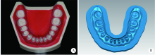
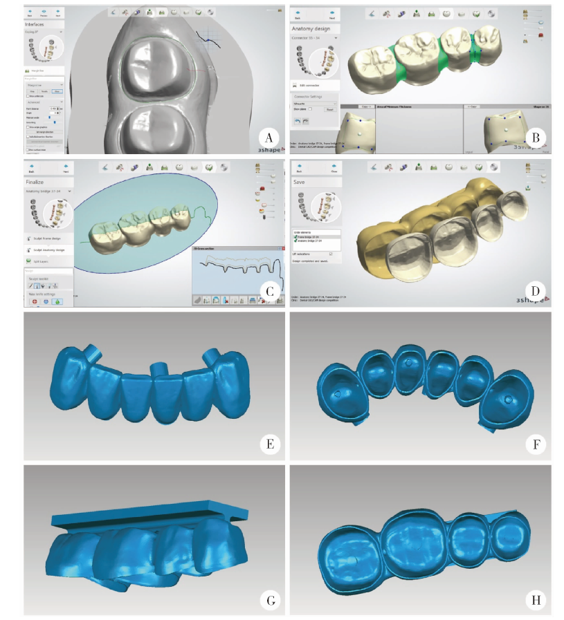
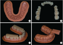
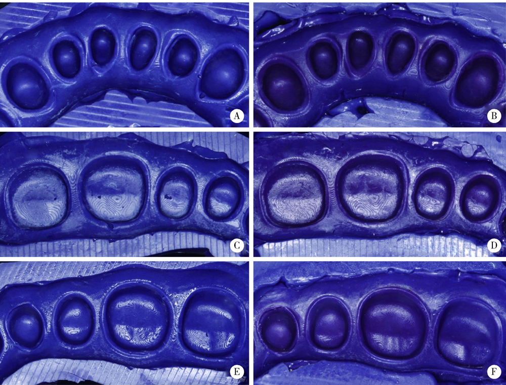
目的: 建立利用三维扫描、计算机辅助设计和三维打印制作数字化个齿托盘的方法,通过体外研究评价数字化个齿托盘法和常规法制取全牙列全冠预备体印模的效果。方法: 在标准下颌牙列模型上进行14颗树脂牙全冠预备,通过三维扫描获取预备体表面数据。利用牙科修复设计软件在每个预备体上确定边缘线位置,设计作为个齿托盘主体部分的解剖全冠和连接体形态,以及作为容纳印模材料空间的基底冠形态。在生成的个齿托盘主体数据外表面增加辅助固位装置,内表面生成组织终止点。设计及打印全牙列预备体模型1副,常规全牙列个别托盘A、B以及数字化个齿托盘各4副。使用个别托盘A采用一步法对全牙列预备体模型进行聚醚印模制取,使用个别托盘B和数字化个齿托盘采用分段取模方式完成聚醚印模制取,各重复制取4次。记录每次印模制取的时间及印模中每个预备体肩台及轴面/牙合面缺陷的数目,对每个预备体印模质量进行评价,比较两种方法制取印模的预备体合格率和预备体质量分布。结果: 常规法制取全牙列全冠预备体印模中肩台缺陷数目显著多于数字化个齿托盘法,轴面/牙合面缺陷数目差异无统计学意义。数字化个齿托盘法制取印模的预备体合格率高于常规法,预备体质量分布优于常规法。结论: 通过数字化方法可以实现个齿托盘的设计和制作,与常规法相比,数字化个齿托盘法制取全牙列全冠预备体印模效果更好。
中图分类号:
- R783.6


| [1] | 冯海兰, 徐军. 口腔修复学 [M]. 2版. 北京: 北京大学医学出版社, 2013: 92-95. |
| [2] | Shillingburg HT, Sather DA, Wilson EL, et al. Fundamentals of fixed prosthodontics[M]. 4th ed. London: Quintessence Publi-shing Company, 2012: 291-325. |
| [3] |
Jung BY, Lee KW. Alternative impression technique for multiple abutments in difficult case to control[J]. J Adv Prosthodont, 2010,2(1):1-3.
doi: 10.4047/jap.2010.2.1.1 pmid: 21165179 |
| [4] | Cannistraci AJ. A new approach to impression taking for crown and bridge[J]. Dent Clin North Am, 1965,29(3):33-42. |
| [5] |
Kimmelman BB, Lerman H. Impressions of single preparations using a copper band-shell[J]. J Prosthet Dent, 1971,26(2):154-158.
doi: 10.1016/0022-3913(71)90047-3 pmid: 4933161 |
| [6] | Hoffman JM. Nontraumatic final impressions for fixed partial dentures[J]. Prosthodont, 1992,1(1):61-64. |
| [7] |
Dimashkieh MR, Morgano SM. A procedure for making fixed prosthodontic impressions with the use of preformed crown shells[J]. J Prosthet Dent, 1995,73(1):95-96.
doi: 10.1016/s0022-3913(05)80277-x pmid: 7699606 |
| [8] |
Matsumoto W, de Almeida Antunes RP, Fernandes RM, et al. Shell tray impression: a technique modification[J]. J Dent Pub H, 2018,9(1):8-16.
doi: 10.17267/2596-3368dentistry.v9i1 |
| [9] |
de Sá AT, de Freitas CA, de Sá FC, et al. Effect of cervical relining of acrylic resin copings on the accuracy of stone dies obtained using a polyether impression material[J]. J Appl Oral Sci, 2008,16(1):7-11.
doi: 10.1590/s1678-77572008000100003 pmid: 19089282 |
| [10] | 林侠, 赵辉. 固定修复印模方法的比较[J]. 现代口腔医学杂志, 2001,15(4):318. |
| [11] | 唐震宇, 钱成明, 王辉, 等. 个齿托盘对硅橡胶印模颈缘清晰度影响的临床研究[J]. 口腔医学研究, 2015,31(6):613-615. |
| [12] | 林侠, 王梅. 个齿托盘法的应用介绍[J]. 大连医科大学学报, 1997,19(4):303. |
| [13] |
Su TS, Sun J. Comparison of repeatability between intraoral digital scanner and extraoral digital scanner: An in-vitro study[J]. J Prosthodont Res, 2015,59(4):236-242.
doi: 10.1016/j.jpor.2015.06.002 pmid: 26211702 |
| [14] |
Kihara H, Hatakeyama W, Komine F, et al. Accuracy and practicality of intraoral scanner in dentistry: A literature review[J]. J Prosthodont Res, 2020,64(2):109-113.
doi: 10.1016/j.jpor.2019.07.010 pmid: 31474576 |
| [15] | 曹悦, 陈俊锴, 赵一姣, 等. 口内三维扫描技术临床应用精度的研究进展[J]. 中华口腔医学杂志, 2020,55(3):201-205. |
| [16] |
Beier US, Kranewitter R, Dumfahrt H. Quality of impressions after use of the Magic FoamCord gingival retraction system: a clinical study of 269 abutment teeth[J]. Int J Prosthodont, 2009,22(2):143-147.
pmid: 19418859 |
| [1] | 于录, 吴灵, 刘筱菁, 李自力. 基于数据库相似性检索的正颌外科手术规划技术流程可行性研究: 随机对照试验[J]. 北京大学学报(医学版), 2026, 58(1): 145-152. |
| [2] | 钱锟, 刘亦洪. 基于直接法和间接法数字印模制作的高嵌体适合性评价的体外研究[J]. 北京大学学报(医学版), 2025, 57(3): 604-609. |
| [3] | 马丽娟, 腾雍辉, 王勇, 赵一姣, 张馨月, 秦庆钊, 尹东. 乳牙缺失数字化丝圈间隙保持器的三维有限元分析[J]. 北京大学学报(医学版), 2025, 57(2): 376-383. |
| [4] | 徐心雨,吴灵,宋凤岐,李自力,张益,刘筱菁. 基于下颌运动轨迹的正颌外科术中下颌骨髁突定位方法及初步精度验证[J]. 北京大学学报(医学版), 2024, 56(1): 57-65. |
| [5] | 李穗,马雯洁,王时敏,丁茜,孙瑶,张磊. 上前牙种植单冠修复体切导的数字化设计正确度[J]. 北京大学学报(医学版), 2024, 56(1): 81-87. |
| [6] | 罗昊,田福聪,王晓燕. 不同椅旁可切削修复材料序列抛光时间及表面粗糙度与光泽度的比较[J]. 北京大学学报(医学版), 2022, 54(3): 565-571. |
| [7] | 冯莎蔚,国慧,王勇,赵一姣,刘鹤. 乳牙数字化参考牙冠模型的初步构建[J]. 北京大学学报(医学版), 2022, 54(2): 327-334. |
| [8] | 李怡,王丽瑜,刘晓强,周倜,吕季喆,谭建国. 不同材料及厚度椅旁CAD/CAM瓷贴面的边缘特征[J]. 北京大学学报(医学版), 2022, 54(1): 140-145. |
| [9] | 王鹃,尉华杰,孙井德,邱立新. 预成刚性连接杆用于无牙颌种植即刻印模制取的应用评价[J]. 北京大学学报(医学版), 2022, 54(1): 187-192. |
| [10] | 邱淑婷,朱玉佳,王时敏,王飞龙,叶红强,赵一姣,刘云松,王勇,周永胜. 姿势微笑位口唇对称参考平面的数字化构建及初步应用验证[J]. 北京大学学报(医学版), 2022, 54(1): 193-199. |
| [11] | 岳兆国,张海东,杨静文,侯建霞. 数字化评估CAD/CAM个性化基台与成品基台影响粘接剂残留的体外研究[J]. 北京大学学报(医学版), 2021, 53(1): 69-75. |
| [12] | 李峥,柳玉树,王时敏,张瑞,贾璐,叶红强,胡文杰,赵文艳,刘云松,周永胜. 数字化方法复制暂时修复体牙合面形态在重度磨耗病例中的应用[J]. 北京大学学报(医学版), 2021, 53(1): 62-68. |
| [13] | 房硕博,杨广聚,康艳凤,孙玉春,谢秋菲. 数字化辅助确定再定位牙合垫颌位方法的探索和精度评价[J]. 北京大学学报(医学版), 2021, 53(1): 76-82. |
| [14] | 罗佳,张宇,崔宏燕,祝宁,沈惠丹,邸萍,林野. 锥度固位结合数字化技术在后牙连续多牙种植即刻修复中的应用[J]. 北京大学学报(医学版), 2020, 52(5): 964-970. |
| [15] | 魏菱,邹东,陈虎,潘韶霞,孙玉春,周永胜. 一种数字化全口义齿的临床疗效评价[J]. 北京大学学报(医学版), 2020, 52(4): 762-770. |
|
||
