北京大学学报(医学版) ›› 2021, Vol. 53 ›› Issue (1): 83-87. doi: 10.19723/j.issn.1671-167X.2021.01.013
应用于无牙颌种植修复设计的三维面部扫描配准方法的对比
国丹妮1,潘韶霞1,Δ( ),衡墨笛2,屈健2,魏秀霞2,周永胜1
- 1.北京大学口腔医学院·口腔医院, 修复科, 国家口腔疾病临床医学研究中心 口腔数字化医疗技术和材料国家工程实验室 口腔数字医学北京市重点实验室,北京 100081
2.北京大学口腔医学院·口腔医院,义齿加工中心 国家口腔疾病临床医学研究中心 口腔数字化医疗技术和材料国家工程实验室 口腔数字医学北京市重点实验室,北京 100081
Comparison of the registration methods for the three-dimensional facial scans applied to the design of full-arch implant supported restoration
GUO Dan-ni1,PAN Shao-xia1,Δ(),HENG Mo-di2,QU Jian2,WEI Xiu-xia2,ZHOU Yong-sheng1
- 1. Department of Prosthodontics, Peking University School and Hospital of Stomatology & National Clinical Research Center for Oral Diseases & National Engineering Laboratory for Digital and Material Technology of Stomatology & Beijing Key Laboratory of Digital Stomatology, Beijing 100081, China
2. Dental Laboratory, Peking University School and Hospital of Stomatology & National Clinical Research Center for Oral Diseases & National Engineering Laboratory for Digital and Material Technology of Stomatology & Beijing Key Laboratory of Digital Stomatology, Beijing 100081, China
摘要:
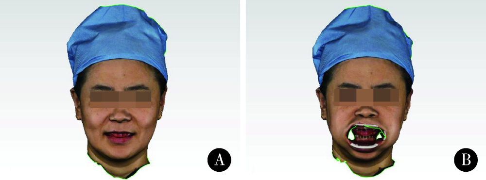
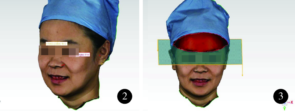
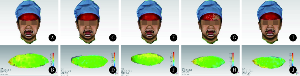
目的: 比较无牙颌种植修复设计三维面部扫描配准的5种方法的配准精度,探究适宜的配准方法。方法: 依据标准共纳入10名受试者,每位受试者戴有具有特征标志点的蜡堤。利用FaceScan三维面部扫描仪获取10位拟行无牙颌种植修复患者的自然大笑位和开口器牵拉暴露口内蜡堤的三维面部扫描数据,将扫描数据导入数字化分析软件Geomagic Qualify 2012中,建立局部坐标系,分别利用面部解剖标志点对齐、面部不动区域对齐、面部点对齐与区域对齐结合、增加面部特征点对齐、增加面部和口内特征标记对齐5种方法将两个面部扫描数据重合,计算同一选定区域的三维偏差,三维偏差越小代表配准精度越高。采用SPSS22.0软件进行统计学分析,面部解剖标志点对齐、面部不动区域对齐、面部点对齐与区域对齐结合3组间差异进行Frideman检验,是否增加面部特征点对齐和是否增加口内特征标记对齐的两组进行配对t检验比较。结果: 直接选择面部解剖标志点对齐[(1.501 2±0.406 1) mm],面上1/3选中区域三维偏差平均值显著大于面部不动区域对齐[(0.629 1±0.150 6) mm]及两种方法相结合[(0.603 5±0.493 4) mm]的偏差(P<0.001);增加面部特征点可显著减小配准后偏差(t=1.001 3, P<0.001),其余组间差异无统计学意义。结论: 面上1/3不动区域应用于无牙颌种植修复设计的三维面部扫描配准是临床可行的,面部扫描操作时可尽量暴露前额区域,增加面部特征点,并利用面部不动区域最佳拟合对齐提高配准精度。
中图分类号:
- R783.4


| [1] |
Wong JY, Oh AK, Ohta E, et al. Validity and reliability of craniofacial anthropometric measurement of 3D digital photogrammetric images[J]. Cleft Palate Craniofac J, 2008,45(3):232-239.
doi: 10.1597/06-175 pmid: 18452351 |
| [2] |
Knoops PG, Beaumont CA, Borghi A, et al. Comparison of three-dimensional scanner systems for craniomaxillofacial imaging[J]. J Plast Reconstr Aesthet Surg, 2017,70(4):441-449.
doi: 10.1016/j.bjps.2016.12.015 pmid: 28161205 |
| [3] |
Artopoulos A, Buytaert JA, Dirckx JJ, et al. Comparison of the accuracy of digital stereophotogrammetry and projection moiré profilometry for three-dimensional imaging of the face[J]. Int J Oral Maxillofac Surg, 2014,43(5):654-662.
doi: 10.1016/j.ijom.2013.10.005 pmid: 24225265 |
| [4] |
Secher JJ, Darvann TA, Pinholt EM. Accuracy and reproducibility of the DAVID SLS-2 scanner in three-dimensional facial imaging[J]. J Craniomaxillofac Surg, 2017,45(10):1662-1670.
doi: 10.1016/j.jcms.2017.07.006 pmid: 28847623 |
| [5] |
Sforza C, de Menezes M, Ferrario V. Soft- and hard-tissue facial anthropometry in three dimensions: what’s new[J]. J Anthropol Sci, 2013,91:159-184.
doi: 10.4436/JASS.91007 |
| [6] |
Zhao YJ, Xiong YX, Wang Y. Three-dimensional accuracy of facial scan for facial deformities in clinics: a new evaluation method for facial scanner accuracy[J]. PLoS One, 2017,12(1):e0169402.
doi: 10.1371/journal.pone.0169402 pmid: 28056044 |
| [7] |
Jang KS, Bayome M, Park JH, et al. A three-dimensional photogrammetric analysis of the facial esthetics of the miss Korea pageant contestants[J]. Korean J Orthod, 2017,47(2):87-99.
doi: 10.4041/kjod.2017.47.2.87 pmid: 28337418 |
| [8] | 刘云松, 叶红强, 谷明, 等. 患者参与的数字化设计在前牙美学修复中的应用[J]. 北京大学学报(医学版), 2014,46(1):90-94. |
| [9] |
Hassan B, Gimenez Gonzalez B, Tahmaseb A, et al. A digital approach integrating facial scanning in a CAD-CAM workflow for complete-mouth implant-supported rehabilitation of patients with edentulism: A pilot clinical study[J]. J Prosthet Dent, 2017,117(4):486-492.
doi: 10.1016/j.prosdent.2016.07.033 pmid: 27881321 |
| [10] |
Coachman C, Calamita MA, Coachman FG, et al. Facially generated and cephalometric guided 3D digital design for complete mouth implant rehabilitation: a clinical report[J]. J Prosthet Dent, 2017,117(5):577-586.
doi: 10.1016/j.prosdent.2016.09.005 pmid: 27836143 |
| [11] |
Hassan B, Greven M, Wismeijer D. Integrating 3D facial scanning in a digital workflow to CAD/CAM design and fabricate complete dentures for immediate total mouth rehabilitation[J]. J Adv Prosthodont, 2017,9(5):381-386.
doi: 10.4047/jap.2017.9.5.381 pmid: 29142646 |
| [12] |
Ritschl LM, Wolff KD, Erben P, et al. Simultaneous, radiation-free registration of the dentoalveolar position and the face by combining 3D photography with a portable scanner and impression-taking[J]. Head Face Med, 2019,15(1):28.
doi: 10.1186/s13005-019-0212-x pmid: 31767030 |
| [13] |
Bohner L, Gamba DD, Hanisch M, et al. Accuracy of digital technologies for the scanning of facial, skeletal, and intraoral tissues: a systematic review[J]. J Prosthet Dent, 2019,121(2):246-251.
doi: 10.1016/j.prosdent.2018.01.015 pmid: 30017156 |
| [14] | 赵一姣, 熊玉雪, 杨慧芳, 等. 2种三维颜面部扫描仪测量精度的定量评价[J]. 实用口腔医学杂志, 2016,32(1):37-42. |
| [15] | 苏莉, 王红梅, 白玉兴. 基于激光扫描的面部软组织三维模型的重叠和分析方法的建立[J]. 北京口腔医学, 2015,23(3):135-140. |
| [16] |
熊玉雪, 杨慧芳, 赵一姣, 等. 两种评价面部三维表面数据不对称度方法的比较[J]. 北京大学学报(医学版), 2015,47(2):340-343.
doi: 10.3969/j.issn.1671-167X.2015.02.030 |
| [17] | Pérez-Giugovaz MG, Park SH, Revilla-León M. Three-dimen-sional virtual representation by superimposing facial and intraoral digital scans with an additively manufactured intraoral scan body [J/OL]. J Prosthet Dent, 2020[2020-09-25]. https://doi.org/10.1016/j.prosdent.2020.07.012. |
| [18] | Lepidi L, Galli M, Grammatica A, et al. Indirect digital workflow for virtual cross-mounting of fixed implant-supported prostheses to create a 3d virtual patient [J/OL]. J Prosthodont, 2020 [2020-8-31]. https://doi.org/10.1111/jopr.13247. |
| [19] | 彭菊香, 江久汇, 赵一姣, 等. 结构光扫描对骨性Ⅲ类错牙合正畸正颌联合治疗前后软组织三维变化的初步评价[J], 北京大学学报(医学版), 2015,47(1):98-103. |
| [20] |
Germec-Cakan D, Canter HI, Nur B, et al. Comparison of facial soft tissue measurements on three-dimensional images and models obtained with different methods[J]. J Craniofac Surg, 2010,21(5):1393-1399.
doi: 10.1097/SCS.0b013e3181ec6976 pmid: 20856027 |
| [21] |
Hajeer MY, Ayoub AF, Millett DT, et al. Three-dimensional imaging in orthognathic surgery: the clinical application of a new method[J]. Int J Adult Orthodon Orthognath Surg, 2002,17(4):318-330.
pmid: 12593004 |
| [22] | 苏莉, 曹丽, 龚宇田. 基于激光扫描的面部软组织三维模型三种重叠方法的对比[J], 北京口腔医学, 2018,26(1):33-36. |
| [23] |
Revilla-León M, Campbell HE, Meyer MJ, et al. Esthetic dental perception comparisons between 2D- and 3D-simulated dental discrepancies[J]. J Prosthet Dent, 2020[2020-01-22]. https://doi.org/10.1016/j.prosdent.2019.11.015.
doi: 10.1016/j.prosdent.2020.09.060 pmid: 33455728 |
| [24] |
Ghoddousi H, Edler R, Haers P, et al. Comparison of three methods of facial measurement[J]. Int J Oral Maxillofac Surg, 2007,36(3):250-258.
doi: 10.1016/j.ijom.2006.10.001 pmid: 17113754 |
| [25] |
Knoops PG, Beaumont CA, Borghi A, et al. Comparison of three-dimensional scanner systems for craniomaxillofacial imaging[J]. J Plast Reconstr Aesthet Surg, 2017,70(4):441-449.
doi: 10.1016/j.bjps.2016.12.015 pmid: 28161205 |
| [1] | 白晓强, 袁芷若, 周永胜, 吕珑薇. 动态牵张促进人骨髓间充质干细胞三维培养的成骨分化[J]. 北京大学学报(医学版), 2026, 58(3): 641-649. |
| [2] | 刘嘉昱, 祝宁, 张育祯, 高贤明, 张宇. 动态导航辅助环钻取骨的准确性[J]. 北京大学学报(医学版), 2026, 58(2): 365-371. |
| [3] | 杨咏涛, 田淯文, 单珅瑶, 李文博, 商相宜, 王艺蓁, 郭殊玮, 高梓翔, 温奥楠, 赵一姣, 王勇. 基于多视图立体视觉的无牙颌种植固定修复软组织数字印模的方法[J]. 北京大学学报(医学版), 2026, 58(1): 126-132. |
| [4] | 刁畅, 王时敏, 李曼, 潘韶霞, 刘洋. 牙列缺失种植覆盖义齿集中𬌗型的临床研究[J]. 北京大学学报(医学版), 2026, 58(1): 133-138. |
| [5] | 温奥楠, 张晓会, 杨咏涛, 高梓翔, 李文博, 单珅瑶, 商相宜, 田淯文, 郭殊玮, 王艺蓁, 王勇, 赵一姣. 基于非刚性配准构建三维颜面微笑仿真序列数据的方法[J]. 北京大学学报(医学版), 2026, 58(1): 139-144. |
| [6] | 孙菲, 王翠, 李思琪, 危伊萍, 余日月, 胡文杰. 赤藓糖醇喷砂与超声治疗对种植体周黏膜炎疗效的随机对照临床研究[J]. 北京大学学报(医学版), 2026, 58(1): 37-42. |
| [7] | 邵梁, 马雯洁, 陈莹, 丁茜, 张磊. 上颌切牙前伸和正中咬合接触解剖特征的数字化测量与分析[J]. 北京大学学报(医学版), 2026, 58(1): 99-106. |
| [8] | 王翠萍, 陈哲, 程永静. 超微血流成像评估与膝骨关节炎临床症状的关联研究[J]. 北京大学学报(医学版), 2025, 57(6): 1096-1100. |
| [9] | 王宇蓝, 曾浩, 张玉峰. 口腔种植中血浆基质的临床转化现状与前沿探索[J]. 北京大学学报(医学版), 2025, 57(5): 836-840. |
| [10] | 于子杨, 郭厚佐, 蒋析, 韩玮华, 林野. 穿颧种植体上颌窦段成骨的影像学研究[J]. 北京大学学报(医学版), 2025, 57(5): 967-974. |
| [11] | 宋凤岐, 徐心雨, 刘筱菁, 李自力. 上颌骨前部和整体顺时针旋转改善骨性Ⅲ类牙颌面畸形患者鼻旁凹陷的对比[J]. 北京大学学报(医学版), 2025, 57(5): 980-988. |
| [12] | 肖宇嘉, 毛渤淳, 周彦恒. 姿势性微笑的三维形态学研究[J]. 北京大学学报(医学版), 2025, 57(5): 989-995. |
| [13] | 王泽远, 于栓宝, 郑浩轲, 陶金, 范雅峰, 张雪培. 基于临床特征和多参数MRI的前列腺癌盆腔淋巴结转移的术前预测模型[J]. 北京大学学报(医学版), 2025, 57(4): 684-691. |
| [14] | 宁圆, 张晓盈, 李雪, 李原, 何菁, 金月波. 干燥综合征并发乳腺淋巴瘤1例[J]. 北京大学学报(医学版), 2025, 57(4): 808-811. |
| [15] | 孙建军, 马千权, 尹晓亮, 杨辰龙, 张嘉, 陈素华, 吴超, 谢京城, 韩芸峰, 林国中, 司雨, 杨军, 邬海博, 赵强. 任意维度重建磁共振对骶管囊肿进行精准分型对于指导微创手术和康复的意义[J]. 北京大学学报(医学版), 2025, 57(2): 303-308. |
|
||
